4954
Open-Source Simulator for Spatial Encoding Effects in Highly Variable B0 and Gradient Fields1Bernard and Irene Schwartz Center for Biomedical Imaging, Department of Radiology, New York University Grossman School of Medicine, New York, NY, United States, 2Center for Advanced Imaging Innovation and Research (CAI2R), Department of Radiology, New York University Grossman School of Medicine, New York, NY, United States, 3Vilcek Institute of Graduate Biomedical Sciences, New York University Grossman School of Medicine, New York, NY, United States
Synopsis
Keywords: Hybrid & Novel Systems Technology, Simulations, spatial encoding
Motivation: Design of accessible scanners may require accounting for substantial magnetic field inhomogeneities, which challenge assumptions used in MRI simulators.
Goal(s): To capture the encoding effects of strong field variations accurately and efficiently, predicting distortions of input images.
Approach: We discretized magnetic fields as usual but extended the MR signal simulation at each grid point from the 0th-order approximation, which assumes a locally-constant field, to a 1st-order approximation, which assumes a locally-linear field.
Results: The 1st-order approximation, which has an analytic solution, better captures strongly varying fields and enables simulations on a coarser grid, greatly improving computational efficiency.
Impact: The simulator enables evaluation of scanner designs with strongly varying magnetic fields. Simulated images can be utilized to generate training data efficiently from existing ground-truth images, enabling exploration of machine-learning techniques prior to the construction of prototype systems.
Introduction
The surge of interest in accessible MRI has driven the development of cost-effective magnet designs1,2. The magnet and gradients are major drivers of a system’s cost and footprint, and, to reduce costs or increase openness, one often sacrifices B0 homogeneity and gradient linearity, leading to challenges in spatial encoding. Therefore, there is a need to evaluate the feasibility of non-traditional systems in silico. We describe the development of an open-source simulation platform that (1) captures strong variations of magnetic fields (2) uses existing, high-quality MR images as input, thus enabling the development of large datasets in populations of actual subjects and proactive evaluation of machine-learning applications for accessible MRI.Methods
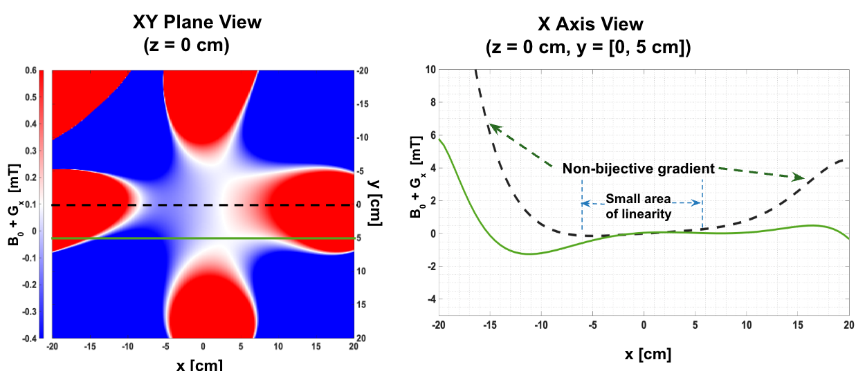
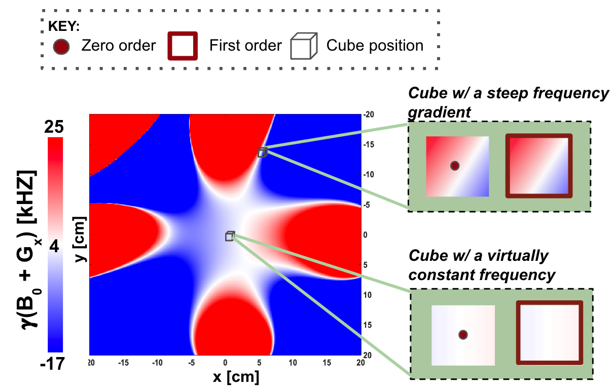
Generally, the MR signal at a timepoint is calculated by an integral of phased transverse magnetization over space. This integral becomes numerically challenging when highly oscillatory complex phases are induced by nonuniform magnetic fields and/or nonlinear gradients3. Therefore, MRI simulators measure the total signal by simulating and summing spin dynamics on a discretized grid over the object and magnetic field4-7. Commonly, the magnetic fields are treated as locally-constant, which is an adequate approximation if the grid resolution is finer than the imaging resolution and, consequently, the magnetic field variations. In the presence of strong B0 inhomogeneities, this relation breaks down and we observe field variations on a scale much finer than the imaging resolution. To combat this, we propose a 1st-order approximation that assumes the magnetic fields to be locally-linear instead of locally-constant (Figure 2). We were able to derive an analytic solution to the integral over a grid-cube assuming: (1) a locally-constant object (2) locally-linear magnetic fields (3) a Gaussian slice profile (4) a perfectly calibrated B1+ field.We demonstrate our simulation platform with the field configuration shown in Figure 1 with a main magnetic field strength of 99mT. The underlying field design results from simulations that (1) prioritize homogeneity over a 5cm sphere at the expense of strong B0 gradients outside of this sphere (2) contain non-linear and non-bijective imaging gradients (Figure 1).
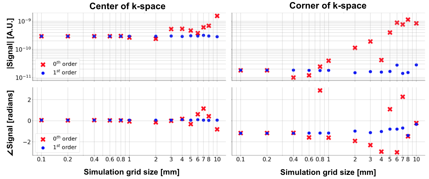
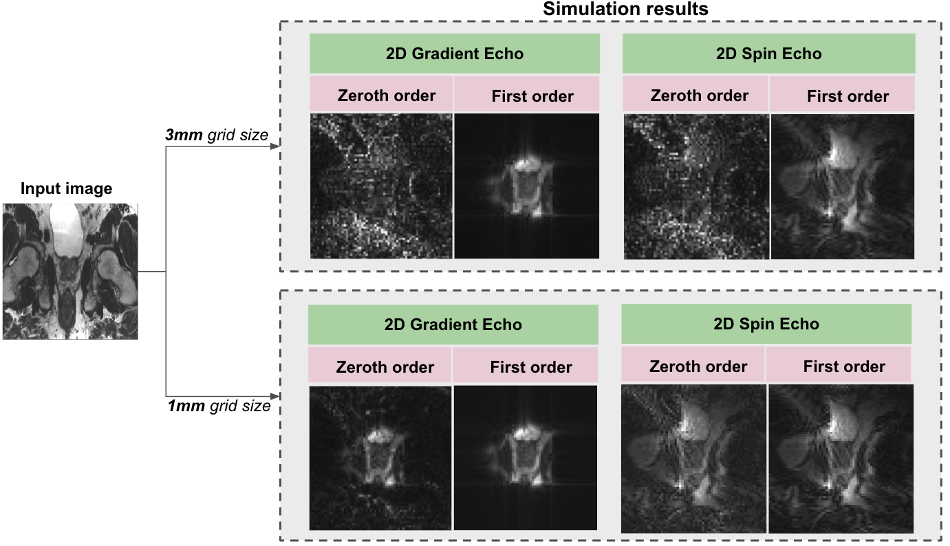
We use clinical standard 3T prostate MR images as inputs to the simulator. Treating the signal intensity as a de-facto spin density at each spatial position, we then calculate MR signal at all k-space points of single-slice gradient- and spin-echo experiments with 3mm imaging resolution, using a simulation grid size of 1mm and 3mm. To compare the effects of 0th and 1st-order approximations, we also calculated the MR signal in both approximations for two k-space points, with grid resolutions ranging from 0.1-10mm.
Results
Figure 3 shows that the k-space signal converges to the same values when approaching a simulation grid resolution of 0.1mm with either approximation. However, the 0th-order approximation exhibits substantial inaccuracies at resolutions of 0.4mm and larger, while the 1st-order approximation has a relative error below 10.8% up to a grid size of 3mm.Figure 4 demonstrates that the image from the 0th-order approximation results in noise-like image artifacts when using a 1mm grid resolution which are further exacerbated at 3mm. In contrast, the 1st-order approximation image is virtually identical at both resolutions, indicating that it is sufficient to simulate on a grid equal to the imaging resolution.
Figure 4 also shows that the field configuration results in outer-volume suppression with a gradient-echo sequence, whereas the spin-echo sequence refocuses the outer-volume signal, which combined with the non-bijective gradients, causes substantial aliasing artifacts.
Discussion and Conclusion
In this work, we expand established MRI simulation concepts in order to capture strong magnetic field variations.This simulator can be used to (1) test and compare different magnet and gradient designs (2) design specialized pulse sequences for specific hardware configurations, and our gradient-echo vs spin-echo comparison provides an example of counterintuitive results emerging from an uncommon hardware design (3) degrade existing clinical MR images and explore machine-learning approaches for the reconstruction, processing, and interpretation of low-cost MR images8.
Our simulator assumes a fixed magnetization magnitude at each point in space (a pre-defined contrast), and accumulates phase during the imaging process, captured on a sub-voxel resolution by a locally-linear field approximation, therefore accounting for concomitant fields and refocusing pulses. Changes in contrast due to field-strength dependent relaxation effects are not captured here.
To derive an analytic solution of the local integral, we assumed a Gaussian RF pulse and perfectly calibrated B1+ field for 2D imaging. However, our general framework also allows simulation of 3D imaging with non-selective RF pulses and a variable B1+. Future work will include a comparison of multi-slice 2D vs. 3D imaging and an extension to diffusion encoding.
Acknowledgements
The work reported in this publication was supported by the Center for Advanced Imaging Innovation and Research (CAI2R), a National Center for Biomedical Imaging and Bioengineering operated by NYU Langone Health and funded by the National Institute of Biomedical Imaging and Bioengineering through award P41EB017183.References
1. Arnold, T. C., Freeman, C. W., Litt, B. & Stein, J. M. Low-field MRI: Clinical promise and challenges. J Magn Reson Imaging 57, 25-44 (2023). https://doi.org:10.1002/jmri.28408
2. Marques, J. P., Simonis, F. F. J. & Webb, A. G. Low-field MRI: An MR physics perspective. J Magn Reson Imaging 49, 1528-1542 (2019). https://doi.org:10.1002/jmri.26637
3. Olver, S. On the quadrature of multivariate highly oscillatory integrals over non-polytope domains. Numerische Mathematik 103, 643-665 (2006).
4. Stocker, T., Vahedipour, K., Pflugfelder, D. & Shah, N. J. High-performance computing MRI simulations. Magn Reson Med 64, 186-193 (2010). https://doi.org:10.1002/mrm.22406
5. Xanthis, C. G. & Aletras, A. H. coreMRI: A high-performance, publicly available MR simulation platform on the cloud. PLoS One 14, e0216594 (2019). https://doi.org:10.1371/journal.pone.0216594
6. Liu, F., Velikina, J. V., Block, W. F., Kijowski, R. & Samsonov, A. A. Fast Realistic MRI Simulations Based on Generalized Multi-Pool Exchange Tissue Model. IEEE Trans Med Imaging 36, 527-537 (2017). https://doi.org:10.1109/TMI.2016.2620961
7. Benoit-Cattin, H., Collewet, G., Belaroussi, B., Saint-Jalmes, H. & Odet, C. The SIMRI project: a versatile and interactive MRI simulator. J Magn Reson 173, 97-115 (2005). https://doi.org:10.1016/j.jmr.2004.09.027
8. Arnold, T. C., Baldassano, S. N., Litt, B. & Stein, J. M. Simulated diagnostic performance of low-field MRI: Harnessing open-access datasets to evaluate novel devices. Magn Reson Imaging 87, 67-76 (2022). https://doi.org:10.1016/j.mri.2021.12.007
9. Campbell-Washburn, A. E. et al. Low-field MRI: A report on the 2022 ISMRM workshop. Magn Reson Med 90, 1682-1694 (2023). https://doi.org:10.1002/mrm.29743
10. Kabil, J. et al. A Review of Numerical Simulation and Analytical Modeling for Medical Devices Safety in MRI. Yearb Med Inform, 152-158 (2016). https://doi.org:10.15265/IY-2016-016
Figures