4952
Utilizing DLP 3D Printing for MRI-Visible Phantoms in Biomedical Applications1Oncology-Pathology, Karolinska Institute, Stockholm, Sweden
Synopsis
Keywords: Phantoms, Interventional Devices, Rapid prototyping, medical devices, phantoms, biopsy
Motivation: This study is motivated by the authors to easily, and cheaply, manufacture phantoms and other accessories that is inherently dimensionally accurate, MRI compatible, and visible in MRI images.
Goal(s): The goal of the study is to develop a manufacturing method to create a MRI-visible localization grid for MR-guided breast biopsies.
Approach: By hollowing out the part without inclusions of any drainage holes, we can ensure liquid resin is trapped inside the part during the printing process.
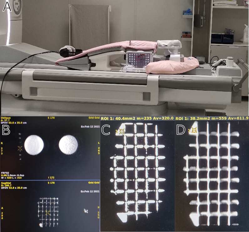
Results: The finished part is shown to be inherently MRI visible as demonstrated on both T1- and T2-weighted images from our MRI biopsy protocol.
Impact: This approach offers an efficient and cost-effective solution for creating MRI-visible objects with high spatial accuracy, which is useful for producing phantoms and other MRI compatible accessories.
Introduction
Digital Light Processing (DLP), a type of 3D printing technology utilizing UV-hardened resin plastics, has gained prominence due to its ability to produce finely detailed objects with inherent liquid-tight properties[1]–[3]. In DLP printing, liquid UV-hardened resin is poured into a vat with a transparent bottom, cured using UV light exposure, and lifted layer by layer to construct the object. DLP technology's accuracy and ability to replicate intricate details make it suitable for printing MRI phantoms. Drainage holes are typically incorporated into the objects to allow unhardened resin to escape during the printing process. If this step is omitted, liquid resin can be trapped inside, making it visible in MRI scans. Leveraging this phenomenon, along with DLP's high precision, this paper presents a cost-effective and efficient method for producing MRI-visible objects with minimal spatial tolerance. One crucial application of this approach is the creation of MRI-visible grid systems for MR-guided breast biopsies. MR-guided biopsies rely on MRI scans for locating the biopsy target, and a grid system made of hard plastic assists in spatial localization. However, the conventional grid, being MRI-invisible, necessitates tight skin contact, which is not always achievable due to breast tissue characteristics and grid design limitations. The inability to establish precise correlation between the biopsy target and the grid can lead to procedure cancellations, which is far from ideal considering that MR-guided biopsies are typically performed as a last resort when other diagnostic methods prove insufficient[4].Purpose
This study focuses on designing, manufacturing, and evaluating the feasibility of using DLP 3D printing technology to create an MRI-visible localization grid for MR-guided breast biopsies.Materials and Methods
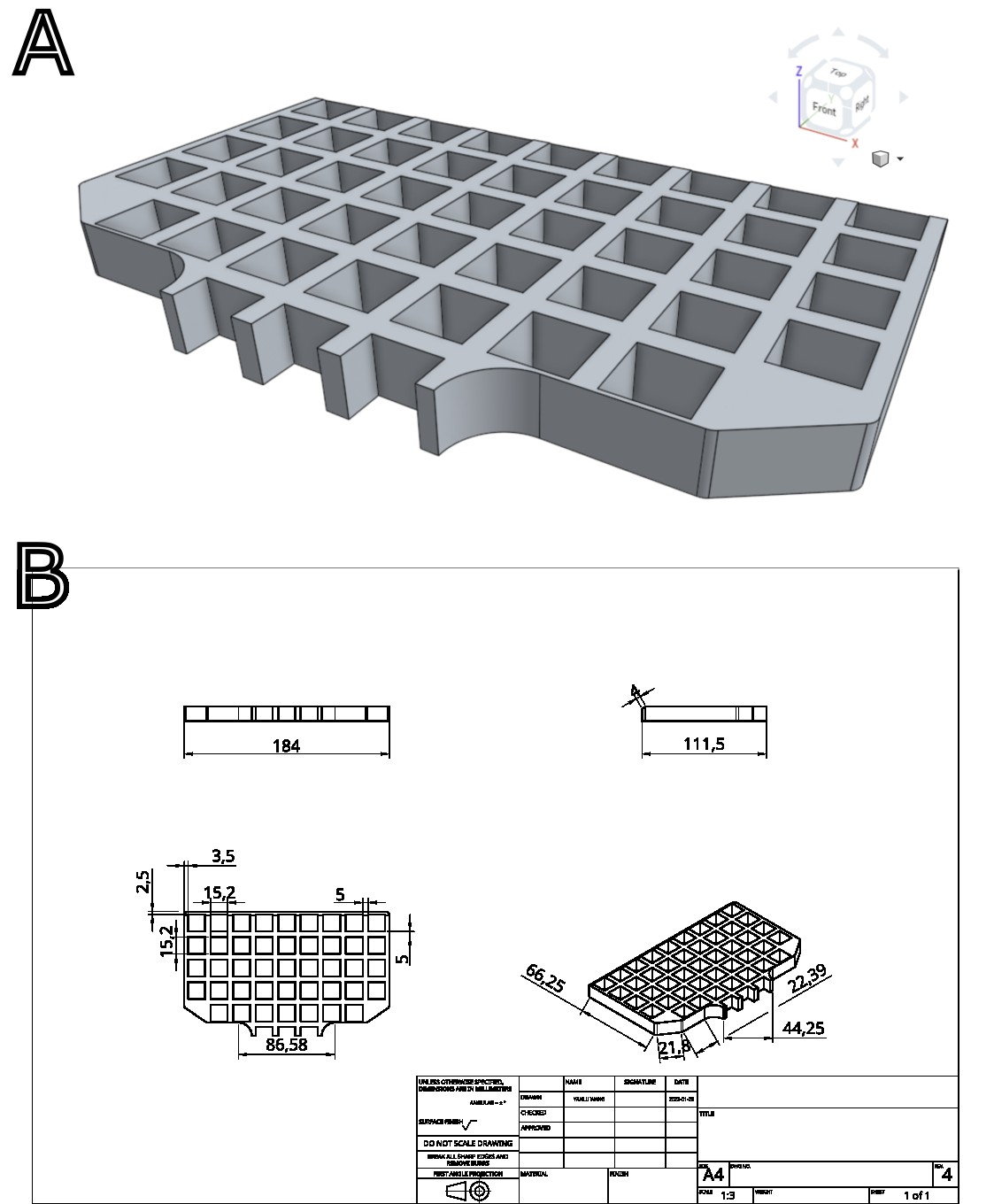
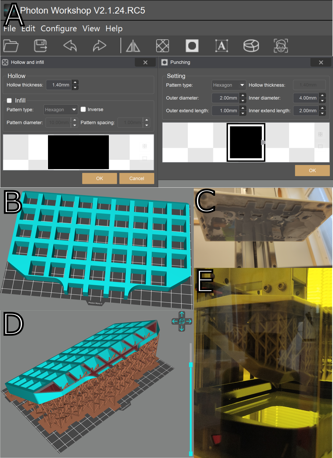
The study employed Digital Light Processing (DLP) 3D printing technology (Anycubic Mono) to create MRI-visible phantoms, with a focus on an MRI-visible grid system for MR-guided breast biopsies. The design of the grid was established using 3D CAD software (Onshape, http://www.onshape.com), ensuring a 1.4mm-thick outer shell to contain liquid resin (Fig. 1). Anycubic's proprietary slicer software (Photon Workshop, v2.1.24), was used to prepare the 3D model for printing, with a critical emphasis on hollowing the part where MR visibility is desired, lack of drainage holes, and adjusting Z-lift height such that the unfinished part never lifts above liquid resin levels in the vat to ensure maximum amount of liquid resin retention inside the finished part (Fig. 2). Post-production steps included the removal of residue liquid resin and UV curing as per standard procedure. The MRI visibility of the grid was evaluated using T1- and T2-weighted MRI sequences used in our MR-guided breast biopsy protocol.Results
We demonstrate the successful use of Digital Light Processing (DLP) 3D printing technology to create MRI-visible phantoms, particularly an MRI-visible grid system for MR-guided breast biopsies. The MRI-visible grid was designed and manufactured with a 1.4mm-thick outer shell to contain UV-hardened resin, ensuring the object's liquid-tight properties. By printing large surfaces at an angle, the printing process's robustness was improved. The finished part was fitted onto our existing MR-guided biopsy setup and the MRI visibility of the grid was confirmed in T1- and T2-weighted MRI sequences in the biopsy MRI protocol (Fig. 3).Discussion
This approach offers an efficient and cost-effective solution for creating MRI-visible objects with high spatial accuracy, which can significantly enhance the accuracy and confidence of MR-guided biopsy procedures. There exist several unique challenges posed by the application of DLP 3D printing technology to create MRI-visible phantoms. Firstly, the necessity of constructing designs that allow for MRI-visible sections to be encased within a hardened outer shell of considerable thickness to ensure the phantom's durability. Tougher materials such as “ABS-like” or engineering resin is preferred as the potential for fractures resulting in resin leakages in parts produced using conventional UV-hardened resins is high. Depending on material color, some background UV light shining through the object and slowly curing the liquid resin inside parts is inevitable, hence long-term MR visibility is questionable. Long-term observations have not yet been thoroughly conducted. Ultimately, the study demonstrated the potential of this method for creating MRI-visible phantoms and markers for biomedical applications.Conclusion
In conclusion, this study highlights the successful application of DLP 3D printing technology in creating dimensionally accurate MRI-visible phantoms for use in MR-guided biopsies. This innovative approach simplifies the spatial localization process, enhancing confidence and accuracy in MR-guided procedures. The method presented here has the potential to revolutionize the creation of MRI-visible objects for a wide range of biomedical applications, offering cost-effective and precise solutions.Acknowledgements
No acknowledgement found.References
[1] M. F. Bieniosek, B. J. Lee, and C. S. Levin, ‘Technical Note: Characterization of custom 3D printed multimodality imaging phantoms’, Med. Phys., vol. 42, no. 10, pp. 5913–5918, 2015, doi: 10.1118/1.4930803.
[2] P. Ramachandran et al., ‘A 3D printed phantom to assess MRI geometric distortion’, Biomed. Phys. Eng. EXPRESS, vol. 7, no. 3, p. 035004, May 2021, doi: 10.1088/2057-1976/abeb7e.
[3] I. A. Tsolakis, W. Papaioannou, E. Papadopoulou, M. Dalampira, and A. I. Tsolakis, ‘Comparison in Terms of Accuracy between DLP and LCD Printing Technology for Dental Model Printing’, Dent. J., vol. 10, no. 10, p. 181, Sep. 2022, doi: 10.3390/dj10100181.
[4] P. Viehweg, A. Heinig, B. Amaya, T. Alberich, M. Laniado, and S. H. Heywang-Köbrunner, ‘MR-guided interventional breast procedures considering vacuum biopsy in particular’, Eur. J. Radiol., vol. 42, no. 1, pp. 32–39, Apr. 2002, doi: 10.1016/s0720-048x(01)00479-x.
Figures