4951
Motion-corrected supine breast MRI using a flexible coil vest and beat pilot tones – preliminary results1High Field MR Center, Center for Medical Physics and Biomedical Engineering, Medical University of Vienna, Vienna, Austria, 2IADI, Université de Lorraine, INSERM U1254, Nancy, France
Synopsis
Keywords: RF Arrays & Systems, Breast
Motivation: This study addresses the challenge of motion artifacts in free-breathing supine breast MRI using a flexible RF coil, aiming to improve image quality and patient comfort.
Goal(s): The primary goal is to demonstrate the feasibility of motion correction using beat pilot tones (BPT) in combination with the GRICS algorithm, requiring no additional on-patient hardware.
Approach: A volunteer underwent supine breast MRI performing various breathing patterns, while BPT motion signals were extracted. The GRICS algorithm was used for retrospective motion correction.
Results: Motion artifacts were effectively reduced for flat abdominal and normal thoracic breathing. However, heavy thoracic breathing artifacts were insufficiently reduced.
Impact: Motion-corrected supine breast MRI with a flexible coil promises improved diagnostic image quality and increased patient comfort. Further optimization of acquisition and motion-correction techniques for clinical supine breast MRI will ultimately aid in early disease detection and management.
Introduction
Flexible multi-channel radio frequency (RF) coils allow for high-resolution breast MRI in a comfortable supine position1. Compared to the standard breast MRI setup with immobilized breasts due to prone positioning on a rigid RF coil, free-breathing supine breast MRI is more susceptible to motion artifacts. In some cases, motion blurring or ghosting results in non-diagnostic image quality. The GRICS2–4 algorithm can reduce these motion artifacts based on signals correlated with subject motion. Pilot tones5 or the more sensitive beat pilot tones (BPT)6–8 acquired concurrently with MRI raw data are attractive for obtaining such motion-correlated time courses without any additional on-patient hardware requirement. The purpose of this work is to provide the proof of principle that motion artifacts during supine breast MRI using a flexible RF coil can be efficiently corrected by combining the BPT approach for motion detection and GRICS algorithm for motion correction.Methods
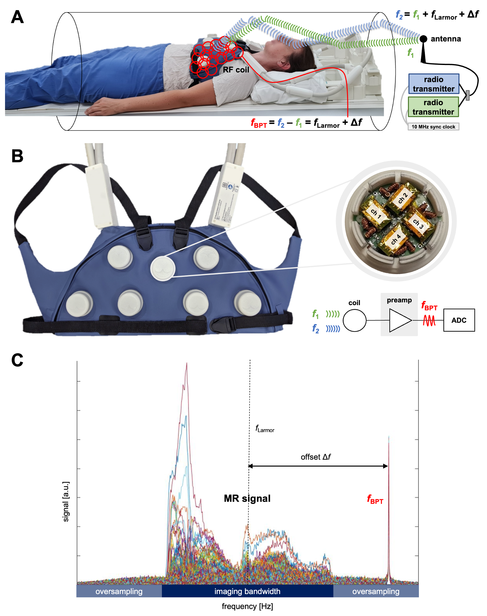
Supine breast MRIOne volunteer (female/26 yrs/bra size 75A) was examined in the supine position using a flexible 28-channel breast RF coil1 (“BraCoil”) at 3 T (Prisma Fit, Siemens Healthineers, Erlangen, Germany). The BraCoil is shown in Fig. 1A+B. Axial T2-weighted TSE data sets were acquired during three different breathing patterns: 1) flat abdominal breathing, 2) normal thoracic breathing, and 3) heavy thoracic breathing, respectively. Sequence parameters are given in Table 1. The study was approved by the Ethics Committee of the Medical University of Vienna (EK2137/2021) and informed written consent was obtained from the volunteer.
Motion detection and correction
The hardware setup is illustrated in Fig. 1A. A commercial Wi-Fi antenna (ANT-5GWWS6-SMA) was positioned at the service end of the MR scanner bore and emitted two signals at frequencies f1=1 GHz (arbitrary choice for preliminary tests) and f2=f1+fLarmor+Δf generated by two software-defined radio (SDR) transmitters (USRP B210). The SDRs were placed outside the scanner room, supplied with the scanner’s 10 MHz system clock and connected to the antenna by a 10 m coaxial cable. Exploiting the non-linearity of the coil preamplifiers, the two signals f1 and f2 produce an intermodulation product at fLarmor + Δf (=f2-f1) which is the BPT frequency fBPT8. On-coil preamplifiers (Microwave Technology, MSM-123281, California, USA) in the BraCoil, allowing the concurrent acquisition of BPT and MR signals are shown in Fig. 1B. Δf was chosen such that the BPT signal would appear outside the imaging bandwidth BWimg but within the acquired bandwidth, as BWimg is oversampled by a factor of 2 by default. BPT signals from all 28 coil channels were extracted from raw data by Fourier transformation along the frequency encoding dimension. The area under the BPT peak was summed for each phase encoding step, resulting in 28 time courses with 11.6 ms time resolution. An example of acquired MR data with the BPT visible in the oversampling bandwidth is shown in Fig. 1C. After low-pass filtering of the BPT signal, retrospective GRICS reconstruction4 for motion correction was performed. The GRICS algorithm assumes the displacement of each pixel to be a product of a static motion model and the motion signal (i.e., the post-processed BPT signal). This allows a joint solution of the motion-corrected MRI reconstruction and a motion model.
Results
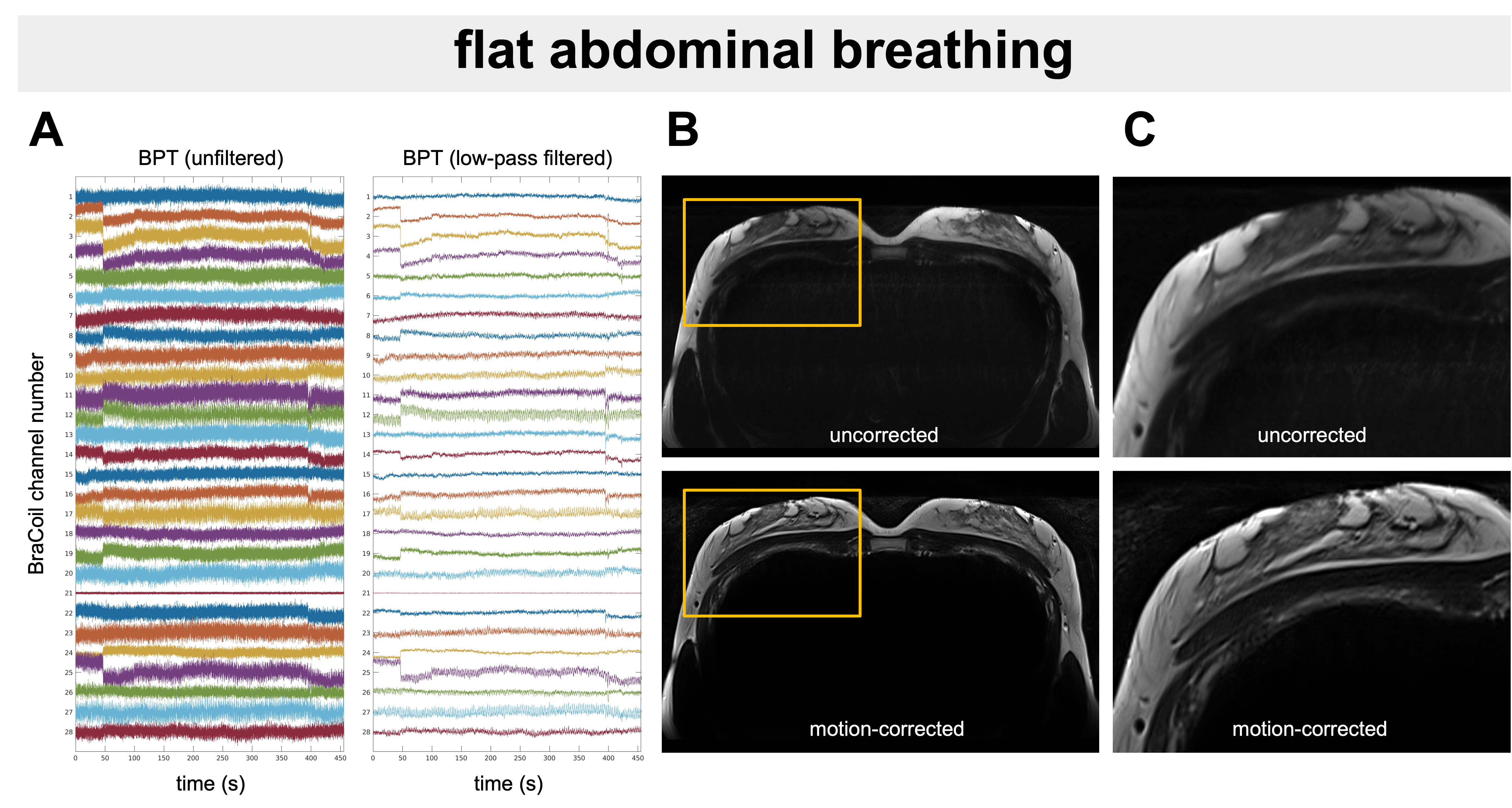
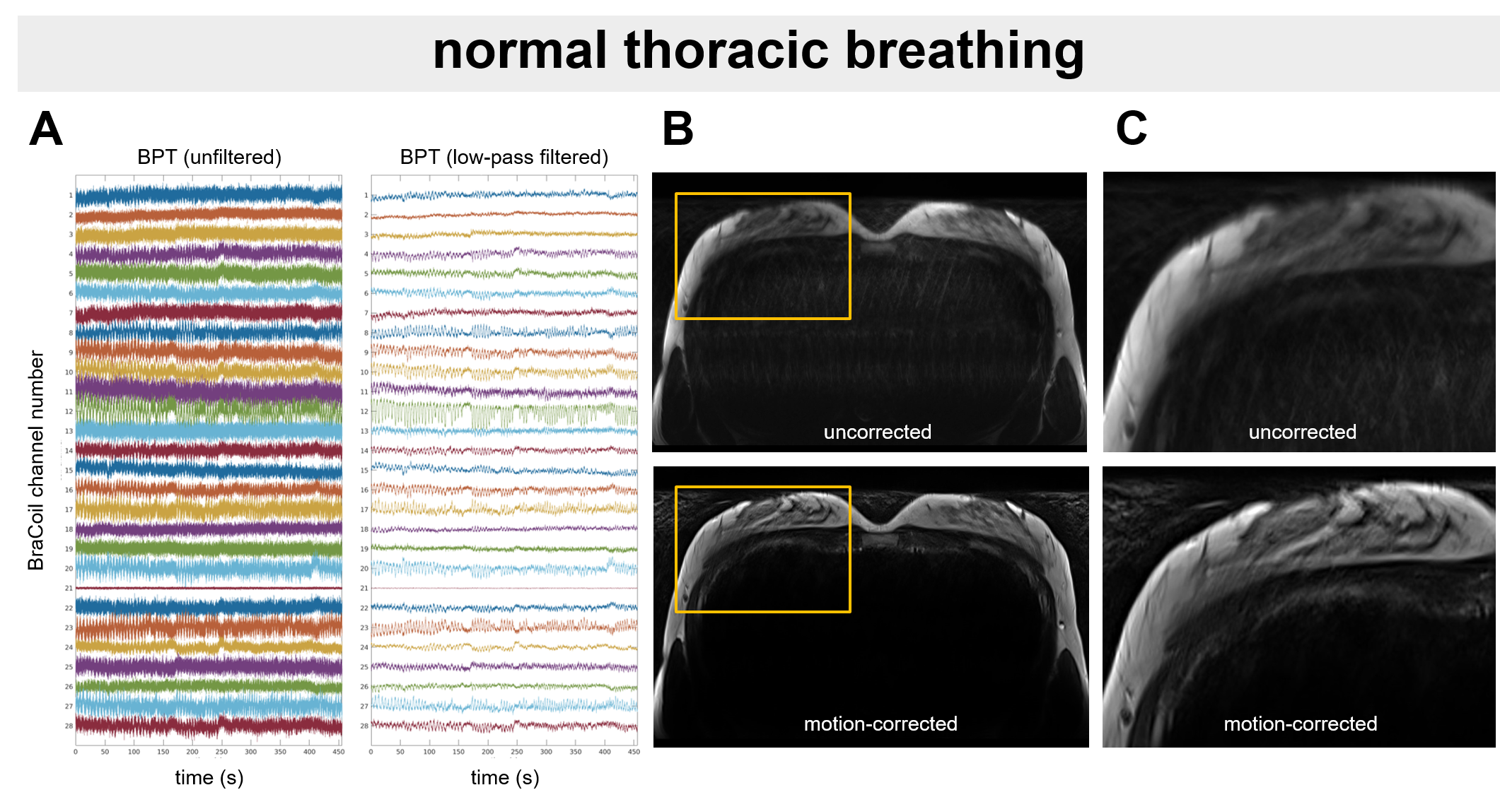
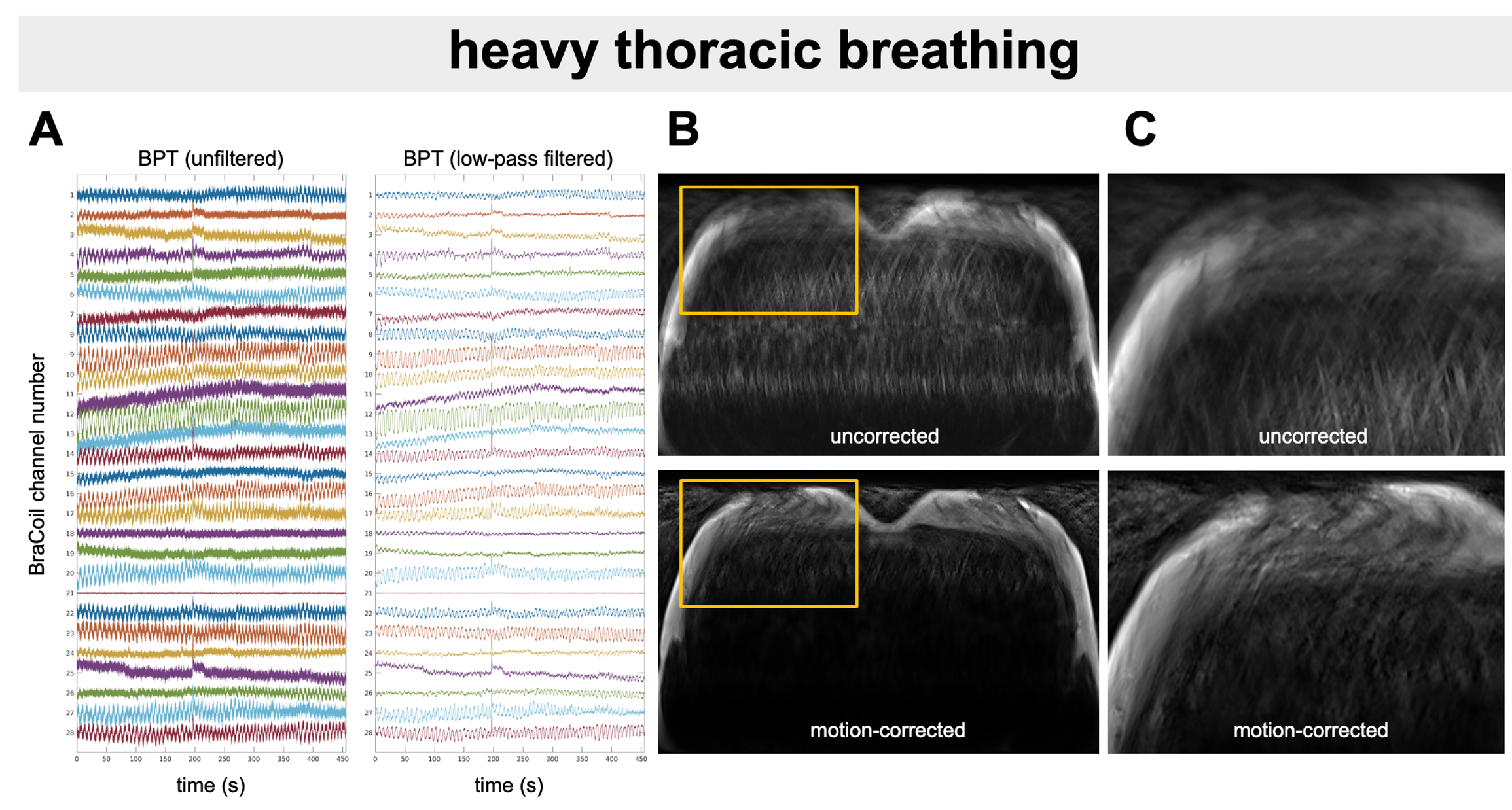
Detected BPT time courses are shown together with supine breast MR images before and after motion correction in Fig. 2 (flat abdominal breathing), Fig. 3 (normal thoracic breathing) and Fig. 4 (heavy thoracic breathing). Artifacts from flat abdominal and normal thoracic breathing could be visibly reduced. It remains challenging to correct for heavy thoracic breathing where the reduction in image blurring is insufficient to yield diagnostic quality.Discussion
From experience, the average breathing pattern usually lies between the instructed normal thoracic and flat abdominal breathing. Presumably, under stress or pain, some patients will perform heavier thoracic breathing. Based on our preliminary results, we therefore assume that for most supine breast MRI cases the proposed motion correction workflow considerably improves image quality. Heavy breathing may lead to increased through-plane motion (spin history artifacts) which cannot be corrected with GRICS-reconstruction of 2D multislice images.In future work, T2-weighted sequences will be accelerated to yield scan times easily feasible in clinical breast MRI protocols and contrast-enhanced T1-weighted images will be included in the motion correction, especially to facilitate lesion conspicuity assessment. Reproducibility tests and setup optimization (antenna position, carrier frequency), will be followed by adaptations of the GRICS algorithm for the proposed workflow.
Conclusion
We have demonstrated the proof of principle for motion-corrected free-breathing supine breast MRI enabled by the combination of a flexible RF coil, beat pilot tones and retrospective GRICS motion correction.Acknowledgements
This work was funded by the joint Austrian/French grant “BRACOIL“ (Austrian Science Fund FWF Nr. I-3618/Agence Nationale de Recherche ANR-17-CE19-0022), Austrian Society for Senology (ÖGS), the Focus Grant “T4MR” from CMPBME MedUni Vienna, and “CITRUS” (Horizon Europe research and innovation programme, grant agreement no 101071008).References
1. Obermann M & Nohava, L. et al. Panoramic magnetic resonance imaging of the breast with a wearable coil vest. Invest. Radiol. 58, 799–810 (2023).2. Odille, F., Vuissoz, P.-A., Marie, P.-Y. & Felblinger, J. Generalized reconstruction by inversion of coupled systems (GRICS) applied to free-breathing MRI. Magn. Reson. Med. 60, 146–157 (2008).
3. Odille, F. et al. Generalized MRI reconstruction including elastic physiological motion and coil sensitivity encoding. Magn. Reson. Med. 59, 1401–1411 (2008).
4. Isaieva, K. et al. Feasibility of online non-rigid motion correction for high-resolution supine breast MRI. Magn. Reson. Med. 90, 2130–2143 (2023).
5. Speier, P. et al. PT-Nav: A novel respiratory navigation method for continuous acquisition based on modulation of a pilot tone on the MR-receiver. in Proc ESMRMB 129:97–98 (2015).
6. Anand, S. & Lustig, M. Beat pilot tone: exploiting preamplifier intermodulation of UHF/SHF RF for improved motion sensitivity over pilot tone navigators. in Proc Intl Soc Mag Reson Med 29 p.568 (2021).
7. Lamar-Bruno, K. Cardiac and Respiratory-Resolved Image Reconstruction with the Beat Pilot Tone. in Proc Intl Soc Mag Reson Med 30 p.4446 (2022).
8. Anand, S. & Lustig, M. Beat Pilot Tone: Versatile, Contact-Free Motion Sensing in MRI with Radio Frequency Intermodulation. arXiv [physics.med-ph] (2023).
Figures