4948
Single port quadrature polarization of two birdcage coils for small animal imaging1Neuroscience Research Institute, Gachon University, Incheon, Korea, Republic of, 2Non-clinical Center, KBIO Osong Medical Innovation Foundation, Chungbuk, Korea, Republic of, 3Department of Health Sciences and Technology, GAIHST, Gachon University, Incheon, Korea, Republic of, 4Department of Biomedical Engineering, Gachon University, Incheon, Korea, Republic of
Synopsis
Keywords: Hybrid & Novel Systems Technology, Prenatal
Motivation: The development of hardware dedicated to pre-clinical applications that can produce high quality images for small animal MRI systems with limited transmission channels.
Goal(s): a double birdcage coil that can deliver uniform field for multiple rats, while having only one transmission channel.
Approach: Employing electromagnetic simulations, we devised a geometric arrangement comprising two birdcage coils with a leg distribution rotated relative to each other. This configuration enables the generation of uniform, high-intensity circularly polarized fields.
Results: Uniform magnetic fields were acquired with double birdcage coil with a single excitation port. These results also showed uniform field and with similar intensity between two rats.
Impact: By employing double birdcage coils with rotated leg distributions, we achieve the generation of uniform and strong circularly polarized magnetic fields. This breakthrough offers significant advancements in pre-clinical applications and has the potential to improve research in small animal MRI.
Introduction
Magnetic resonance imaging (MRI) is a great tool not only for diagnostic and treatment on clinical human patients, but also for pre-clinical analysis on small animals, such as neo-natal rats. Pre-clinical studies on neo-natal rats have been of interest to study the evolution of disease, drug administration and to understand anatomical development [1]. The use of small animals also allows the imaging of multiple subjects at the same time, this results useful for control measurements and comparison between protocols. However, the use of MRI for small animals often requires the use of dedicated systems that have smaller bore compared to the systems used on hospitals. These systems often have hardware limitations in which often a single transmission channel is available, and the most common volume coil used is the birdcage (BC) coil. While the use of a single channel BC coil can be used for linear polarization, the field uniformity and intensity can be greatly improved by using double transmission channels and create a circular polarization. While the use of BC coils the field intensity is focused at the center, the placement of multiple samples would experience different field intensities, thus compromising the image quality and the study at hand. In this work explore the use of double birdcage coils to provide localized and same field pattern and intensity for two neo-natal rats, we propose the combination of two BC coils by modifying the geometry structure to generate circular polarization (CP) by using a single excitation port.Methods
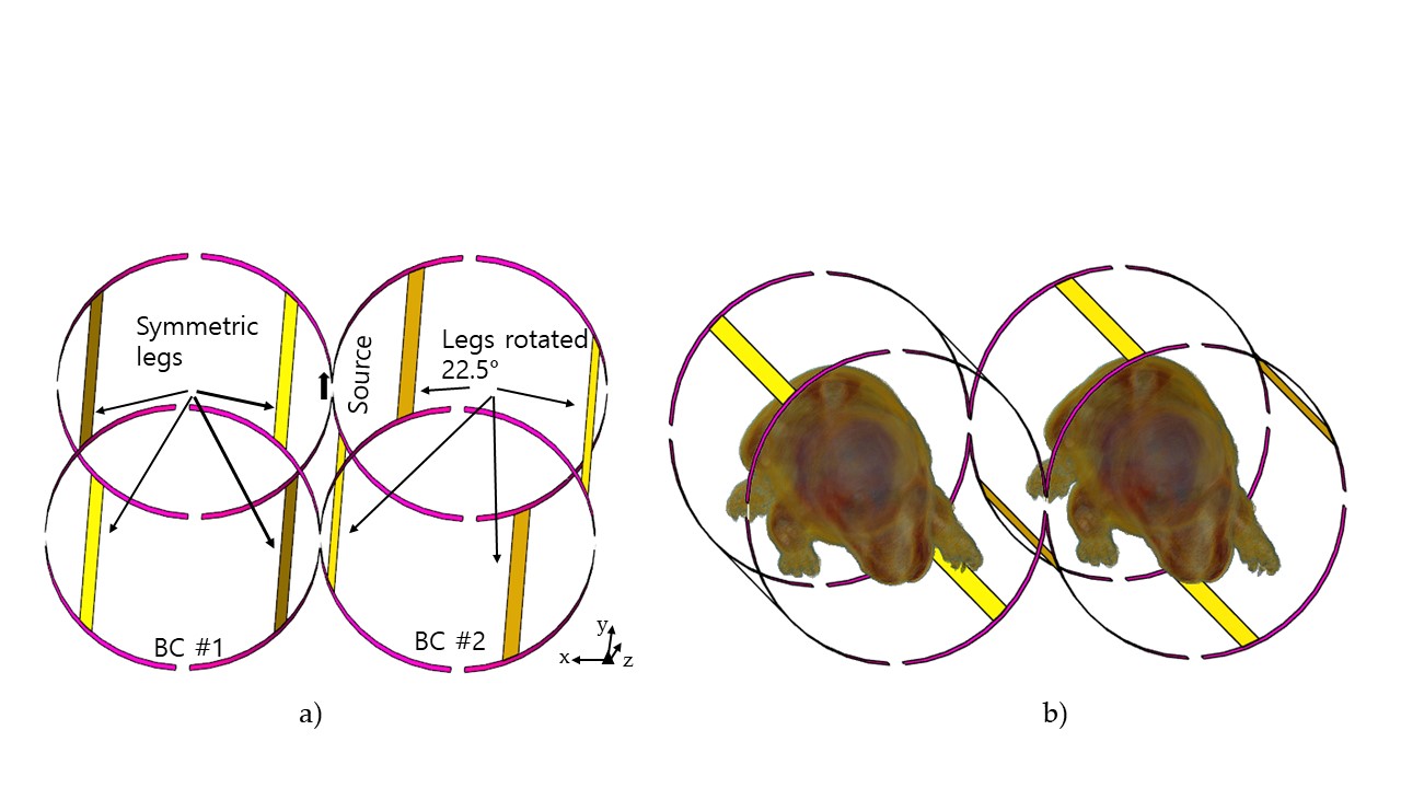
We designed two BC coils based on high-pass type, as shown in Fig. 1a, the radius of the ring was 20mm and length of 72.5 mm, and four legs, the BC coils are connected to next to each other, the source is placed in between the BC coils, and capacitor are used for tuning, the position of the legs of the BC #2 were rotated by 22.5°, this angle was found empirically by rotating the legs and checking the current and B1 field distribution, to have uniform magnetic B1 field, similar to the CP mode. Electromagnetic (EM) simulations were performed by using FDTD software, Sim4life, (Zurich, Switzerland), with an excitation signal of 300 MHz and bandwidth of 600 MHz. The magnetic field was measured within two neo-natal rats [2], of approximate 90 mm length and weight of 14g. The permittivity and conductivity for a total of 82 tissues were automatically assigned according to the values for 300MHz.Results
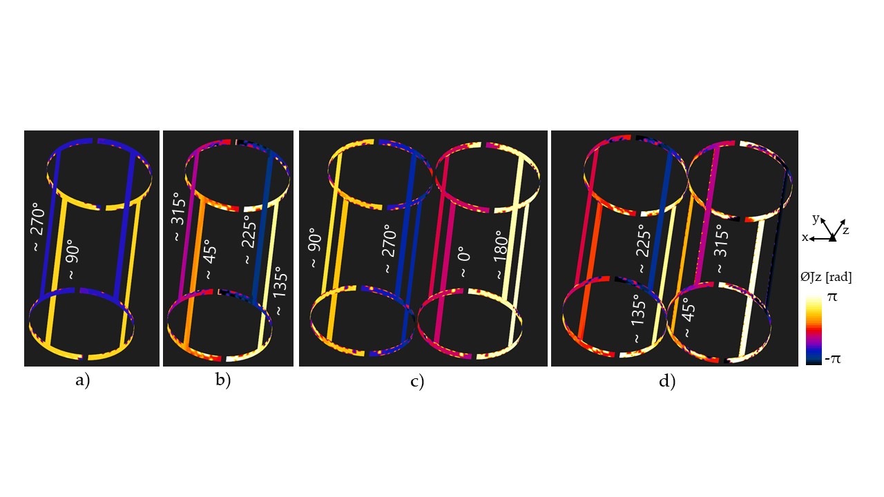
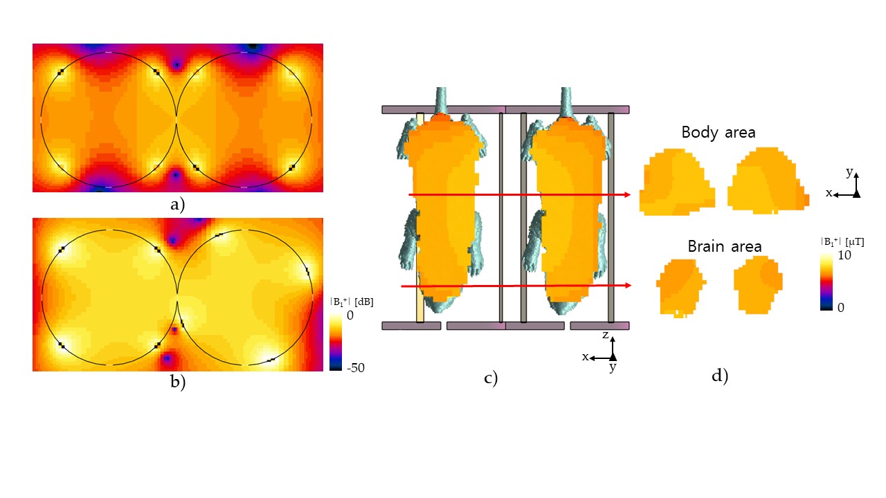
We investigate first the performance of the proposed geometry by analyzing the phase of the surface current density (J) distribution on the conductor lines of the BC coils, in particular the z component (Jz). Figure 2a shows the phase of Jz for the case for when a BC coil is excited with only one port, in which case the phase shows a of 90° and 270°, with a difference of 180°, whereas, the case of a BC coil in CP mode would have a phase Jz as shown in Fig. 2b, with phase of 45, 135, 225 and 315° at each leg, with a phase difference of 90° for each leg. For the case when two BC coils are set together, in the symmetrical geometry, the phase Jz is shown in Fig. 2c, which resembles the linear mode, in which each BC coil also has a 180° difference between left and right. In the proposed coil structure, the legs have been rotated by 22.5° and the corresponding phase Jz is shown in Fig. 2d, which shows also an approximate 90° shift between each leg. Although there are a few degrees offset, it can be solved by further optimal capacitor value tuning.To compare the performance of the transmitted magnetic B1field in empty space, Fig. 3a shows the case for when the two coils have symmetric and same leg position, which clearly shows a linear mode, whereas, in Fig. 3b the B1 field for the rotated legs in BC #2, show a more uniform field focused on the center of each BC coil. We further tested the double BC coil configuration with the neo-natal rat, as shown in Fig. 3c in the z-x plane, and the x-y plane for the brain and body area. The mean value for the B1 field on the brain was 5.8 and 5.6uT and standard deviation of 1.7 and 1.4uT, for BC #1 and BC#2, respectively. The proposed geometry configuration can provide a uniform B1 field for double BC coil, while also deliver a localize and strong field.
Acknowledgements
This research was supported by a grant from the Osong Medical innovation Foundation R and D Project funded by the Republic of Korea’s Ministry of Health and Welfare (No: HI22C1989)References
1) Beckmann, N., Mueggler, T., Allegrini, P.R., Laurent, D. and Rudin, M., 2001. From anatomy to the target: contributions of magnetic resonance imaging to preclinical pharmaceutical research. The Anatomical Record: An Official Publication of the American Association of Anatomists, 265(2), pp.85-100.
2) itis.swiss/vip-animals
Figures