4942
Investigating the robustness of estimating B1+maps by a neural network at different UHF sites.1Physikalisch-Technische Bundesanstalt (PTB), Braunschweig and Berlin, Germany, 2School of Computation, Information and Technology, Technical University of Munich, Munich, Germany, 3Medical Physics in Radiology, German Cancer Research Center (DKFZ), Heidelberg, Germany, 4Faculty of Physics and Astronomy, Heidelberg University, Heidelberg, Germany, Heidelberg, Germany, 5Center for Magnetic Resonance Research, University of Minnesota, Minneapolis, MN, United States, Minneapolis, MN, United States, 6Center for Magnetic Resonance Research, University of Minnesota, Minneapolis, MN, United States, Minneapolis, MN, United States, Minneapolis, Germany
Synopsis
Keywords: High-Field MRI, RF Pulse Design & Fields, High-Field MRI, pTx, B1+ mapping, neural networks
Motivation: B1+ -maps needed for subject-specific pTx can be derived from localizers using a neural network (NN), omitting separate B1+-mapping. Ideally, a single, general NN applicable to all UHF sites is highly desired.
Goal(s): Investigate the robustness of a neural network predicted B1+-field maps. Utilizing receive profiles from an 8Tx/32Rx head coil as neural network input.
Approach: Comparing the performance on data from identical and different commercial head coils across multiple MRI sites.
Results: Achieving SSIMs as high as 96% and RMSEs as low as 2.7%, with error mapping predominantly localizing discrepancies at the cranial margins, suggesting that larger datasets could enhance Gaussian convergence.
Impact: The study suggests that a single NN trained by a large B1+ library for one type of pTx head coil may be disseminated to other UHF sites that use the same coil. This will enable a fast, streamlined pTx calibration.
Introduction
A central challenge in ultra-high field (UHF) MRI is the heterogeneous flip angle (FA), which can be mitigated using parallel transmission (pTx). However, subject-specific pTx requires $$$B^{+}_{1}$$$-mapping of each transmit (Tx) channel, which is usually time-consuming. To omit the necessity for additional B1+ mapping, recent work in high-field MRI of the body1 and the brain2 showed that $$$B^{+}_{1}$$$-maps of 8 Tx channels can be estimated using a neural network (NN) from a single localizer scan that is acquired with 8 or 32 receive (Rx) elements. Since this NN estimates the clip_image004.png"> profiles (Tx) from the B1- (Rx) weighted localizer images, the NN depends on the coil structure. Thus, the question arises if a single NN trained at site A can be used at site B. Therefore, we tested a NN-based B1+ mapping approach using two versions of a commercial 8Tx32Rx head coil (Nova Medical, USA) at three different 7T sites and report initial results of this study.Methods
Absolute, Tx-channel-wise axial $$$B^{+}_{1}$$$-maps were acquired using two 8Tx/32Rx head of the same model coils (Nova Medical, USA) at three UHF sites: A: BUFF Berlin, Germany; B: DKFZ, Heidelberg, Germany; C: CMRR, Minneapolis, USA. Measurements A+B were performed with an older 7T scanner (both Magnetom 7T, Siemens) scanner using an identical coil (coil I). In contrast, at C, the second 8Tx/32Rx head coil (coil II) was used on an upgraded 7T system (Terra Fit, Siemens).The $$$B^{+}_{1}$$$-maps were generated through a hybrid method3, combining 8 Tx-channel-wise low-FA GRE images (TR=100ms, TE=2.9ms, resolution=2x2x4mm3, FOV=256x192mm2, 15 transverse slices) with a single 3D actual flip-angle image (AFI) (TR=75ms, TE=1.9 ms, resolution=2x2x4mm3, FOV=256x192mm2, FA=60°) acquired in CP+ mode. A ninth GRE image with all Tx channels transmitting in CP+ is obtained, which serves as a localizer (c.f. Fig1A).
Localizer and $$$B^{+}_{1}$$$ data of 12 subjects of site A+Coil I were split into real and imaginary components, normalized (c.f.1) and used for training a UNet4 with a symmetric loss function5 (c.f. Fig1B). The NN was validated i) by two further volunteers from Site A+Coil I, ii) three subjects from Site B+ Coil I, and one from Site 3+Coil II. Training was conducted using the ADAM optimizer on a 24 GB NVIDIA Titan RTX: learning rate $$$B^{+}_{1}$$$, 4000 epochs, batch size=2. Ground truth (GT) B1+-maps were compared with the network's predicted B1+-maps (PR) using the structural-similarity-index measure (SSIM) and root-mean-squared-error (RMSE) as evaluation metrics. Voxels with a magnitude error >|±10%| were marked to illustrate high difference regions for the PR.
Results and Discussion
Fig.2 illustrates GT and PR |$$$B^{+}_{1}$$$|-maps for all 8 Tx-channels, their absolute difference, and resulting error map for test data from Sites A-C. Site A&B show qualitatively the best agreement with the GT data. Tx5 shows the largest deviation for Site A&B, while the largest errors are near the skull. In comparison Site C shows larger deviations from GT and more centrally-located errors.Same qualitative observations are evident for the relative phases (c.f. Fig.3). Here, Tx8 shows pronounced deviations for Site A, and Tx5 shows an offset in the phase. Mean absolute phase differences for Sites A&B are (0.014±0.043)rad and (0.019±0.084)rad, whereas (0.029±0.073)rad for Site C.
Quantitative values in Fig.4 support this observation: Mean RMSE values for the magnitude of 2.7%, 3.5%,4.9% were found for Sites A,B,C, and corresponding mean SSIM scores of 96%, 93%,86%. Regarding amplitude and phase, SSIM scores were 63.7%, 86.6%,49.9%. Thus, excellent amplitude prediction fidelity is obtained when the identical coil is used at different sites, which is reduced when a duplicate coil was used.
Investigating the Gaussian behavior is relevant for this study to identify subjects less represented by the NN. Therefore, a SSIM value analysis for amplitude PRs includes histograms, kernel distribution curves (KDE), and Gaussian fits to discern the distribution of prediction accuracies across different sites (c.f. Fig5). For Site B&C, the KDEs approximate their respective Gaussian distribution, whereas Site A showcases some discrepancies stemming from intrinsic dataset deviation. Nevertheless, if all datasets are merged, Sites A&B form a more normalized distribution, and the mean value of Site C deviates due to the lowest data count.
Conclusion
The findings of this study suggest that estimating 7T channel-wise, multi-sliceAcknowledgements
We gratefully acknowledge funding from the German Research Foundation (GRK2260, BIOQIC).
References
1) Krueger F, Aigner CS, Hammernik K, Dietrich S, Lutz M, Schulz-Menger J, Schaeffter T, Schmitter S. Rapid estimation of 2D relative B1+-maps from localizers in the human heart at 7T using deep learning", Magn Reson Med. 2023;83:1002–1015.
2) Krueger F, Aigner CS, Lutz M, Riemann LT, Degenhardt K, Ittermann B, Schaeffter T, Hammernik K. Schmitter S. Rapid multi-slice whole-brain B1+-mapping at 7T using deep learning. In: Proceedings of the 31st Annual Meeting of ISMRM, Toronto, Canada: Abstract 6318; 2023.
3) Van de Moortele PF, Snyder C, DelaBarre L, Adriany G, Vaughan T, Ugurbil K. Calibration Tools for RF Shim at Very High Field with Multiple Element RF Coils: from Ultra Fast Local Relative Phase to Absolute Magnitude B1+ Mapping. In: Proceedings of the 15th Annual Meeting of ISMRM, Berlin, Germany: Abstract 1676; 2007.
4) Ronneberger O, Fischer P, Brox T. U-Net: Convolutional Networks for Biomedical Image Segmentation. MICCAI. 2015;9351:234-241.
5) Terpstra ML, Maspero M, Sbrizzi A, van den Berg CAT. ⊥-Loss: a Symmetric Loss Function for Magnetic Resonance Imaging Reconstruction and Image Registration With Deep Learning. Med. Image Anal. 2022;80:102509.
Figures
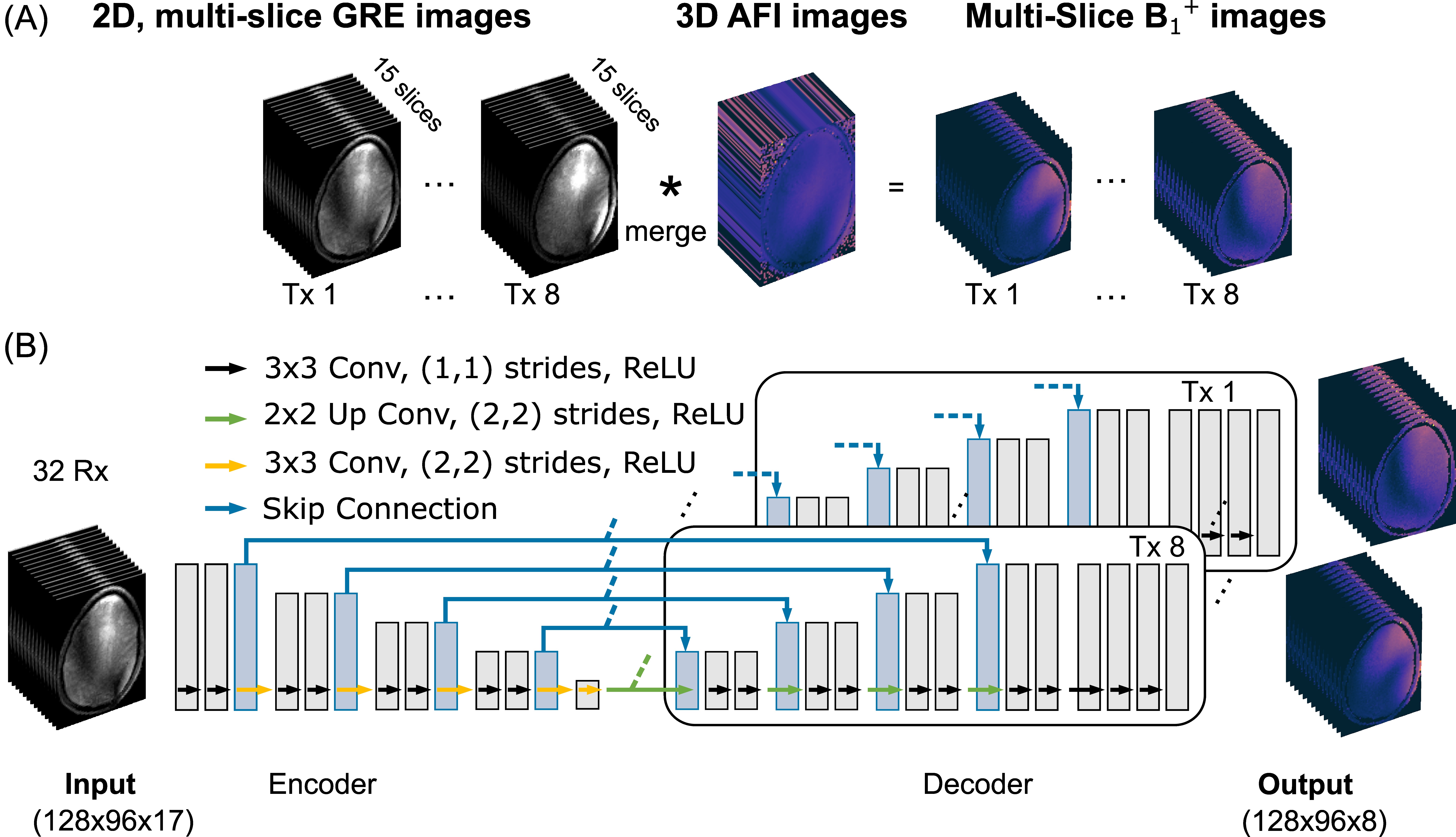
Figure 1:
(A) demonstrates the hybrid approach to generate Tx-wise multi-slice absolute B1+ - maps from multi-slice GRE images and a 3D actual flip angle image.
(B) illustrates the UNet architecture. The network uses the complex, multi-slice (GRE) localizer image of all 32 Rx channels with all Tx transmitting in CP mode as an input and is trained on Tx-wise multi-slice B1+ maps as GT. Every Tx-Channel utilizes a separate decoder pipeline.
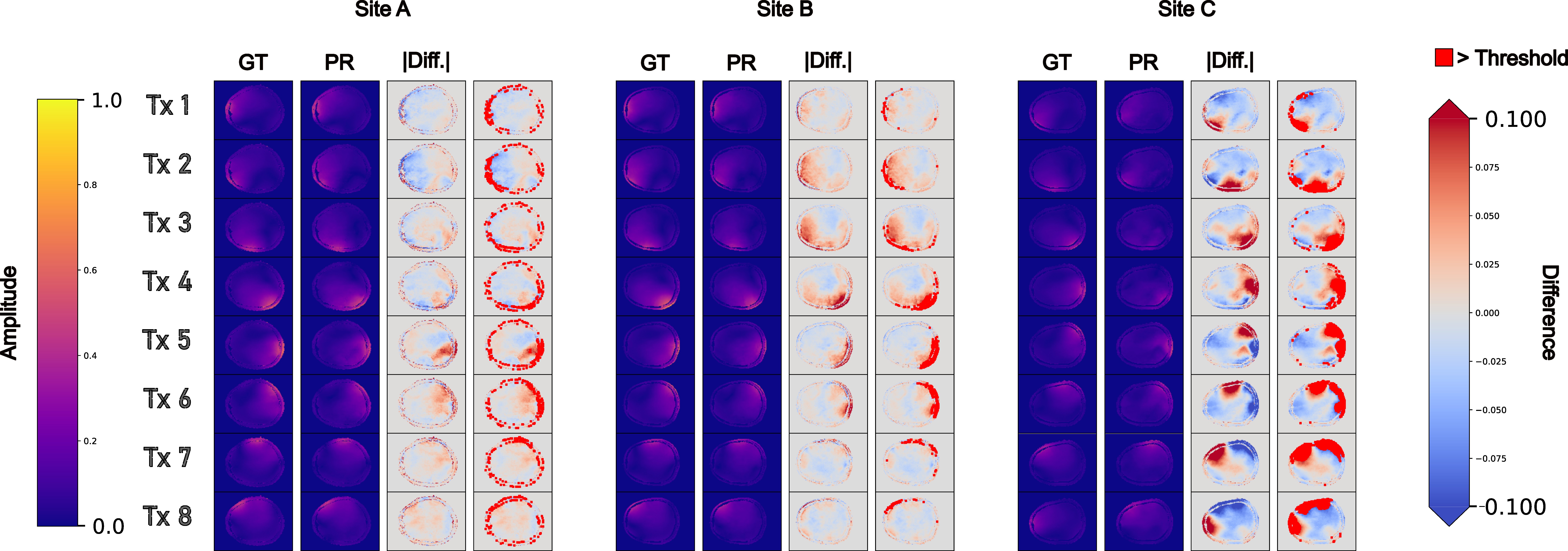
Figure 2: a three-site comparison of ground truth (GT) versus predicted (PR) B+1 amplitudes of all eight Tx channels in a single slice. The first two columns reveal a high degree of visual concordance, while the third column measures absolute differences, and the fourth highlights voxels with errors of >10%. Sites A and B show smaller deviations than Site C, with most errors near coil regions and skull boundaries, suggesting scanner and coil consistency contributes to prediction accuracy.
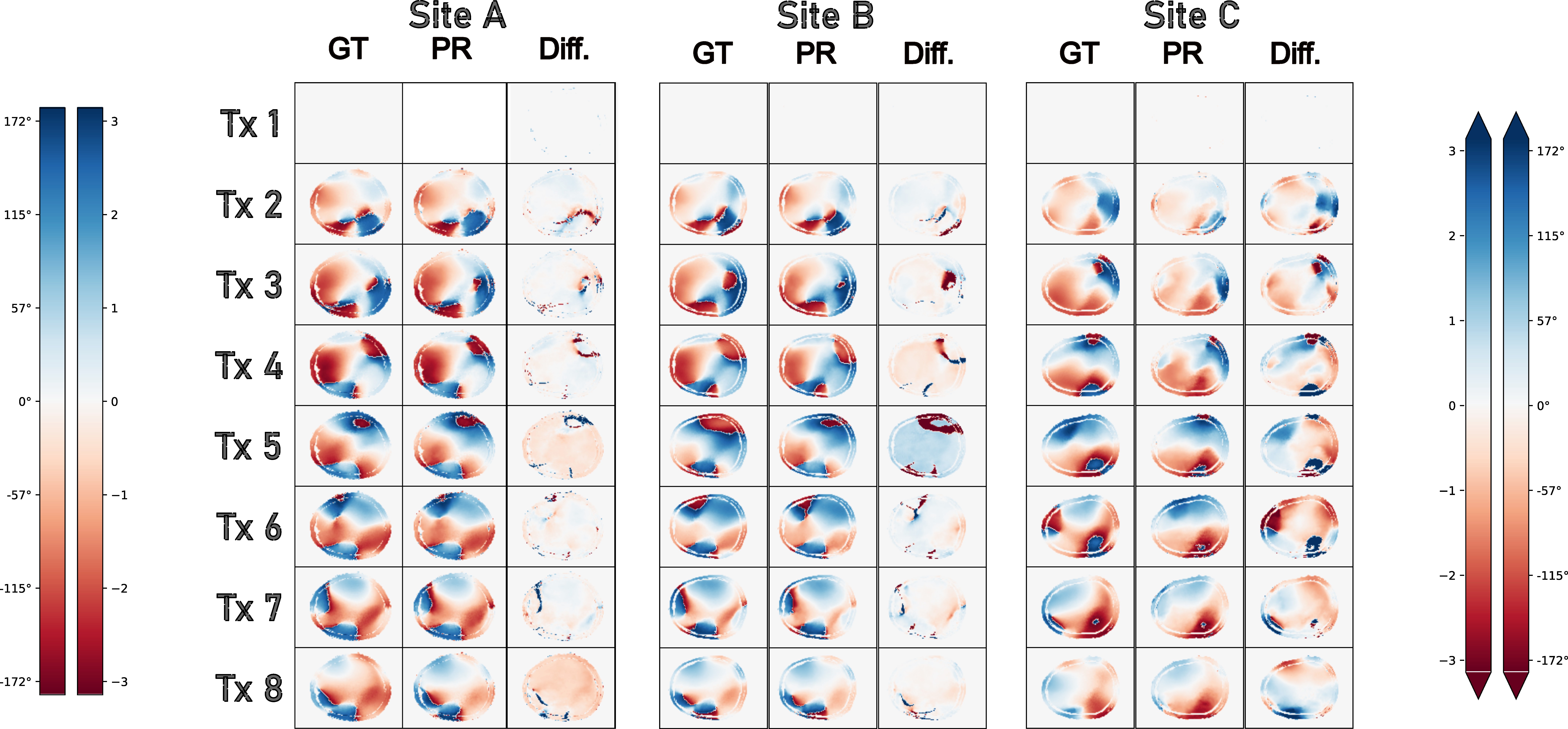
Figure 3: ground truth (GT) vs. predicted (PR) MRI phase images corresponding to Fig 2 across the eight Tx channels for the three sites . Columns show GT phases, PR phases, and absolute as well as relative differences. Sites A and B align closely, with Site C displaying more errors, particularly near phase wraps, likely due to varied scanner and coil types.
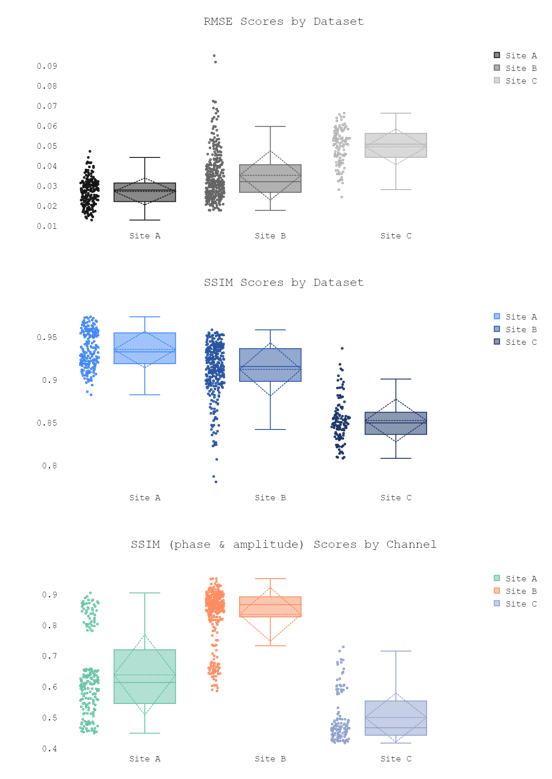
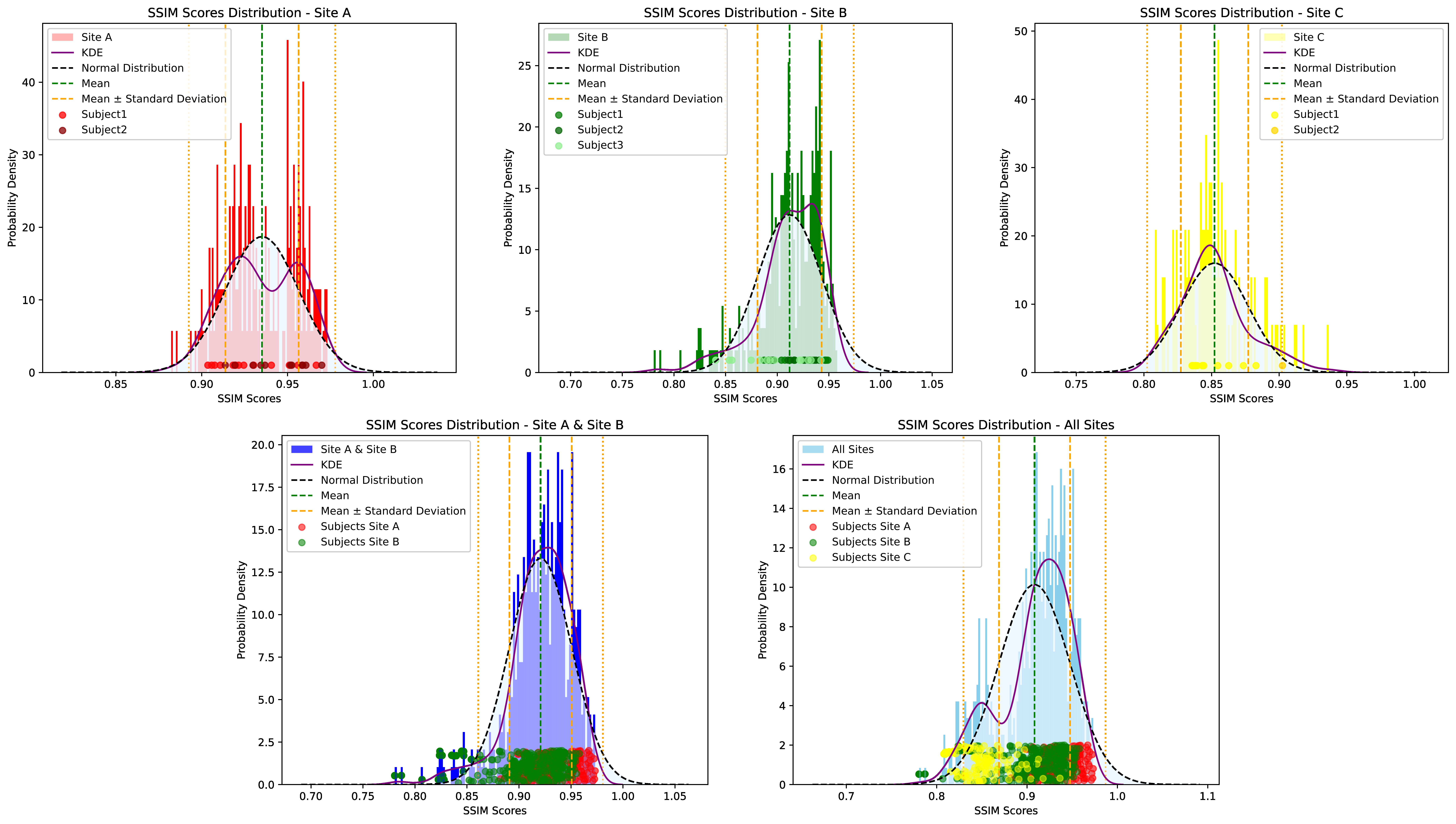
Figure 5: SSIM histograms for MRI amplitudes by site, with scatters per slice and subject variance color-coded. Site A shows bimodal variance, Site B and Site C fit Gaussian curves well. Combined data analysis reveals Site C as an outlier, affecting Gaussian fit due to its distinct scanner and coil setup.