4936
Tissue-Mimicking Phantoms with Tunable Acoustic and Mechanical Properties for Visualizing MRgFUS Cavitation Lesions1Electrical Engineering, Brigham Young University, Provo, UT, United States, 2University of Utah, Salt Lake, UT, United States
Synopsis
Keywords: MR-Guided Focused Ultrasound, Phantoms, histotripsy, cavitation, lesioning, HIFU
Motivation: Develop tissue-mimicking phantoms for MR-guided focused ultrasound (MRgFUS) procedures, enabling precise cavitation lesion visualization.
Goal(s): Phantoms with tunable acoustic and mechanical properties, capable of producing MR image contrast when exposed to cavitation lesioning. Previous designs lacked tunable parameters or MR contrast, hindering comprehensive study of cavitation behavior. This project seeks to fill this gap by developing versatile phantoms with MR contrast.
Approach: Combine evaporated milk, saline, agarose, and live red blood cells to create versatile phantoms. Conduct systematic experiments to validate tunable acoustic attenuation, mechanical stiffness, and lesion contrast for MRgFUS.
Results: Successfully produced customizable phantoms with excellent lesion contrast in MRgFUS.
Impact: Researchers can use the results presented here to construct MR visible phantoms that interrogate acoustic cavitation.
Introduction
The focus of this project is to develop phantoms with customizable acoustic attenuation and mechanical stiffness properties that can also produce MR image contrast when exposed to acoustic cavitation lesioning. Phantoms with tunable acoustic attenuation and mechanical stiffness permit interrogation of cavitation properties1,2. Meanwhile, MR image contrast permits visualization of cavitation lesioning. Previous phantom designs either only had red blood cells (RBCs), used to create image contrast, as a single layer, or they didn’t have tunable parameters3. [ Tunable phantoms with 3D lesion contrast were produced according to the following methods.Materials and Methods
Evaporated milk and deionized water were combined in ratios of 0, 25, 50, 75, and 100% into a 300mL volume while agarose (A-204, GoldBio) was added in w/v ratios of .5%. Making each gel also required[30mL of live RBCs, and 3g of phosphate buffered saline powder (BSP017, Albert Bio) to help keep the RBCs alive so that their hemoglobin would stay intact and provide contrast.To obtain the RBCs, 500mL of whole bovine blood was purchased from a local butcher shop and mixed with 55mL of anticoagulant and then centrifuged in 15mL tubes at 1250rfc for 10 minutes3 before extracting the plasma and white buffy coat. The RBCs were refrigerated until used. All gels were made within one week of obtaining whole blood to ensure freshness.
Everything but the RBCs was mixed together and then heated to 80 oC. After heating, the mixture was stirred and then placed in a vacuum chamber at 20psi[until cooled to 40 oC. Then, RBCs were carefully added and thoroughly stirred. Finally, the gel was syringed into one of two molds. Gels for acoustic and mechanical testing were formed using a cylindrical, acrylic container (inner diameter 5.08-cm, height 2.54-cm) was silicon-sealed with ultrasound transparent, clear, mylar plastic on each end. A hemispherical mold was used to create gels for testing MRI contrast due to cavitation lesioning [SA7] .
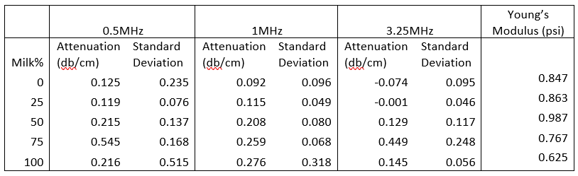
Attenuation was measured using a through transmission setup4 as seen in Figure 1b. Three separate 5-cycle bursts at 0.5, 1, and 3 MHz were transmitted through the gel and recorded. The experiment was repeated with water as a control. Each gel variation had 3 phantoms which were measured at 4 locations, ninety degrees apart, generating a total of 12 measurements per gel variation.
Young’s modulus for each gel was obtained using a tensile tester (1kip mini, Instron) [in unconfined compression on four gels from each type of gel4, see Figure 1c. The gels were compressed at a rate of 0.08in/s for 2s. The tensile tester’s reported force values and displacement values were then used to calculate the average Young's Modulus4 .
Phantom MR properties including T1, T2, and T2*, were obtained on 4 gels (held at 20 oC) from each gel type using a 3T MRI scanner with a 64 channel head coil (Vida, Siemens, Erlangen, DE) using 2D inversion recovery, multi-echo spin echo, and multi-echo gradient echo sequences, respectively. Parameters were TI: 50, 200, 400, 600, 800, 1200; TE?TR: 12/5000ms, Pixel Bandwidth: 130 Hz/px.
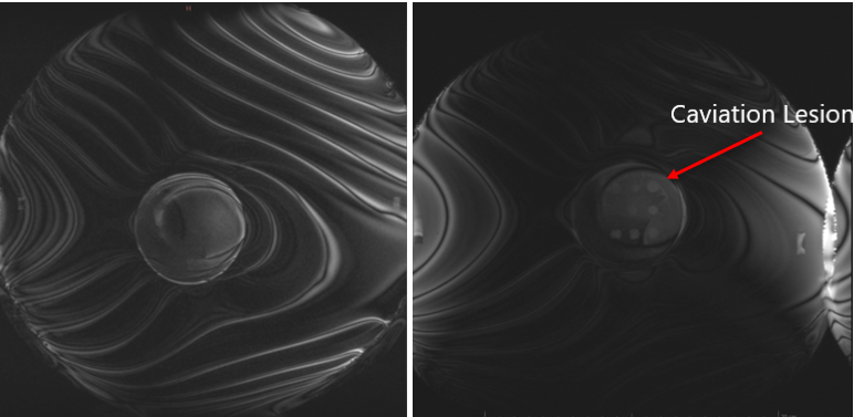
One gel sample[(0% milk, .5% agarose) was placed at the center of hemispherical focused ultrasound device (Exablate Neuro, Insightect, Haifa, Israel), and insonated with a requested power of 340 acoustic Watts for 0.075 s. Afterwards, the gel was scanned using a 3T MR Scanner (Skyra, Siemens) and a 3D bSSFP sequence. Parameters were TE/TR: 5.35/10.7 ms; Resolutiou: 0.47x0.47x1 mm; Pixel Bandwidth: 130 Hz/px.
Results
The average and standard deviation of the attenuation of each type of gel is shown in Table 1. Figure 2 shows the Young’s Modulus of each type of gel. Lastly, Figure 3 shows the results of successful histotripsy lesioning in a gel.Discussion & Conclusion
In general, the agarose gels had an increase in attenuation as the amount of evaporated milk increased (see Table 1). They also provided excellent lesioning contrast for visualizing MRgFUS lesions (see Figure 3). Future work could include studying the properties of phantoms with different percentages of agarose powder, as well as trying different kinds of gelatins.Acknowledgements
This work was supported by the Focused Ultrasound Foundation and R21 EB033117.References
1. Hendley, S. A., Bollen, V., Anthony, G. J., Paul, J. D., & Bader, K. B. (2019). In vitro assessment of stiffness-dependent histotripsy bubble cloud activity in gel phantoms and blood clots. Physics in Medicine and Biology, 64(14). https://doi.org/10.1088/1361-6560/ab25a6
2. Vlaisavljevich, E., Lin, K. W., Warnez, M. T., Singh, R., Mancia, L., Putnam, A. J., Johnsen, E., Cain, C., & Xu, Z. (2015). Effects of tissue stiffness, ultrasound frequency, and pressure on histotripsy-induced cavitation bubble behavior. Physics in Medicine and Biology, 60(6), 2271–2292. https://doi.org/10.1088/0031-9155/60/6/2271
3. Maxwell AD, Wang TY, Yuan L, Duryea AP, Xu Z, Cain CA. A tissue phantom for visualization and measurement of ultrasound-induced cavitation damage. Ultrasound Med Biol. 2010 Dec;36(12):2132-43. doi: 10.1016/j.ultrasmedbio.2010.08.023. Epub 2010 Oct 28. PMID: 21030142; PMCID: PMC2997329.
4. Farrer, A. I., Odéen, H., de Bever, J., Coats, B., Parker, D. L., Payne, A., & Christensen, D. A. (2015). Characterization and evaluation of tissue-mimicking gelatin phantoms for use with MRgFUS. Journal of Therapeutic Ultrasound, 3(1). https://doi.org/10.1186/s40349-015-0030-y
Figures
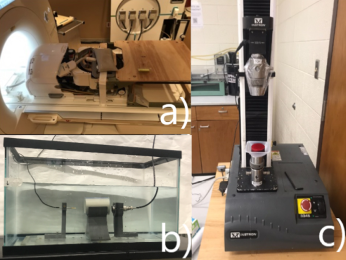
Experimental setup: a) shows the the phantom holder and focused ultrasound transducer at the MR imaging site where we did MRgFUS; b) shows the through-transmission setup for measuring acoustic attenuation; c) shows the tensile tester setup for measuring mechanical stiffness properties