4930
Glymphatic function as a predictor of mild cognitive impairment to dementia conversion in Parkinson’s disease1the first hospital of China Medical University, Shenyang, China, 2MR Research Collaboration, Siemens Healthineers, Beijing China, Beijing, China
Synopsis
Keywords: Diffusion Analysis & Visualization, Diffusion Tensor Imaging, DTI-ALPS
Motivation: Impaired glymphatic function is reported to be associated with cognitive function in Parkinson’s disease (PD). However, an evaluation of the role of the glymphatic dysfunction contributing to the dementia conversion of mild cognitive impairment (MCI) with use of MRI remains lacking.
Goal(s): To evaluate the value of glymphatic function to predict the dementia conversion in PD with MCI.
Approach: We calculated the water diffusion in the periventricular white matter using DTI-ALPS to evaluate the glymphatic function in PD with cognitive impairment during the follow-up.
Results: The left DTI-ALPS is significantly lower in dementia converters, and is valuable for prediction of PD dementia conversion.
Impact: The reduction of glymphatic activity, assessed by DTI-ALPS, was more severe in PD with MCI convert to dementia. Therefore, DTI-ALPS may assist in identifying PD with MCI at a high risk of dementia conversion before the onset of dementia symptoms.
Introduction
The glymphatic function contributes to the vital clearance of the metabolic byproducts and pathological proteins, such as a-synuclein, amyloid-β and tau [1]. Impairments in the glymphatic function may contribute to the abnormal accumulation of a-synuclein, which underlie the pathology of the Parkinson’s disease (PD) [2]. Recently, a non-invasive method known as Diffusion tensor image analysis along the perivascular space (DTI-ALPS) has emerged as a promising tool for glymphatic function evaluation. Water diffusivity in the periventricular white matter is calculated along x,y, and z axes using diffusion tensor images to derive the DTI-ALPS index. Specially, the DTI-ALPS index quantifies water diffusivity along the x-axis, aligned with the perivascular space (PVS) direction, offering a means to partially assess the activity of the glymphatic system. Studies have reported correlation between DTI-ALPS and cognitive function in PD and Alzheimer’s disease [3,4]. A lower DTI-ALPS index was correlated with increased severity of cognitive impairment in neurodegenerative diseases [5]. Yet, so far, no study has evaluated the value of glymphatic function to predict the cognitive conversion in PD patients. Thus, in this study, we aimed to investigate the potential of DTI-ALPS as a tool for predicting the conversion from mild cognitive impairment to dementia in patients with PD. Additionally, we aim to explore the associations between DTI-ALPS index and cognitive function.Methods
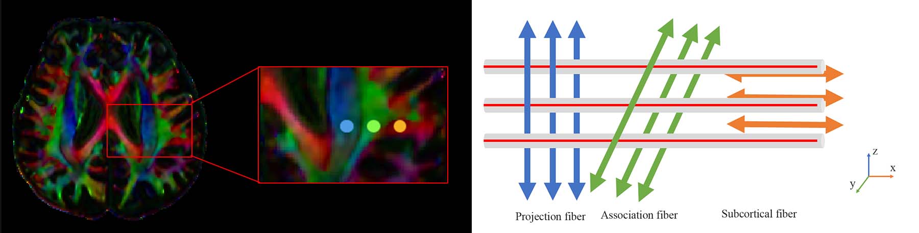
We retrospectively included 36 patients with PD meeting criteria for PD-MCI into either PD with dementia (PDD) converters (n=15) or nonconverters (n=21), depending on whether they developed dementia within 18 months of PD diagnosis. Meanwhile, 20 age- and gender-matched healthy controls (HC) were included. All participants underwent MRI scans and comprehensive neuropsychological assessments at baseline. Diffusion imaging was acquired using a 3T MRI scanner (MAGNETOM Verio, Siemens Healthcare, Erlangen, Germany). The DTI were obtained with b values of 0 and 1000 s/mm2, utilizing echo planer imaging sequence with TR=10300 ms, TE= 94 ms, 64 diffusion directions, FOV= 256×256 mm, matrix = 128×128, slice thickness = 2 mm. Susceptibility weighted images were performed as well. The DTI-ALPS were processed using FSL (version 6.0.23, FMRIB Software, Library; http://www.fmrib.ox.ac.uk/fsl). This included essential processing steps including brain extraction, denoising, eddy current correction and DTIFIT. Color-coded fractional anisotropy (FA) maps and diffusivity maps in directions of the x-, y-, and z axes (Dx, Dy, Dz) were generated. On the color-coded FA map, spherical region of interests (ROIs) with a 5mm diameter were placed within the projection fibers, association fibers, and subcortical fibers respectively in bilateral hemispheres. Specifically, we calculated the diffusivities along the x-axis of ROIs within projection (Dxproj) and association (Dxassoc) fibers areas, which partially represented water diffusivity along the PVS with minimal influence from the nerve fibers. Additionally, diffusivites along the y-axis of projection fibers (Dyproj) and along the z-axis of association fibers (Dzassoc) were extracted. The disparities between mean diffusivity along the x-axis in projection fibers (Dxproj and Dxassoc) and mean diffusivity along the y-axis in projection fibers (Dyproj) and along the z-axis of association fibers (Dzassoc) might be indicative of PVS involvement. DTI-ALPS index is defined as follows: ALPS index = mean (Dxproj, Dxassoc)/mean (Dyproj, Dzassoc) [6] DTI-ALPS index was compared among HC, PDD converters and nonconverters using one-way analysis of variance (ANOVA), followed by posthoc tests with false discovery rate (FDR) correction between pairs of groups. Additionally, binary logistic regression was performed to analyze the effect of ALPS indexes and diffusivities in the fibers on the cognitive conversion. Spearman correlation was also conducted to investigate the relationships between DTI-ALPS index and cognitive function after controlling for age, sex, and years of education.Results
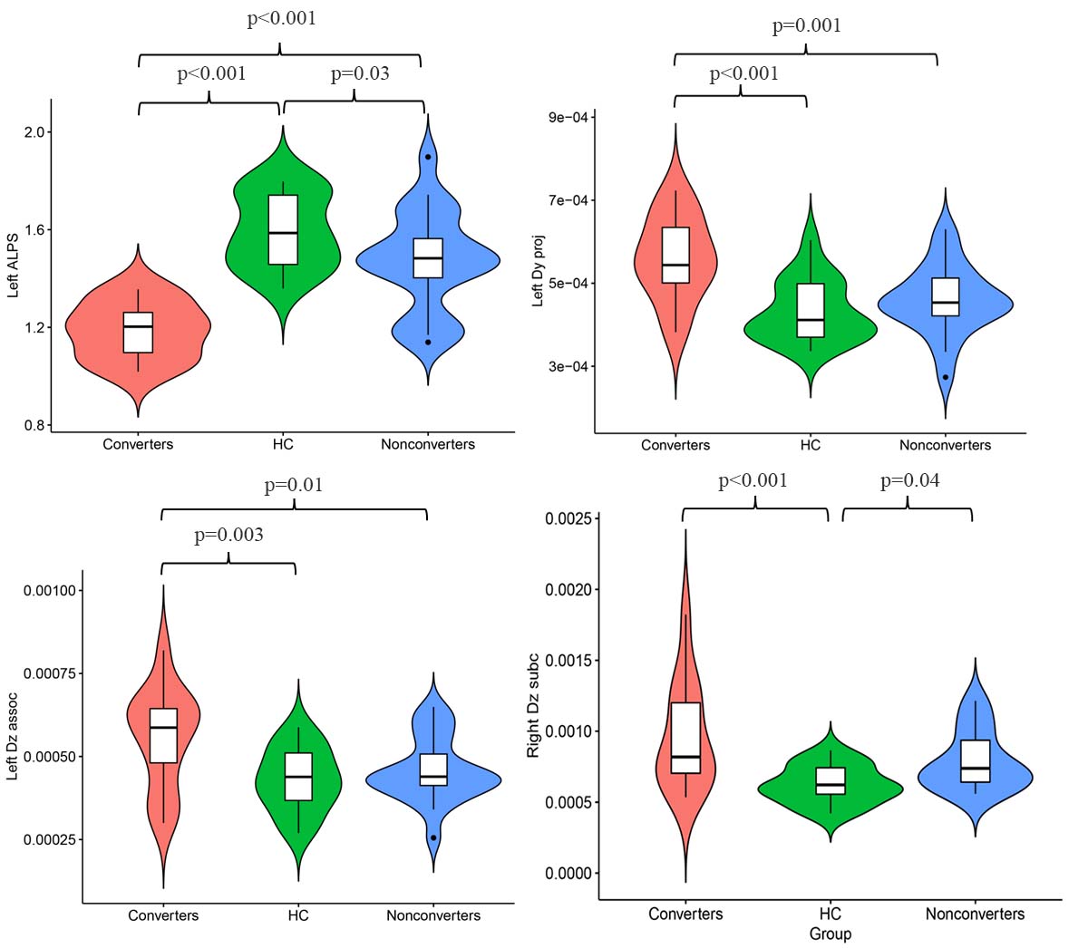
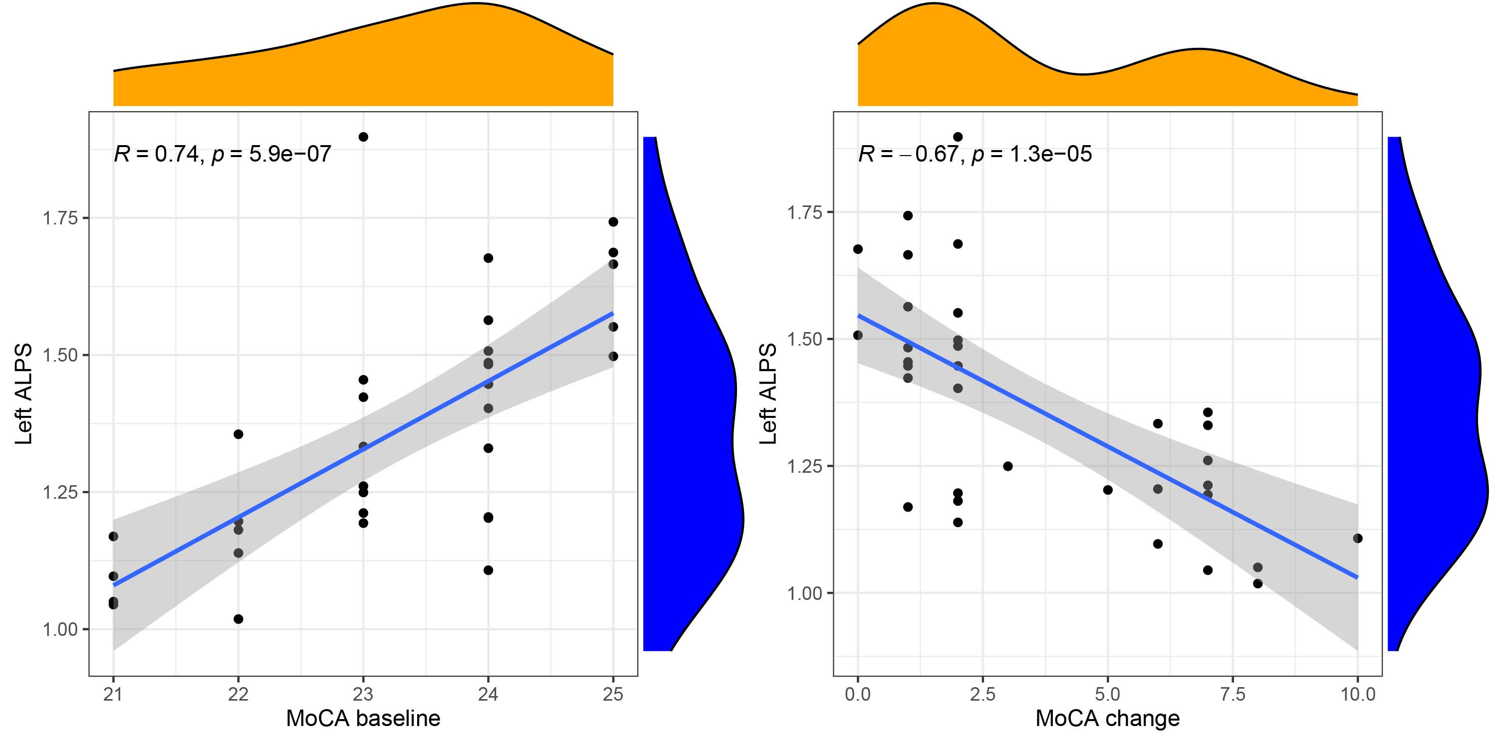
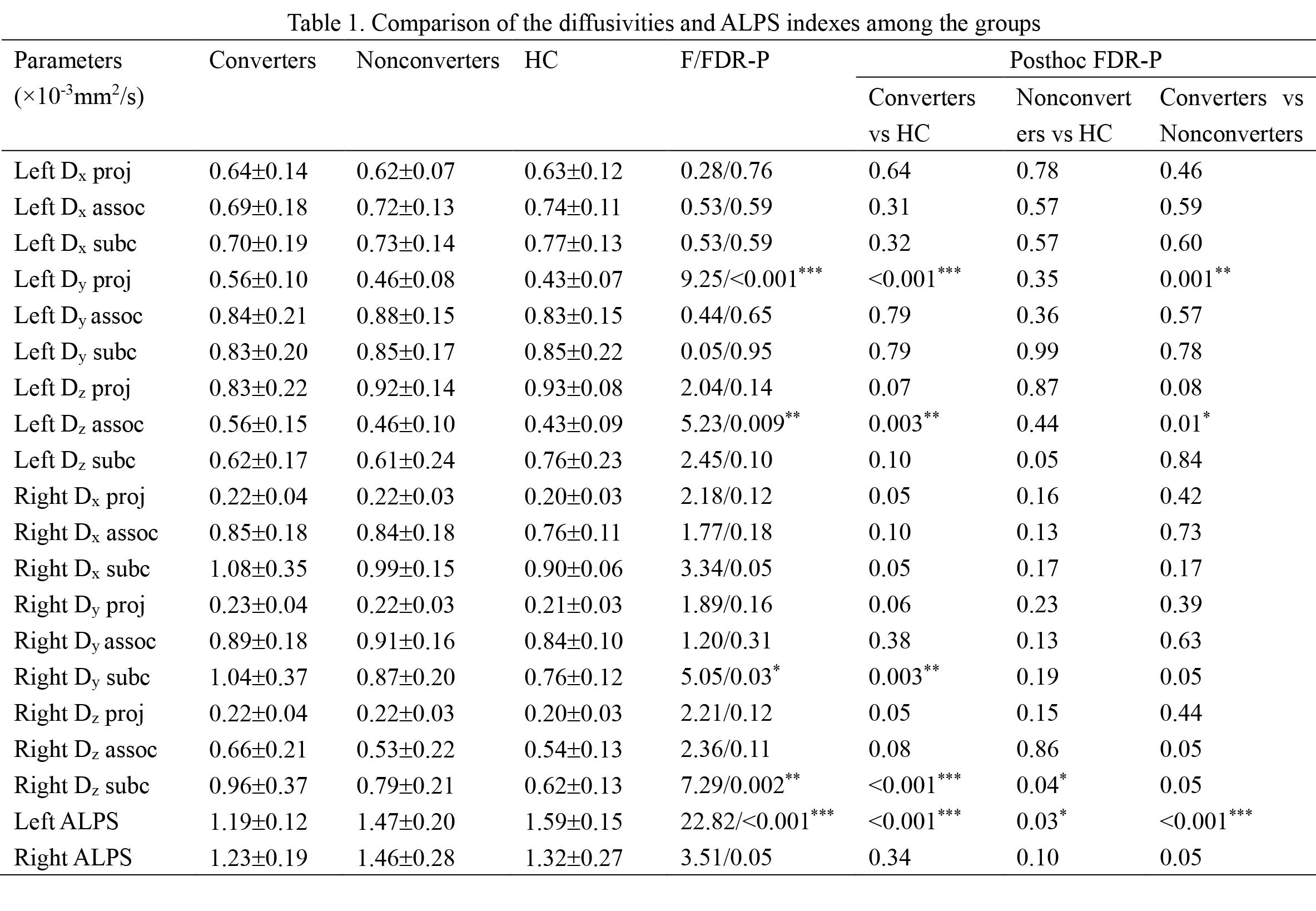
Fifteen converters (mean age, 72.75 years±12.55; 9 women), twenty-one nonconverters (mean age, 69.60 years±10.42; 13 women), and twenty HC (mean age, 68.53 years±9.72; 10 women) were included. Significant lower DTI-ALPS was observed in converters group compared with nonconverters, as well as HC (P<0.001). Besides, the diffusivity along the y-axis in the association fibers was increased in dementia converters in comparison with nonconverters and HC (P<0.001). Binary logistic regression showed that a lower left ALPS index could predict the prevalence of PDD conversion (OR= 33.80(95%CI:13.3,54.3), P<0.01). Besides, the left DTI-ALPS index showed significant correlation with MoCA scores at the baseline and the MoCA score change in the follow-up (r=0.74, P<0.001; r=-0.67, P<0.001, respectively).Discussion/Conclusion
This was a preliminary study of exploring the value of glymphatic function for dementia conversion prediction in PD. The results suggest that the lower left DTI-ALPS index in dementia converters.The DTI-ALPS in PD patients with mild cognitive impairment demonstrated a more severe reduction of glymphatic activity in individuals with dementia conversion.Acknowledgements
No acknowledgement found.References
[1] Wardlaw JM, Benveniste H, Nedergaard M, et al. Perivascular spaces in the brain: anatomy, physiology and pathology. Nat Rev Neurol. 2020;16(3):137-153. doi:10.1038/s41582-020-0312-z
[2] Rasmussen MK, Mestre H, Nedergaard M. The glymphatic pathway in neurological disorders. Lancet Neurol. 2018;17(11):1016-1024. doi:10.1016/S1474-4422(18)30318-1
[3] Zhang X, Wang Y, Jiao B, et al. Glymphatic system impairment in Alzheimer's disease: associations with perivascular space volume and cognitive function [published online ahead of print, 2023 Aug 23]. Eur Radiol. 2023;10.1007/s00330-023-10122-3. doi:10.1007/s00330-023-10122-3
[4] Bae YJ, Kim JM, Choi BS, et al. Glymphatic function assessment in Parkinson's disease using diffusion tensor image analysis along the perivascular space. Parkinsonism Relat Disord. 2023; 114:105767. doi: 10.1016/j.parkreldis.2023.105767
[5] Bae YJ, Kim JM, Choi BS, et al. Altered Brain Glymphatic Flow at Diffusion-Tensor MRI in Rapid Eye Movement Sleep Behavior Disorder. Radiology. 2023;307(5): e221848. doi:10.1148/radiol.221848
[6] Taoka T, Masutani Y, Kawai H, et al. Evaluation of glymphatic system activity with the diffusion MR technique: diffusion tensor image analysis along the perivascular space (DTI-ALPS) in Alzheimer's disease cases. Jpn J Radiol. 2017;35(4):172-178. doi: 10.1007/s11604-017-0617-z.
Figures