4929
Glymphatic dysfunction in patients with Chronic kidney disease is associated with cognitive decline1Department of Radiology, Beijing Friendship Hospital, Capital Medical University, Beijing, China, 2GE HealthCare, MR Research, Beijing, China, Beijing, China
Synopsis
Keywords: fMRI Analysis, Kidney, glymphatic system
Motivation: The relationship between CSF inflow, which is an essential physiological activity of the glymphatic system, and cognitive decline in patients with chronic kidney disease (CKD) is unclear.
Goal(s): Our goal was to explore the patterns of CSF inflow changes in patients with CKD and its relationship with cognitive decline.
Approach: The CSF in the the subarachnoid space along the middle cerebral were analyzed with ultra-long echo time and low-b diffusion tensor imaging.
Results: Disturbed CSF inflow patterns is associated with cognitive decline in patients with CKD.
Impact: A novel approach to measure the CSF inflow of the glymphatic system exhibits the potential for detecting brain glymphatic dysfunction in patients with CKD, which may be one of the pathogenic mechanisms of cognitive decline.
Introduction
Chronic kidney disease (CKD), defined as the presence of impaired renal function (estimated glomerular filtration rate(eGFR) <60 mL/min/1.73m2 ) or proteinuria (urine albumin-to-creatinine ratio < 30 mg/g), has become a globally recognized public health problem1. Patients with CKD experience cognitive decline early in life, which is particularly severe in CKD stage 5. The glymphatic system is a recently discovered extracellular clearance system in the brain that facilitates the recirculation of cerebrospinal fluid (CSF) through the brain parenchyma and supports the clearance of interstitial solutes, including tau and amyloid-β2. Considering that patients with dementia or cerebrovascular disease exhibit impaired functionality of the glymphatic system3, it can be expected that dysfunction of glymphatic system may be present in patients with CKD. The inflow of CSF from the subarachnoid space (SAS) into the periarterial space is regarded as an essential physiological activity of the glymphatic system. It has been proposed to measure the CSF inflow in the SAS around the middle cerebral artery (MCA) using diffusion-weighted magnetic resonance imaging method with ultra-long echo time (TE), low b-value, and multi-directionality (DTIlow-b )4. However, the relationship between CSF inflow and cognitive decline in patients with CKD has not been investigated. This study also aims to explore the pattern of glymphatic function changes and its relationship with cognitive decline in patients with CKD based on the DTIlow-b method.Methods
The study was approved by the Ethics Committee of Beijing Friendship Hospital of Capital Medical University and implemented in accordance with the Declaration of Helsinki. A total of 26 patients with CKD and 16 sex-, age-, and education-matched healthy controls (HCs) were enrolled in the study. All participants underwent imaging on a 3T MRI system (Discovery MR750W, General Electric, Milwaukee, Wisconsin, USA) with an eight-channel phased array coil. DWI imaging with long TE and low b values was performed using a single-shot spin-echo echo planar imaging sequence (TE/TR=133ms/4000ms; acquisition matrix=128´128; number of slices=20; voxel size=1.875´1.875´3mm3; b value=0 and 130 s/mm2 with 30 noncolinear directions). The preprocessing of each participant’s DWI data was performed using the following steps: image denoising (mrtrix’s dwidenoise), removing Gibbs ringing artefacts (mrtrix’s mrdegibbs), motion and eddy current distortion correciton (FSL’s eddy). The ROIs in the SAS at the M1 stage of both left and right MCA (MCA SAS) were carefully delineated by one neuroradiologist with with 3-years of experience and further checked by a senior neuroradiologist with 10-years of experience, both of whom were blind to the group information (Figure 2). For each ROI, voxels were averaged to construct the diffusion tensor, from which the axial diffusivity (AD) and fractional anisotropy (FA) were subsequently derived. The mean AD and FA value of the bilateral MCA SAS ROIs were taken for further analysis. Intergroup comparisons were accomplished using two-sample t-tests. The Pearson correlation test was used to analyze the correlation between the DTIlow-b index and MoCA scores.Results
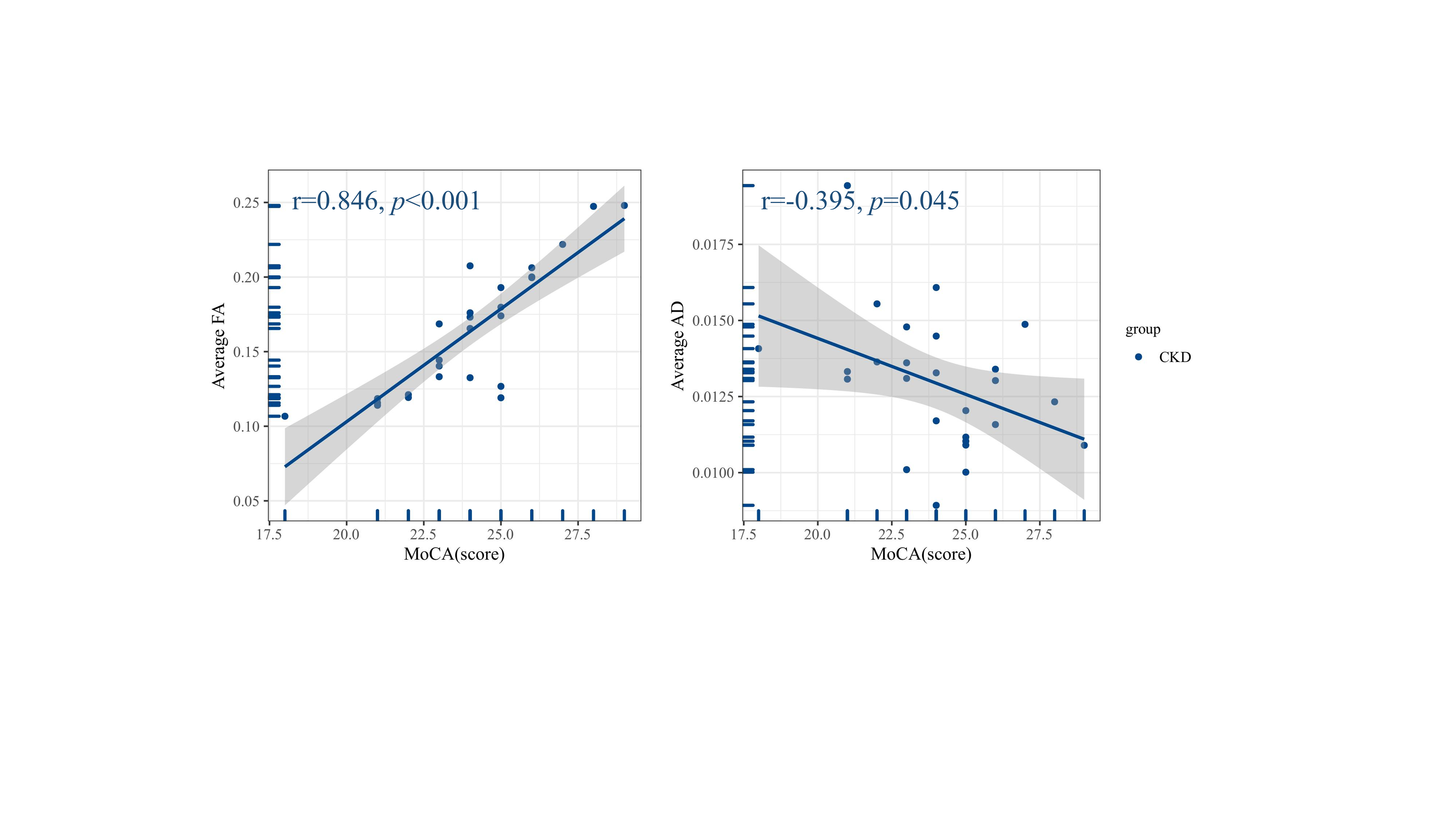
Patients with CKD showed significantly lower FA compared to HCs (P = 0.008). However, no significant difference was observed in AD values between the two groups (P = 0.301) (Figure 3). As for the correlation analysis, there was a positive correlation between FA values and MoCA scores (r = 0.846; P < 0.001) and a significant negative correlation between AD values and MoCA (r = -0.395; P = 0.045) in patients with CKD (Figure 4).Discussion
FA in this study represents CSF anisotropy, similar to FA in conventional DTI. AD has advantages in measuring CSF flow along the vascular direction, reflecting CSF inflow activity in glymphatic system pathways5. In the present study, the CKD group exhibited lower FA, which was significantly positively correlated with MoCA scores. Although there was no statistical difference in AD between the two groups, it is interesting to note that, in terms of trend, AD was higher in the CKD group than in the HCs, with a significant negative correlation with MoCA scores. This may suggest that the faster the flow rate of CSF inflow within the MCA SAS, the more severe the cognitive decline of the patients. This suggests that impaired activity of the glymphatic system is prevalent in patients with CKD, which may provide new evidence to unravel the underlying mechanisms by which cognitive decline occurs in patients with CKD.Conclusion
Patients with CKD exhibit circulatory abnormalities in the glymphatic system, and these changes are associated with cognitive decline. This suggests that abnormalities in glymphatic system may be a potential mechanism for cognitive decline in patients with CKD, and that DTIlow-b will be a potential new tool for revealing cognitive decline in patients with CKD.Acknowledgements
This work was supported by the National Natural Science Foundation of China (No. 82202099), the Beijing Municipal Administration of Hospitals Clinical Medicine Development of Special Funding Support (No. ZYLX201824 and ZYLX202101), the Beijing Municipal Administration of Hospital’s Mission Plan (No. SML20150101), the Beijing Scholars Program (No. [2015] 160), the Beijing Friendship Hospital, Capital Medical University (seed project No. YYZZ202129), and the Training Fund for Open Projects at Clinical Institutes and Departments of Capital Medical University (No. CCMU2022ZKYXY011).References
1. Wang, L.; Xu, X.; Zhang, M.; Hu, C.; Zhang, X.; Li, C.; Nie, S.; Huang, Z.; Zhao, Z.; Hou, F. F.; Zhou, M., Prevalence of Chronic Kidney Disease in China: Results From the Sixth China Chronic Disease and Risk Factor Surveillance. JAMA internal medicine 2023, 183 (4), 298-310.
2. Boespflug, E. L.; Iliff, J. J., The Emerging Relationship Between Interstitial Fluid-Cerebrospinal Fluid Exchange, Amyloid-β, and Sleep. Biological psychiatry 2018, 83 (4), 328-336.
3. van der Thiel, M. M.; Backes, W. H.; Ramakers, I.; Jansen, J. F. A., Novel developments in non-contrast enhanced MRI of the perivascular clearance system: What are the possibilities for Alzheimer's disease research? Neuroscience and biobehavioral reviews 2023, 144, 104999.
4. Harrison, I. F.; Siow, B.; Akilo, A. B.; Evans, P. G.; Ismail, O.; Ohene, Y.; Nahavandi, P.; Thomas, D. L.; Lythgoe, M. F.; Wells, J. A., Non-invasive imaging of CSF-mediated brain clearance pathways via assessment of perivascular fluid movement with diffusion tensor MRI. eLife 2018, 7.
5. Han, G.; Zhou, Y.; Zhang, K.; Jiao, B.; Hu, J.; Zhang, Y.; Wang, Z.; Lou, M.; Bai, R., Age- and time-of-day dependence of glymphatic function in the human brain measured via two diffusion MRI methods. Frontiers in aging neuroscience 2023, 15, 1173221.Figures
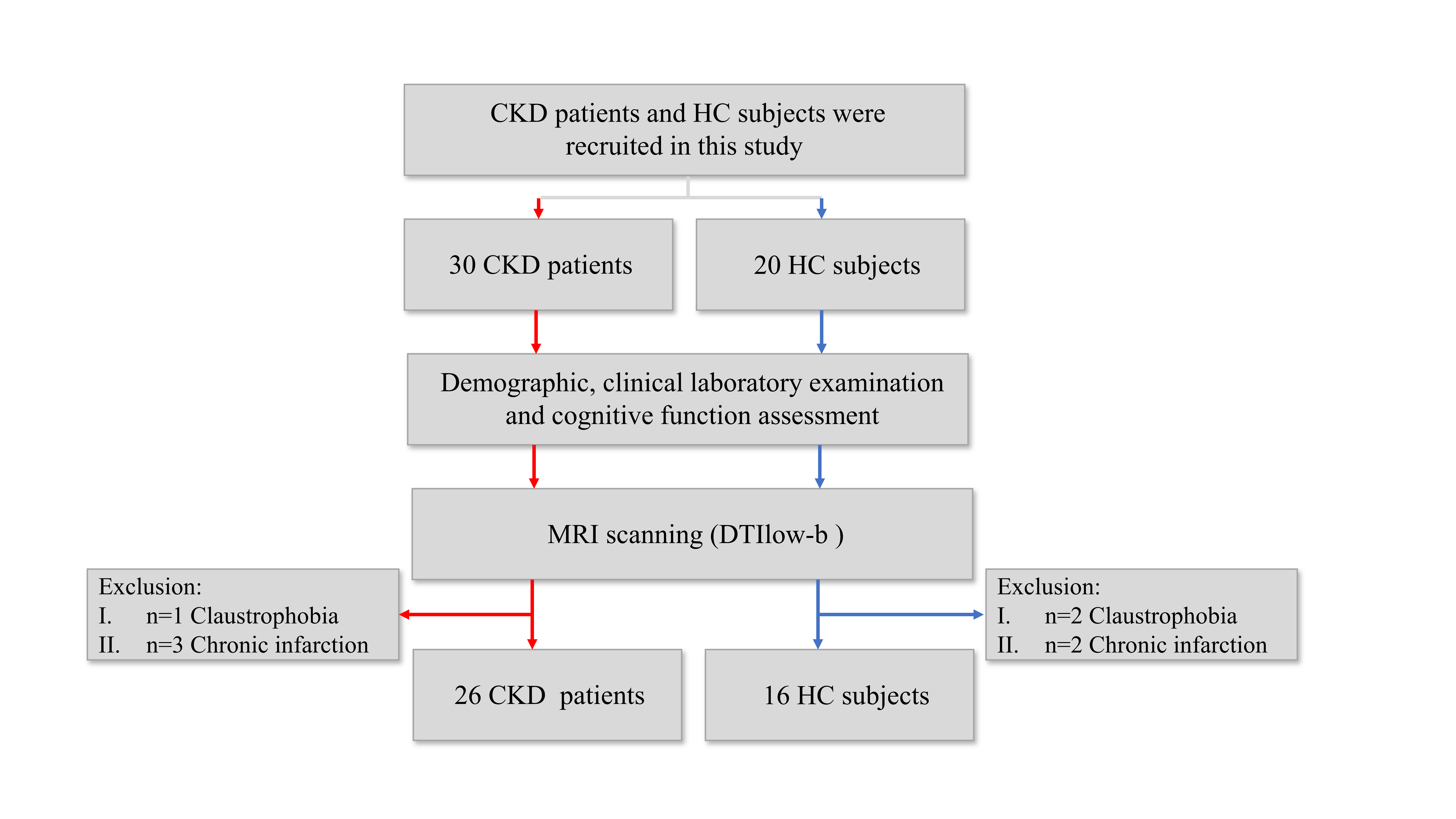
Figure1. Summary of patient recruitment and exclusions. CKD, chronic kidney disease; HC, healthy control; MRI, magnetic resonance imaging; DTIlow-b, diffusion-weighted magnetic resonance imaging method with ultra-long echo time, low b-value, and multi-directionality.
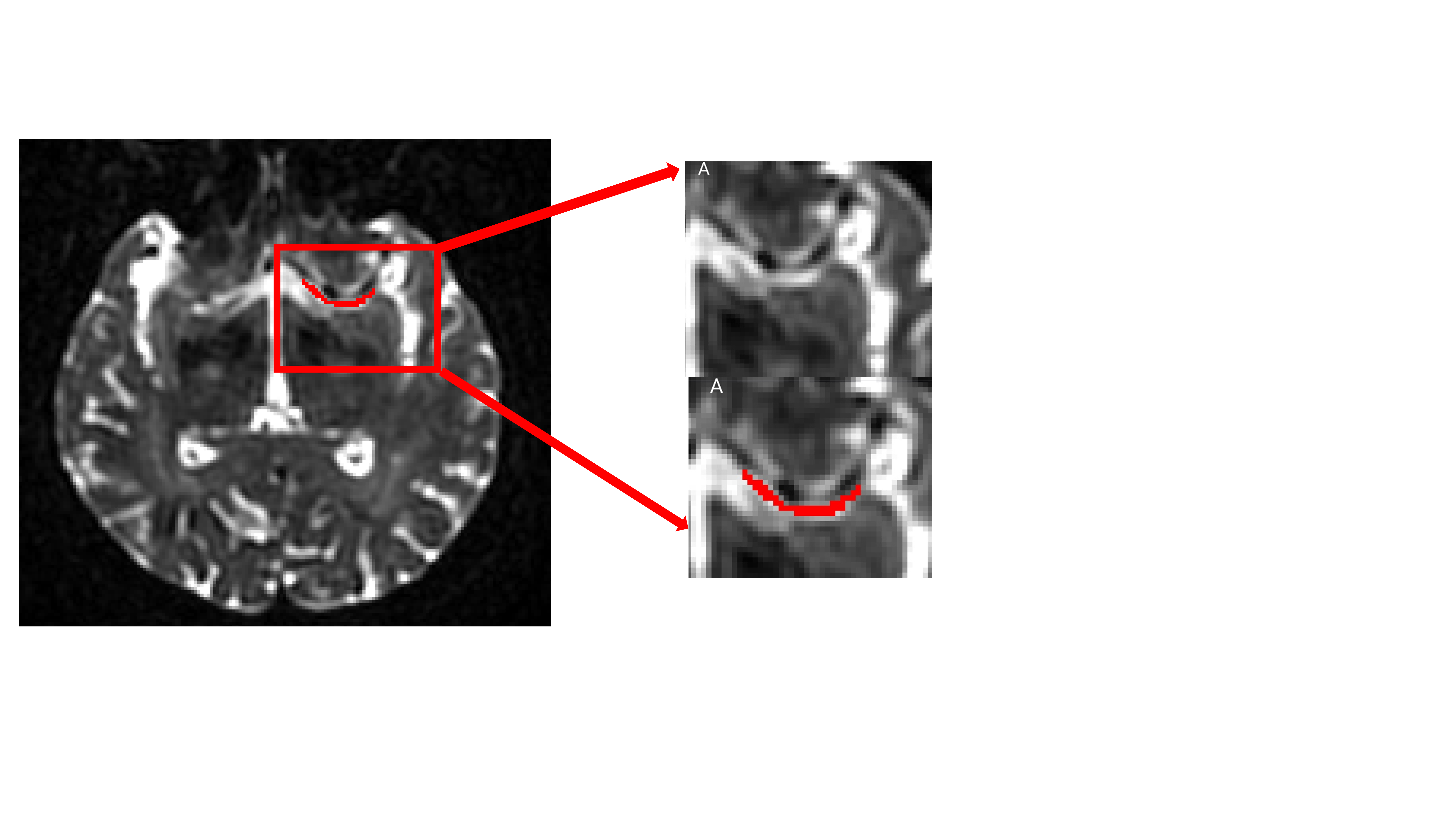
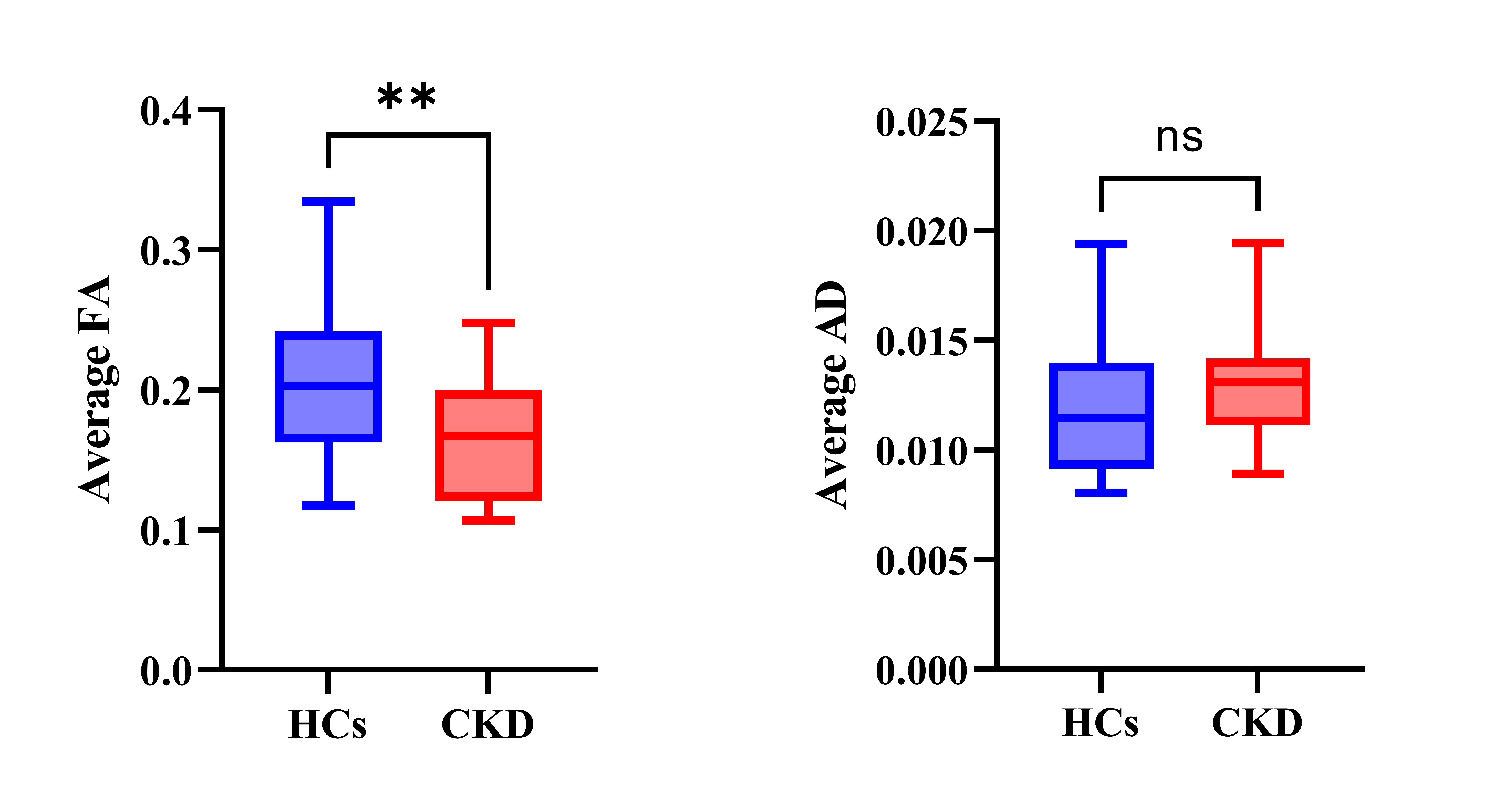
Figure 3. Between-group detection of DTIlow-b metrics in MCA SAS. SAS, subarachnoid space; MCA, middle cerebral artery; AD, axial diffusivity, FA, fractional anisotropy; CKD, chronic kidney disease; HC, healthy control.