4928
Comparing cerebral blood flow and cerebrospinal fluid flow during breath-holding and motor tasks in the human brain.1KAIST, Daejeon, Korea, Republic of
Synopsis
Keywords: Neurofluids, Neurofluids
Motivation: Recent studies highlight the significant impact of arterial pulsation on CSF movements in animal studies, but direct comparison in awake humans is still limited.
Goal(s): This study aims to concurrently measure CSF and CBF, excluding the influence of breathing, to analyze their correlation. Additionally, it investigates changes in CSF movement during the motor task.
Approach: Simultaneously measuring CBF and CSF by applying pCASL, and comparing how they change during breath-holding and motor tasks.
Results: During breath-holding, we observed a positive correlation between CBF and CSF. Furthermore, we confirmed reduced CSF inflow during the motor task compared to the resting state.
Impact: The relationship between CSF movement and CBF was analyzed during breath-holding and motor tasks in humans for the first time. This study offers a new way to study CBF and CSF movement, providing a better understanding of CBF-CSF physiology.
Introduction
Several recent fMRI studies have developed techniques to compare brain activation and cerebrospinal fluid (CSF) flow dynamics, demonstrating a close correlation between the BOLD signal and CSF movement1-3. The correlation between BOLD and CSF movement is mainly explained by changes in cerebral blood flow (CBF)4. When neurons are activated, the surrounding blood vessels expand, allowing arterial blood to flow in, which in turn affects the nearby CSF movement5. To simultaneously compare the CBF and CSF in the human brain, previous studies used phase contrast MRI (PC-MRI) to measure CBF and CSF at the craniospinal junction, or employed EPI-fMRI, which indirectly reflects CBF and CBV through the BOLD signal6. However, these methods had limitations in directly comparing the changes in CBF perfusing into the brain with CSF movement. Recently, a technique was introduced to measure the flow dynamics of CSF using pCASL in reference to PC-MRI7. However, the relationship between CSF and CBF was not investigated well. In this study, we aimed to simultaneously measure changes in CBF and CSF movement using pCASL and analyzed their relationship during breath holding. We also investigated the evolution of CBF and CSF during motor tasks.Method
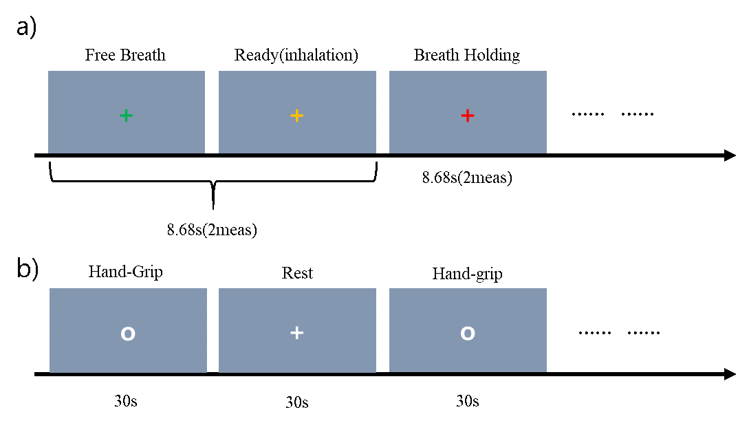
This study includes breath-holding (BH) and motor tasks. The BH task was carried out to measure changes in CSF and CBF by excluding the impact of respiration which greatly affects CSF movement8. The motor task was performed with repeated hand-grip (RHG) to measure changes in CSF-CBF in the behavior task (see Figure 1). We scanned 18 participants on a SEIMENS Trio 3T-MRI. Among them, 14 subjects participated in the BH task, and 9 subjects participated in the RHG task. We used a 1.5s post-labeling delay (PLD), 1.8s labeling duration, 67.5mm label plane offset, and background suppression for the pCASL preparation. We employed a 2D multi-slice echo planar imaging (EPI) readout with FOV = 230mm, voxel size = 3.6x3.6x6 mm, TR/TE = 4340/14ms, focusing first on the cortical region (slices 6th~23rd) and then the ventricular part7. The CSF flow dynamics can be divided into overall unidirectional movement and bidirectional pulsation according to the cardiac cycle. In this study, standard deviation of the CSF signal changes extracted from the pCASL label plane slice was used as an indicator of bidirectional CSF pulsation (CSF Pulsation). The average signals from slices above and below the label plane center were used to measure overall CSF convective inflow and outflow (CSF Inflow/outflow), respectively.Results
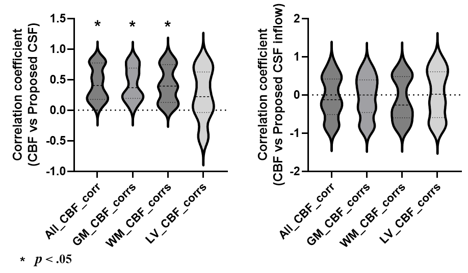
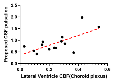
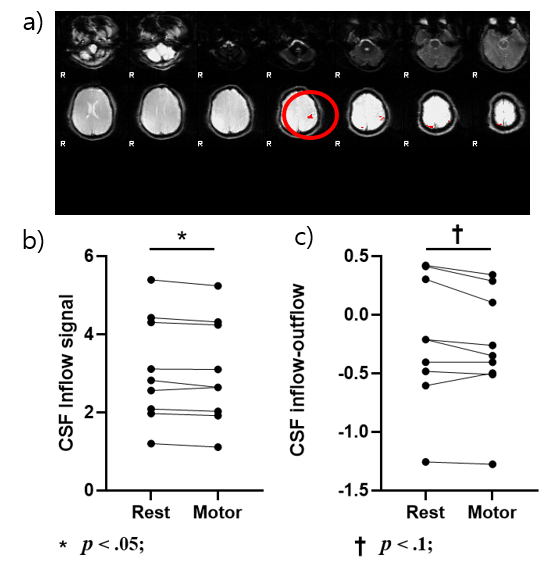
For the BH task, the CSF pulsation showed significant positive correlations with the CBF values (GM, WM, etc) across subjects (p<.05; Figure 2). However, CSF inflow and outflow showed no significant correlation with CBF (p>.05). It should be noted that the CSF pulsation was significantly correlated with the pCASL CBF signal in the lateral ventricle, which contains choroid plexus (r=0.539, p<.05; n=14, figure 3). Finally, for the RHG task, it was confirmed that functional CBF signal changes were induced in the motor cortex as expected (Figure 4a). The CSF inflow and the subtraction of the CSF outflow from inflow, both measured at the craniospinal junction, were significantly lower during the RHG task periods compared to the rest periods (Figure 4. b&c; p<.05 and p=.06, respectively).Discussion & Conclusion
Recent studies have demonstrated that arterial pulsation is a major source of power for CSF flow dynamics in animal models9. In our study, we used pCASL to simultaneously measure CBF and CSF dynamics (bidirectional pulsation, convective inflow and outflow), enabling the comparison of CSF flow dynamics and CBF changes in the in vivo human brain. This study showed a strong positive correlation between bidirectional CSF pulsation and CBF, excluding the effect of breathing. However, overall CSF inflow/outflow did not show any significant correlation with CBF, which is consistent with previous studies showing that CSF pulsation occurs at around the same time as arterial pulsation, but overall CSF inflow/outflow appears 2–4 s later than the CBF changes1.In addition, in this study, the pCASL CBF signal in the lateral ventricle showed positive correlation with the proposed CSF pulsation, possibly reflecting the CSF pulsation levels. The lateral ventricle includes Choroid plexus, responsible for the CSF generation. In this sense, our observation is consistent with previous studies showing that the perfusion signal in Choroid plexus is related to CSF10. Finally, during the motor task, the overall CSF inflow decreased, and the CSF inflow subtracted by the outflow also tended to decrease. This is consistent with previous studies1,11. The proposed method offers specific functional CSF flow dynamics information along with CBF with high temporal resolution, warranting further investigation.Acknowledgements
No acknowledgement found.References
1 Kim, J. H., Im, J. G. & Park, S. H. Measurement of CSF pulsation from EPI-based human fMRI. Neuroimage 257, 119293, doi:10.1016/j.neuroimage.2022.119293 (2022).
2 Yang, H.-C. et al. Coupling between cerebrovascular oscillations and CSF flow fluctuations during wakefulness: An fMRI study. Journal of Cerebral Blood Flow & Metabolism 42, 1091-1103, doi:10.1177/0271678X221074639 (2022).
3 Fultz, N. E. et al. Coupled electrophysiological, hemodynamic, and cerebrospinal fluid oscillations in human sleep. 366, 628-631, doi:doi:10.1126/science.aax5440 (2019).
4 Wang, Y. et al. Cerebrovascular activity is a major factor in the cerebrospinal fluid flow dynamics. Neuroimage 258, 119362, doi:10.1016/j.neuroimage.2022.119362 (2022).
5 Kedarasetti, R. T., Drew, P. J. & Costanzo, F. Arterial pulsations drive oscillatory flow of CSF but not directional pumping. Sci Rep 10, 10102, doi:10.1038/s41598-020-66887-w (2020).
6 Lagana, M. M. et al. Blood and cerebrospinal fluid flow oscillations measured with real-time phase-contrast MRI: breathing mode matters. Fluids and barriers of the CNS 19, 100, doi:10.1186/s12987-022-00394-0 (2022).
7 Jae-Geun Im, J.-H. K., Sung-Hong Park. Estimation of CSF pulsations at the craniospinal kunction using pseudo-continuous arterial spin labeling. ISMRM Abstracts (2023).
8 Yamada, S. et al. Influence of respiration on cerebrospinal fluid movement using magnetic resonance spin labeling. Fluids and barriers of the CNS 10, 36, doi:10.1186/2045-8118-10-36 (2013).
9 Holstein-Ronsbo, S. et al. Glymphatic influx and clearance are accelerated by neurovascular coupling. Nat Neurosci 26, 1042-1053, doi:10.1038/s41593-023-01327-2 (2023).
10 Lee, H. et al. Choroid plexus tissue perfusion and blood to CSF barrier function in rats measured with continuous arterial spin labeling. NeuroImage 261, 119512, doi:https://doi.org/10.1016/j.neuroimage.2022.119512 (2022).
11 Tarumi, T. et al. Brain blood and cerebrospinal fluid flow dynamics during rhythmic handgrip exercise in young healthy men and women. The Journal of physiology 599, 1799-1813, doi:https://doi.org/10.1113/JP281063 (2021).
Figures