4926
Exploring the development of glymphatic system function in very preterm infants through the diffusion along perivascular space index1Department of Radiology, The First Affiliated Hospital of Xi'an Jiaotong University, Xi'an, China, 2Shaanxi Engineering Research Center of Computational Imaging and Medical Intelligence, Xi'an, China
Synopsis
Keywords: Normal Development, Neonatal
Motivation: Despite white matter perivascular spaces visual grades in preterm was not different from term infants, whether the glymphatic system function differently with preterm and term infants remains unclear.
Goal(s): Exploring development of glymphatic system function in very preterm (VP) infants, comparing with term infants.
Approach: The diffusion along perivascular space index (ALPS) was used to evaluate the glymphatic system function via diffusion tensor images (DTI).
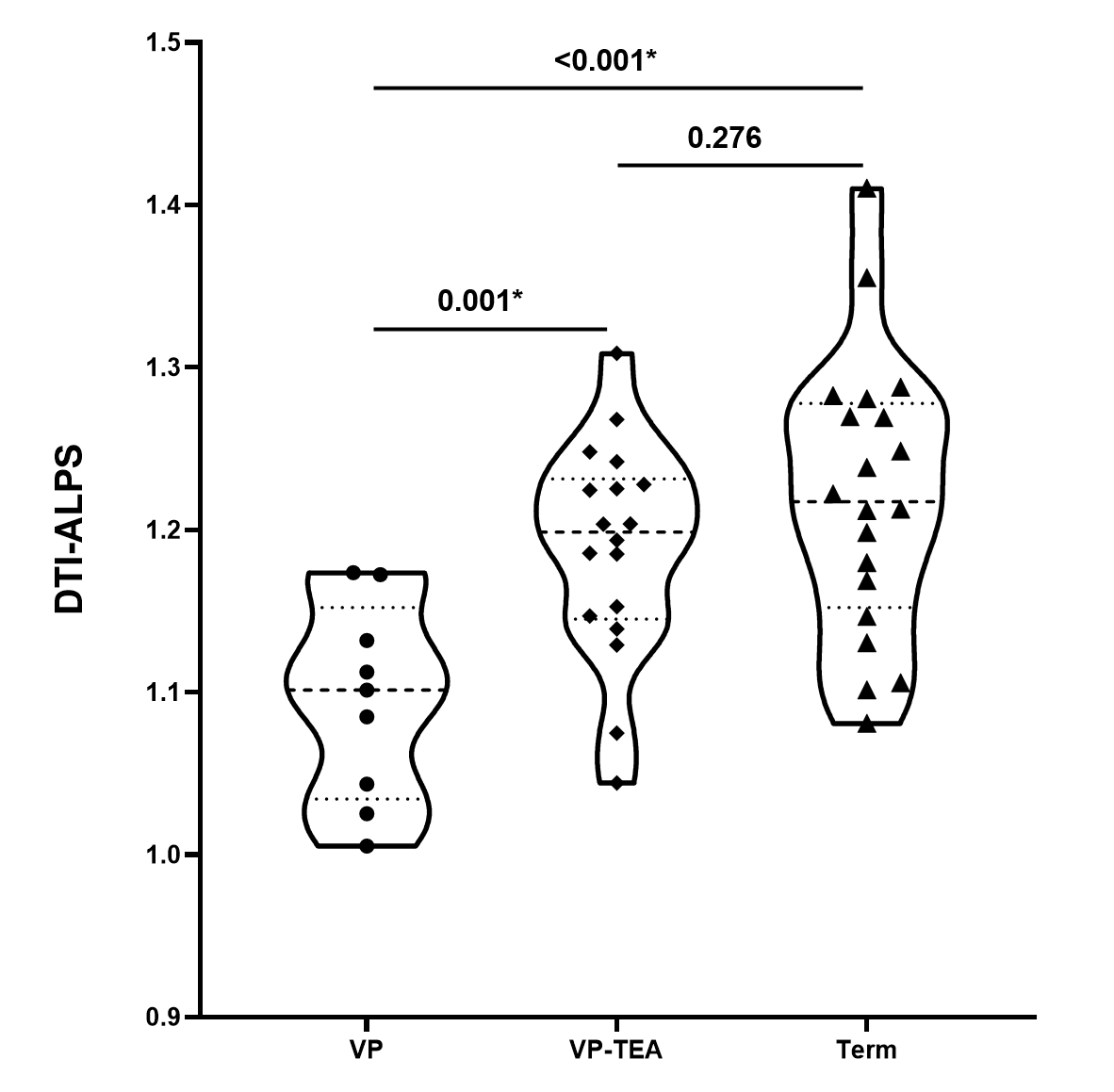
Results: The DTI-ALPS index was significantly lower in VP neonates than VP infants at term equivalent age (TEA) or term neonates. However, the DTI-ALPS index in VP infants at TEA was not found differences with term neonates.
Impact: The glymphatic system function was developing with gestational age, while very preterm infants presented “catch-up” growth pattern up to TEA.
Introduction
The glymphatic system is of critical importance in clearance of brain interstitial solutes, with perivascular spaces (PVSs) as one of the main anatomical pathways1. While the basal ganglia PVSs volume of preterm neonates was smaller than term neonates, PVSs assessment of white matter was not different between them2. However, whether the glymphatic system function differently with preterm and term infants remain unclear. Additionally, studies evaluating glymphatic system function in neonates using brain MRI are lacking. Diffusion-weighted MRI has been proposed as a less invasive alternative to evaluate glymphatic function by calculating the diffusion along perivascular space (DTI- ALPS) index3. Therefore, we aimed to exploring the development of glymphatic system function in very preterm infants comparing with term infants.Methods and Materials
The Institutional Review Broad of the first author’s affiliation approved this study and written informed consent was obtained from parents of the children.Participants Very preterm infants with gestational age <32 weeks (group1), very preterm infants at term equivalent age (TEA) (group 2) and term neonates (group 3) were included and underwent diffusion tensor images (DTI). All the subjects were without abnormalities on conventional MRI.
MR Protocols DTI was performed on a 3.0T MRI scanner (Signa HDxt, General Electric Medical System, Milwaukee, WI, USA) with an 8-channel head coil. Parameters: directions, 30; b values, 0 and 600 s/mm2; TR/TE, 11000/69.5 ms; slice thickness, 2.5 mm with 2.5 mm spacing; FOV, 240 mm; and matrix size = 256 × 256.
Data and statistical analysis DTI raw data were preprocessed by FMRIB software library (FSL; http://www.fmrib.ox.ac.uk/fsl) and diffusion tensor was estimated by linear regression using diffusion-weighted imaging data at b = 600 s/mm2. Then we selected for each subject the axial slices where veins run perpendicular to the lateral ventricle to accurately select brain areas where veins and PVSs run over the x-axis3. On the selected slice, we used the diffusion direction map, and drew two 3 × 3 × 3 mm cubic regions of interest (ROI): one region of interest was drawn over the area of projection fibers (ROI proj) and the other over the area of associative fibers (ROI assoc) in the left hemisphere. Then DTI-ALPS index was originally calculated according to the formula: mean(Dx proj, Dx assoc)/mean(Dy proj, Dz assoc)3,4. Mann-Whitney U test was used to explore the differences of DTI-ALPS index among the above groups.
Results
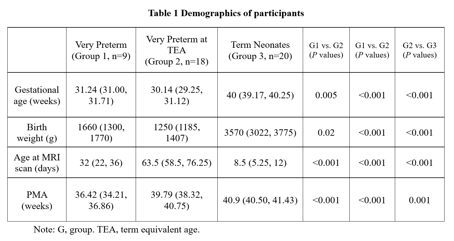
Forty-seven infants were included in this study and nine were very preterm infants, 18 were very preterm infants at TEA, and 20 term neonates. Demographics of those participants were showed in the Table 1. DTI-ALPS index was observed lower in very preterm infants than very preterm infants at TEA (P=0.001), and term neonates (P<0.002). However, DTI-ALPS index in very preterm neonates at TEA was not found differences with term neonates (P=0.276) (Figure 1).Discussion
Exploring the development of glymphatic system function is contribute to better understand and identify the impairment of glymphatic system. Very preterm infants are of lower glymphatic system function that may be associated with AQP4 expression alterations due to preterm births5. And previous animal studies suggested that PVSs develop mainly at the postnatal period which the CSF tracer penetration into the parenchyma only in the newborn mice other than the fetal period6. Furthermore, when very preterm infants are up to TEA, the DTI-ALPS index was increased and was not observed obviously differences with term neonates. The results indicating that glymphatic system function is of age-related development, and very preterm infants may “catch up” with term neonates. But in developing brain, the relationships of DTI-ALPS and age and the neurodevelopmental outcomes were needed to further study.Conclusion
The glymphatic system function of very preterm infants was immaturity than term neonates, but the glymphatic system function was developing with gestational age, and very preterm infants may present the “catch-up” growth pattern up to TEA.Acknowledgements
This work was supported by the National Natural Science Foundation of China (81901516, 82272618, 81971581). Please address correspondence to Jian Yang, e-mail: yj1118@mail.xjtu.edu.cn and Xianjun Li, e-mail: xianj.li@mail.xjtu.edu.cn.References
1. Li X, Lin Z, Liu C, Bai R, Wu D, Yang J. Glymphatic Imaging in Pediatrics. J Magn Reson Imaging. 2023.
2. Kim JY, Nam Y, Kim S, Shin NY, Kim HG. MRI-visible Perivascular Spaces in the Neonatal Brain. Radiology. 2023;307(2):e221314.
3.Taoka T, Masutani Y, Kawai H, et al. Evaluation of glymphatic system activity with the diffusion MR technique: diffusion tensor image analysis along the perivascular space (DTI-ALPS) in Alzheimer's disease cases. Jpn J Radiol. 2017;35(4):172-178.
4. Carotenuto A, Cacciaguerra L, Pagani E, Preziosa P, Filippi M, Rocca MA. Glymphatic system impairment in multiple sclerosis: relation with brain damage and disability. Brain. 2022;145(8):2785-2795.
5. Eidahl JML, Stray-Pedersen A, Rognum TO, Opdal SH. Aquaporin 4 expression in the hippocampus in sudden infant death syndrome and sudden unexplained death in childhood. J Chem Neuroanat. 2021;115:101962.
6. Munk AS, Wang W, Bèchet NB, et al. PDGF-B Is Required for Development of the Glymphatic System. Cell Rep. 2019;26(11):2955-2969.e2953.
Figures