4925
Interaction of neurofluids flow dynamics studied by PC-MRI1Medical Image Processing, University Hospital, Amiens, France, 2CHIMERE UR 7516, Jules Verne University of Picardy, Amiens, France, 3Radiology, University Hospital, Amiens, France, 4Neurosurgery, University Hospital, Amiens, France
Synopsis
Keywords: Neurofluids, Neurofluids
Motivation: Cervical-level is often chosen to estimate Cerebral Blood-Volume Change (CB-VC) during cardiac cycle. Due to the heterogeneity in extracranial cerebral veins anatomy and their high compliance, we hypothesize that the intracranial level could be a better choice to investigate blood and CSF interactions.
Goal(s): To determine the best level for studying the interaction of neurofluids flow dynamics.
Approach: Using PC-MRI, CB-VC and CSF-Volume Change (CSF-VC) were calculated in 36 volunteers at intracranial and extracranial levels, and their interactions were compared by linear regressions.
Results: The interaction between CSF-VC and CB-VC dynamics at intracranial level (R2:0.82±0.16) was higher (p<0.001) than at extracranial level (R2:0.46±0.36).
Impact: This study highlights the greater consistency of spinal CSF-VC response to vascular volume dynamics measured intracranially rather than at the cervical level. These findings are valuable to consider for studying cranio-spinal neurofluids flow dynamics interactions, pressure and compliance.
Introduction
Following the Monro-Kellie doctrine, Cerebral Blood Volume Changes (CB-VC) are mirrored by Cerebrospinal Fluid Volume Changes (CSF-VC) at the spinal canal. Using cine phase-contrast MRI (PC-MRI), previous studies1–3 demonstrated a strong correlation between spinal CSF and arteriovenous (AV) flow waveforms during the cardiac cycle.Nevertheless, to calculate CB-VC, most studies4–6 consider arterial and venous flows measured extracranially at the cervical level. Due to the significant heterogeneity in extracranial cerebral veins anatomy7 and higher compliance, we hypothesize that intracranial and extracranial vascular levels interact differently with spinal CSF.
This study aims to determine which level, intracranial or extracranial, is more suitable for measuring arterial and venous flows to study the interactions of cerebral blood and CSF dynamics.
Methods
-Image acquisition and processingThirty-six healthy young volunteers (age:19–32; 16 women) underwent 3T MRI investigation.
Sagittal 3D phase-contrast cranio-cervical angiography was used to study arterial and venous trees (TE/TR=3ms/5ms; FOV=350*350*350 mm3; spatial resolution=1.5*1.5*3 mm3; flip angle=12°). Sagittal 3D balanced sequence was used to investigate CSF morphology.
2D PC-MRI sequence quantified CSF flows at C2-C3 level and blood flows at cervical and intracranial levels (Fig.1-A). The intracranial plane included three arteries (ICAR, ICAL: right and left internal carotid arteries, BA: basilar artery) and two sinuses (SS: straight sinus, SSS: superior sagittal sinus). The extracranial plane included four arteries (VAR, VAL: right and left vertebral arteries, ICAR, ICAL) and two veins (RJ, LJ: right and left internal jugular veins).
A finger plethysmograph was used as cardiac gating. The sequence parameters are detailed in Fig.1-B.
PC-MRI processing was performed using in-house software–Flow2.08 (Fig.1-C) to segment and automatically reconstruct CSF and vascular flow curves along 32 temporal points of the cardiac cycle.
-CB-VC and CSF-VC dynamics
As shown in Fig.2, total cerebral arterial blood flow dynamics at both intracranial and extracranial levels were derived by simply summing the flows of individual arterial vessels at each level. Similarly, for total cerebral venous flow dynamics. Venous flow curves were then corrected to account for unconsidered peripheral venous drainage, ensuring that the mean total venous flows are equal to mean arterial flows.
Intracranial and extracranial AV flows were calculated by subtracting venous outflow from the arterial inflow. Through integration over time, we calculated the CB-VC dynamics at the two levels.
The CSF-VC dynamics was calculated by integrating the CSF flow measured at C2-C3 and compared to the two CB-VC dynamics calculated at intracranial and extracranial planes (Fig.3).
-Statistical analysis
Linear regressions were performed to evaluate the relationship (R2 and slope) between the 32 points of the CSF-VC curve and the 32 points of the two CB-VC curves (intracranial and extracranial planes). The differences between extracranial and intracranial measurements were assessed using either a paired Student’s t-test or Wilcoxon’s test, depending on the normality of the data distribution. The threshold for significance was set to p<0.05.
Results
Fig.4-A demonstrates significantly higher (p<0.001) R2 and linear regression slope values at the intracranial level (R2:0.82±0.16; slope: -0.73±0.18) compared to the extracranial level (R2:0.46±0.36; slope: -0.36±0.33).Fig. 4-A also reveals significant (p<0.001) differences between intracranial and extracranial CB-VC measurements.
Fig.4-B provides examples of the results obtained from three subjects. In case a, the subject presented a strong negative linear relationship between intracranial CB-VC and the CSF-VC, as well as between extracranial CB-VC and the CSF-VC. Conversely, case c exhibited a strong negative linear relationship at the intracranial level, while the linear relationship was weakly positive at the extracranial level.
Discussion
Linear regression parameters presented higher variability at the extracranial level (Fig.4-A). These findings suggest that the CSF response to vascular volume variation within the craniospinal system is more consistent with the arterial and venous measurements acquired at the intracranial plane. Notably, the differences observed between both levels are mainly attributed to the venous compartment, as extracranially, the internal jugular veins show greater inter-subject variability in vessel morphology and flow pulsatility. Rapidly, because of the compliance of the internal jugular veins, blood volume can easily expand outside the cranium, modifying the dynamics of the cerebral blood volume curve. Consequently, intracranial pressure should be more closely related to intracranial AV than extracranial AV measurements.Conclusion
Arterial and venous flow measurements obtained by PC-MRI at the intracranial plane are more relevant for analyzing the interaction between blood and CSF compared to the extracranial level, where significant individual venous heterogeneity exists.Acknowledgements
This research was supported by EquipEX FIGURES (Facing Faces Institute Guilding Research), Hanuman ANR-18-CE45-0014 and Region Haut de France. Thanks to the staff members at the Facing Faces Institute (Amiens, France) for technical assistance. Thanks to David Chechin from Phillips industry for his scientific support and Héléna Freulet, Garance Arbeaumont-Trocmé, Vilhem Marion and Julien Van Gysel (MRI research technicians) for assistance with the acquisition of high-quality images.References
1. Bhadelia RA, Bogdan AR, Kaplan RF, Wolpert SM. Cerebrospinal fluid pulsation amplitude and its quantitative relationship to cerebral blood flow pulsations: a phase-contrast MR flow imaging study. Neuroradiology. 1997;39(4):258-264. doi:10.1007/s002340050404
2. Balédent O, Henry-Feugeas MC, C ´eCILE, Idy-Peretti I. Cerebrospinal Fluid Dynamics and Relation with Blood Flow: A Magnetic Resonance Study with Semiautomated Cerebrospinal Fluid Segmentation. Invest Radiol. 2001;36(7):368.
3. Zhu DC, Xenos M, Linninger AA, Penn RD. Dynamics of lateral ventricle and cerebrospinal fluid in normal and hydrocephalic brains. J Magn Reson Imaging. 2006;24(4):756-770. doi:10.1002/jmri.20679
4. Wåhlin A, Ambarki K, Hauksson J, Birgander R, Malm J, Eklund A. Phase contrast MRI quantification of pulsatile volumes of brain arteries, veins, and cerebrospinal fluids compartments: Repeatability and physiological interactions. J Magn Reson Imaging. 2012;35(5):1055-1062. doi:10.1002/jmri.23527
5. Sakhare AR, Barisano G, Pa J. Assessing test–retest reliability of phase contrast MRI for measuring cerebrospinal fluid and cerebral blood flow dynamics. Magn Reson Med. 2019;82(2):658-670. doi:10.1002/mrm.27752
6. Laganà MM, Di Tella S, Ferrari F, et al. Blood and cerebrospinal fluid flow oscillations measured with real-time phase-contrast MRI: breathing mode matters. Fluids Barriers CNS. 2022;19(1):100. doi:10.1186/s12987-022-00394-0
7. Stoquart-Elsankari S, Lehmann P, Villette A, et al. A phase-contrast MRI study of physiologic cerebral venous flow. J Cereb Blood Flow Metab Off J Int Soc Cereb Blood Flow Metab. 2009;29(6):1208-1215. doi:10.1038/jcbfm.2009.29
8. Liu P, Fall S, Balédent O. Flow 2.0 -a flexible, scalable, cross-platform post-processing software for realtime phase contrast sequences. Published online July 26, 2022. doi:10.48550/arXiv.2207.12712
2. Balédent O, Henry-Feugeas MC, C ´eCILE, Idy-Peretti I. Cerebrospinal Fluid Dynamics and Relation with Blood Flow: A Magnetic Resonance Study with Semiautomated Cerebrospinal Fluid Segmentation. Invest Radiol. 2001;36(7):368.
3. Zhu DC, Xenos M, Linninger AA, Penn RD. Dynamics of lateral ventricle and cerebrospinal fluid in normal and hydrocephalic brains. J Magn Reson Imaging. 2006;24(4):756-770. doi:10.1002/jmri.20679
4. Wåhlin A, Ambarki K, Hauksson J, Birgander R, Malm J, Eklund A. Phase contrast MRI quantification of pulsatile volumes of brain arteries, veins, and cerebrospinal fluids compartments: Repeatability and physiological interactions. J Magn Reson Imaging. 2012;35(5):1055-1062. doi:10.1002/jmri.23527
5. Sakhare AR, Barisano G, Pa J. Assessing test–retest reliability of phase contrast MRI for measuring cerebrospinal fluid and cerebral blood flow dynamics. Magn Reson Med. 2019;82(2):658-670. doi:10.1002/mrm.27752
6. Laganà MM, Di Tella S, Ferrari F, et al. Blood and cerebrospinal fluid flow oscillations measured with real-time phase-contrast MRI: breathing mode matters. Fluids Barriers CNS. 2022;19(1):100. doi:10.1186/s12987-022-00394-0
7. Stoquart-Elsankari S, Lehmann P, Villette A, et al. A phase-contrast MRI study of physiologic cerebral venous flow. J Cereb Blood Flow Metab Off J Int Soc Cereb Blood Flow Metab. 2009;29(6):1208-1215. doi:10.1038/jcbfm.2009.29
8. Liu P, Fall S, Balédent O. Flow 2.0 -a flexible, scalable, cross-platform post-processing software for realtime phase contrast sequences. Published online July 26, 2022. doi:10.48550/arXiv.2207.12712
Figures
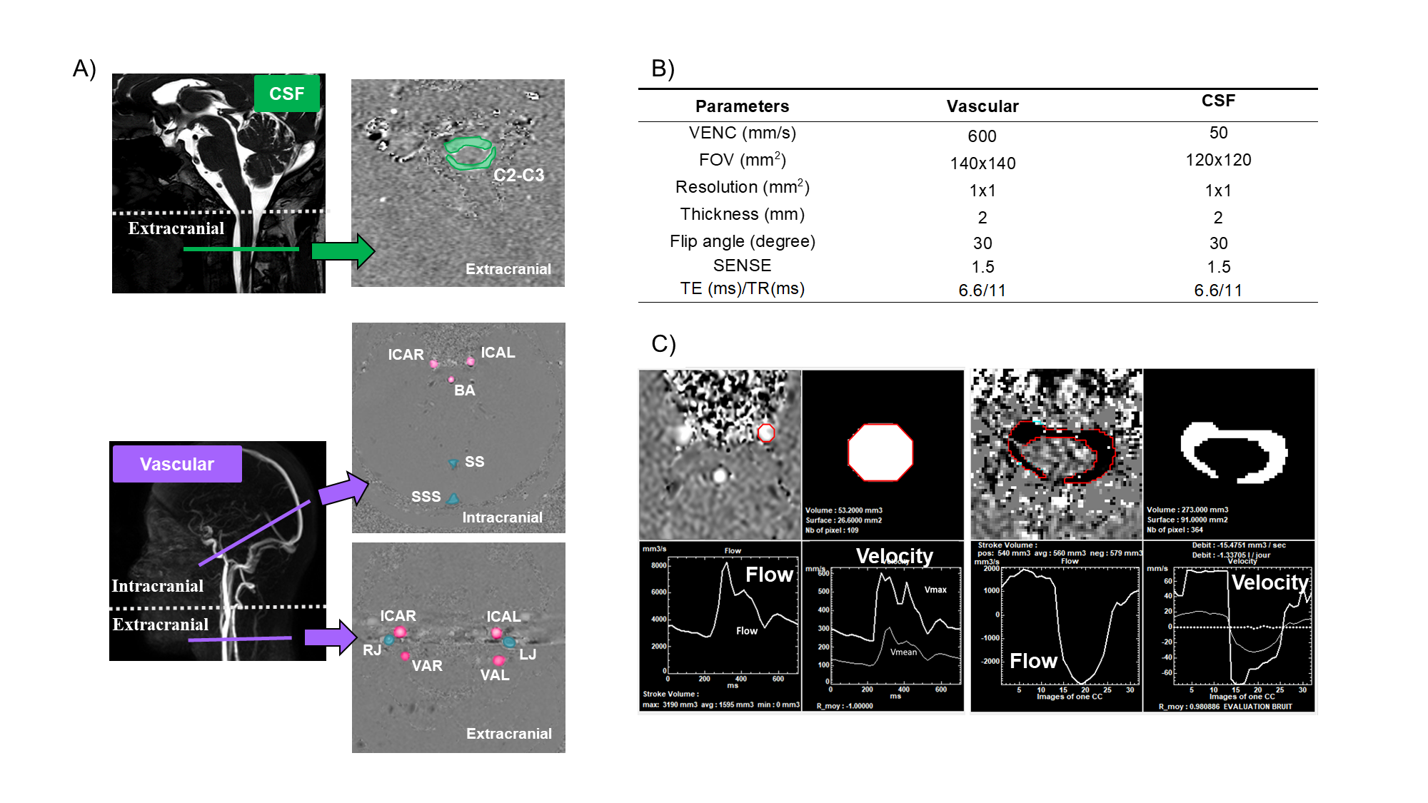
Figure 1: PC-MRI image acquisition and processing. A) Quantification of CSF flow at C2-C3 level (green). Arterial (pink) and venous (blue) flow measured at intracranial and extracranial planes (purple). B) Main cine PC-MRI parameters. C) "Flow" software semi-automatically segments, computes ROI area, fluid velocity, and generates a fluid flow curve over 32 temporal points of the cardiac cycle. Example: left internal carotid artery (left) and cervical CSF flow (right).
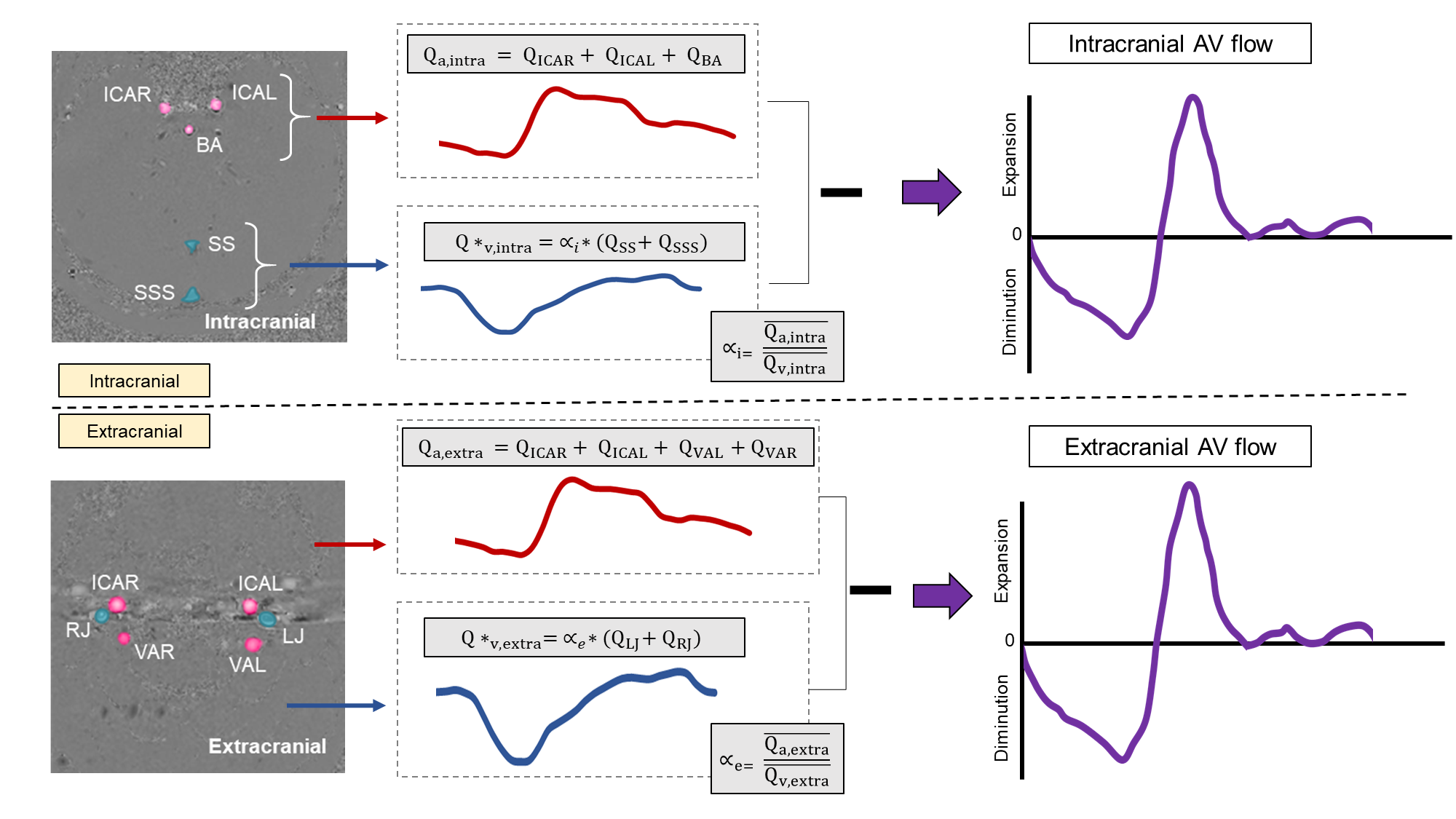
Figure 2: Arteriovenous flows at intracranial and extracranial planes. The total venous flow was adjusted by αi and αe, which are calculated by dividing the mean value of the arterial flow by the mean value of the venous flow. Intracranial and extracranial AV flows were calculated by subtracting venous outflow from the arterial inflow.
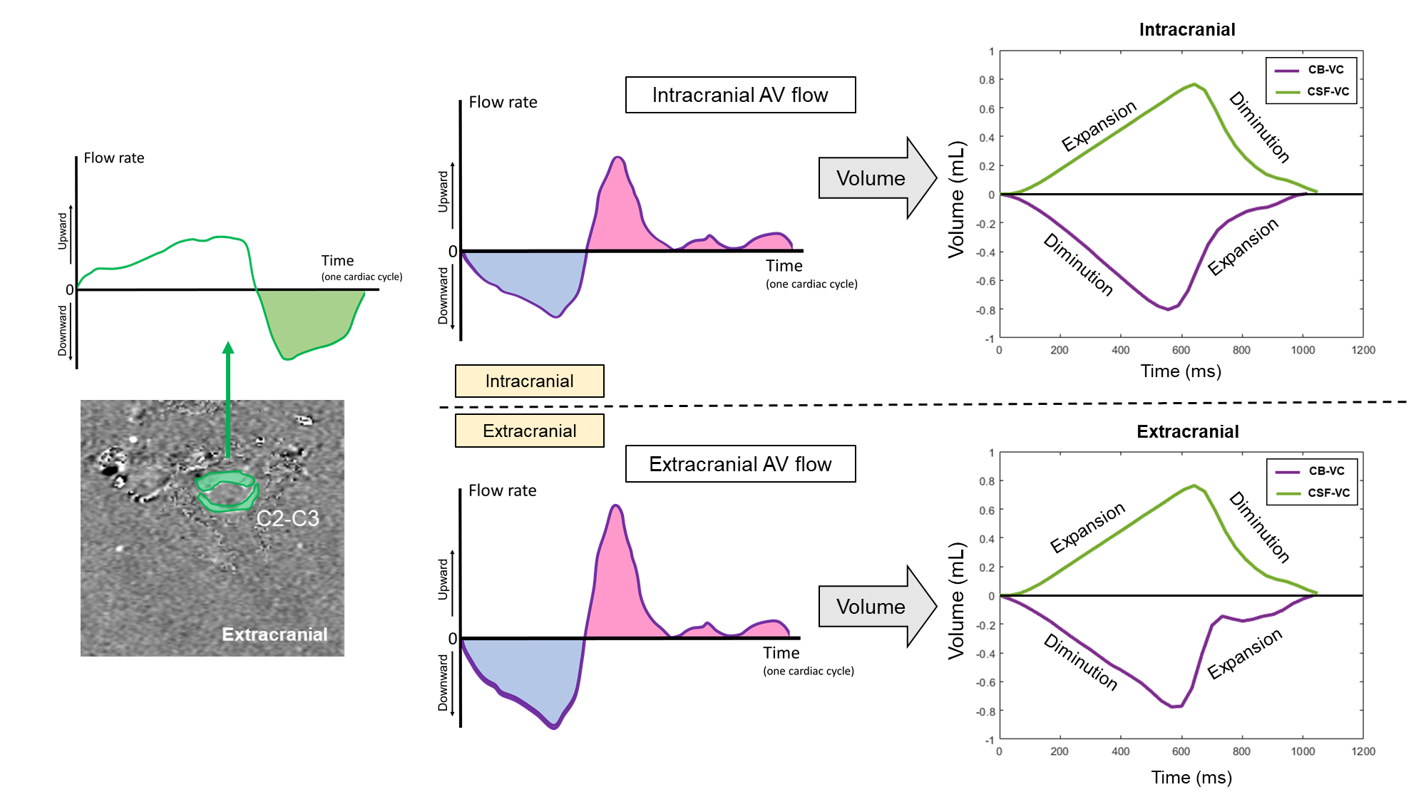
Figure 3: CB-VC and CSF-VC at intracranial and extracranial planes. The CSF-VC measured at C2-C3 served as a reference in both intracranial and extracranial planes. CB-VC was calculated at both levels based on AV flows.
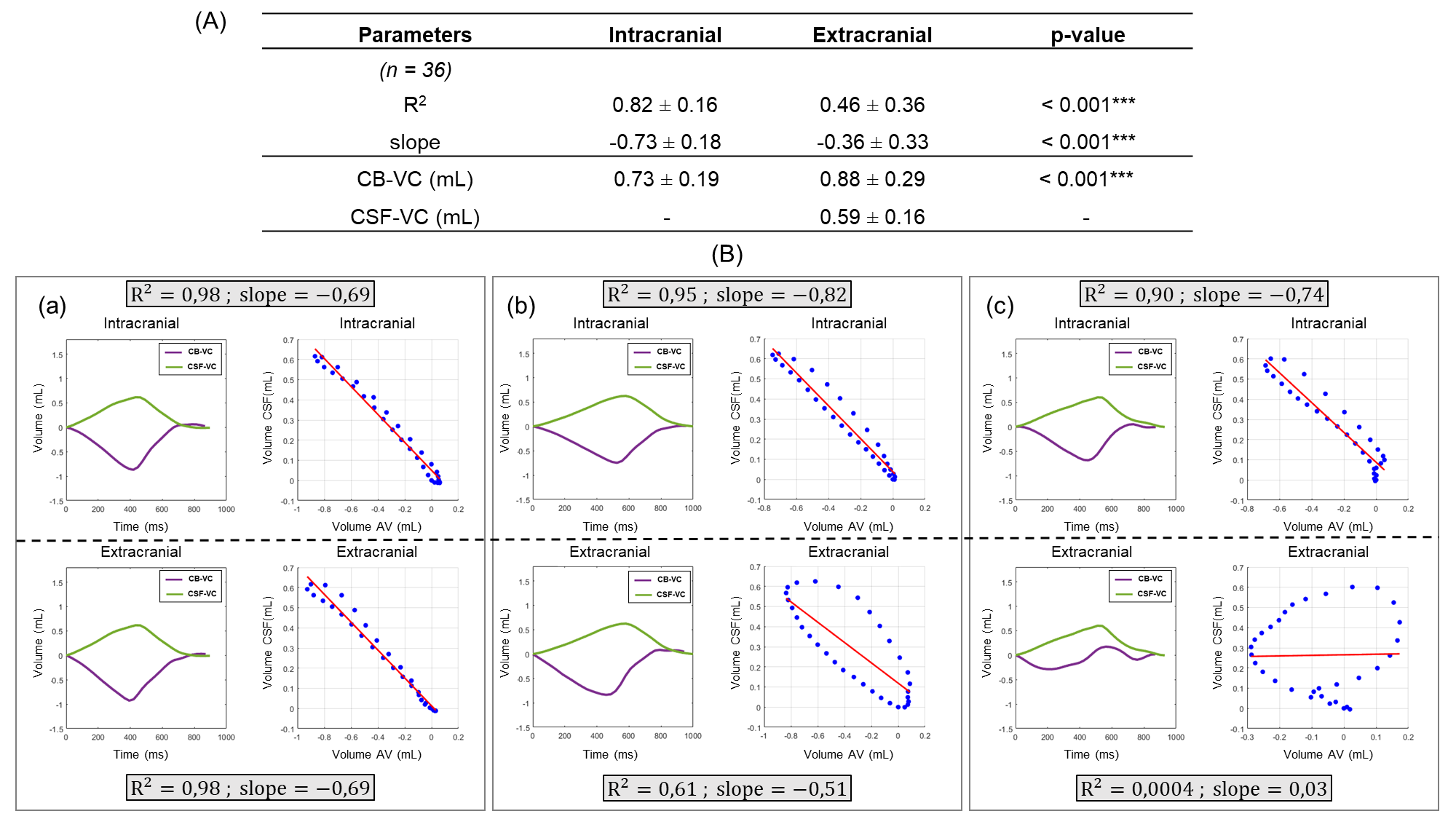
Figure 4. Results from statistical analysis. A) Linear regression results for extracranial and intracranial planes. CB-VC and CSF-VC measurements. *** indicates p < 0.001. B) Results from three different subjects. CB-VC (purple) and CSF-VC (green) curves with intracranial and extracranial linear regressions. The results of R2 and slope values are presented for each case. a) Strong negative linear relationship in both planes; b) satisfactory extracranial relationship; c) weak extracranial relationship.