4924
MR Elastography as a Potential Imaging Biomarker and Non-Invasive Method for Diagnosing Spontaneous Intracranial Hypotension1Radiology, Mayo Clinic, Rochester, MN, United States, 2Mayo Clinic, Rochester, MN, United States
Synopsis
Keywords: Neurofluids, Elastography, SIH; CSF Leak
Motivation: Spontaneous intracranial hypotension (SIH) is a debilitating disease with a normal brain MRI in 20% of cases.
Goal(s): Our goal was to use MR elastography (MRE) to identify unique viscoelastic patterns in SIH patients.
Approach: We performed brain MRE in 15 SIH patients and compared their mechanical properties to 65 healthy controls.
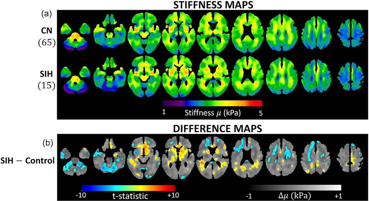
Results: SIH patients exhibited significant differences in both stiffness and damping ratio compared to healthy controls.
Impact: We demonstrate MRE as a potential imaging biomarker and a non-invasive method for diagnosing SIH, including patients with a normal brain MRI.
INTRODUCTION
Spontaneous Intracranial Hypotension (SIH) is a condition, often presenting with debilitating headache, resulting from a leak of cerebrospinal fluid from the spinal canal arising independently of a medical procedure or trauma. SIH can present with normal brain MRI findings and non-specific symptoms, leading to the underdiagnosis in some patients and unnecessary invasive myelography in others who are found not to have the condition. Given the likelihood that SIH alters intracranial biomechanics, the goal of this study was to evaluate MR elastography (MRE) as a potential non-invasive test to diagnose the condition.METHODS
We performed MRE in 15 confirmed SIH patients from September 2022 to April 2023. Age, sex, symptom duration, and brain MRI Bern score were collected. MRE data were used to compute stiffness and damping ratio maps, and voxel-wise modeling was performed to detect clusters of significant differences in mechanical properties between SIH patients and healthy control participants. To evaluate diagnostic accuracy, each exam was summarized by two spatial pattern scores (one each for stiffness and damping ratio) and group-wise discrimination was evaluated by receiver operating characteristic curve analysis.RESULTS
Of the 15 SIH patients enrolled and imaged with Brain MRE, 9 patients (60%) were female. The mean age was 53.2 years old (range 35-70). The mean Bern score was 4.7 (range 0-9). Four patients had a Bern score of 0. Symptom duration ranged from 1 month to 6 years. All 15 patients reported headache. Thirteen (86.7%) had orthostatic headache, while the remaining 2 (13.3%) had headaches that worsened with exertion. Five patients (33.3%) had photophobia and 10 (66.7%) patients reported tinnitus.SIH patients exhibited significant differences in both stiffness and damping ratio (false discovery rate corrected Q<0.05). Pattern analysis discriminated SIH patients from healthy controls with an area under the curve (AUC) of 0.97 overall, and AUC was 0.97 in those without MRI findings.
CONCLUSION
Results from this pilot study demonstrate MRE as a potential imaging biomarker and a non-invasive method for diagnosing SIH, including patients with a normal brain MRI.Acknowledgements
No acknowledgement found.References
Schievink WI, Maya MM, Jean-Pierre S, Nuño M, Prasad RS, Moser FG. A classification system of spontaneous spinal CSF leaks. Neurology. Aug 16 2016;87(7):673-9. doi:10.1212/wnl.0000000000002986
Schievink WI. Spontaneous Intracranial Hypotension. N Engl J Med. Dec 2 2021;385(23):2173-2178. doi:10.1056/NEJMra2101561
Cogswell PM, Murphy MC, Madhavan AA, et al. Features of Idiopathic Intracranial Hypertension on MRI With MR Elastography: Prospective Comparison With Control Individuals and Assessment of Postintervention Changes. AJR Am J Roentgenol. Dec 2022;219(6):940-951. doi:10.2214/ajr.22.27904
Murphy MC, Huston J, 3rd, Jack CR, Jr., et al. Measuring the characteristic topography of brain stiffness with magnetic resonance elastography. PLoS One. 2013;8(12):e81668. doi:10.1371/journal.pone.0081668
Figures