4923
How Breathing and Cardiac functions interact with Cerebral arterio-venous blood flows: Origin of CSF oscillations1Amiens Picardy University Hospital, CHIMERE UR.7516, Amiens, France, 2Jules Verne University of Picardy, CHIMERE UR 7516, Amiens, France, 3Amiens Picardy University Hospital, Neurosurgery Department, Amiens, France, 4Amiens Picardy University Hospital, Radiology Department, Amiens, France
Synopsis
Keywords: Neurofluids, Neurofluids, Real time, phase contrast, CSF, CBF
Motivation: The main drivers of CSF oscillations are currently controversial.
Goal(s): To investigate whether the breathing or cardiac regulated Cerebral blood flow are the major driver of CSF dynamics.
Approach: Investigate Cerebral blood and CSF flows through the intracranial compartment during free and deep breathing using real-time phase-contrast sequence. To quantify the neurofluids volume displacement along the cardiac cycle.
Results: Both cardiac and breathing cycles influenced neurofluids volume displacements. CSF dynamics is significantly correlated with intracranial blood volume change. CSF dynamic acts as a compensatory mechanism of intracranial blood volume dynamics.
Impact: This study confirms that intracranial blood volume change due to cardiac and breathing activities are the main drivers of CSF dynamic. This study provides valuable insights for understanding CSF circulation's complex mechanism and investigating idiopathic cerebral diseases.
Introduction
Although changes in cerebral blood volume (CBV) were initially recognized as the major driver of cerebrospinal fluid (CSF) oscillations1,2, recent advances in real-time phase-contrast MRI (RT-PC) have revealed the influence of breathing on CSF dynamics3-5. New mechanistic hypotheses challenging the established Monro-Kellie doctrine have emerged, such as CSF flow is driven by the thoracic and lumbar spina6.Therefore, quantifying both CBV and CSF displacement volume (CSFV) under the influence of breathing will provide a better understanding of CSF dynamics. The aim of this study was to measure the CBV and CSFV during free- and deep-breathing patterns using RT-PC to investigate whether the breathing-modulated CBV fluctuations are the main driving force of CSF oscillations.
Methods
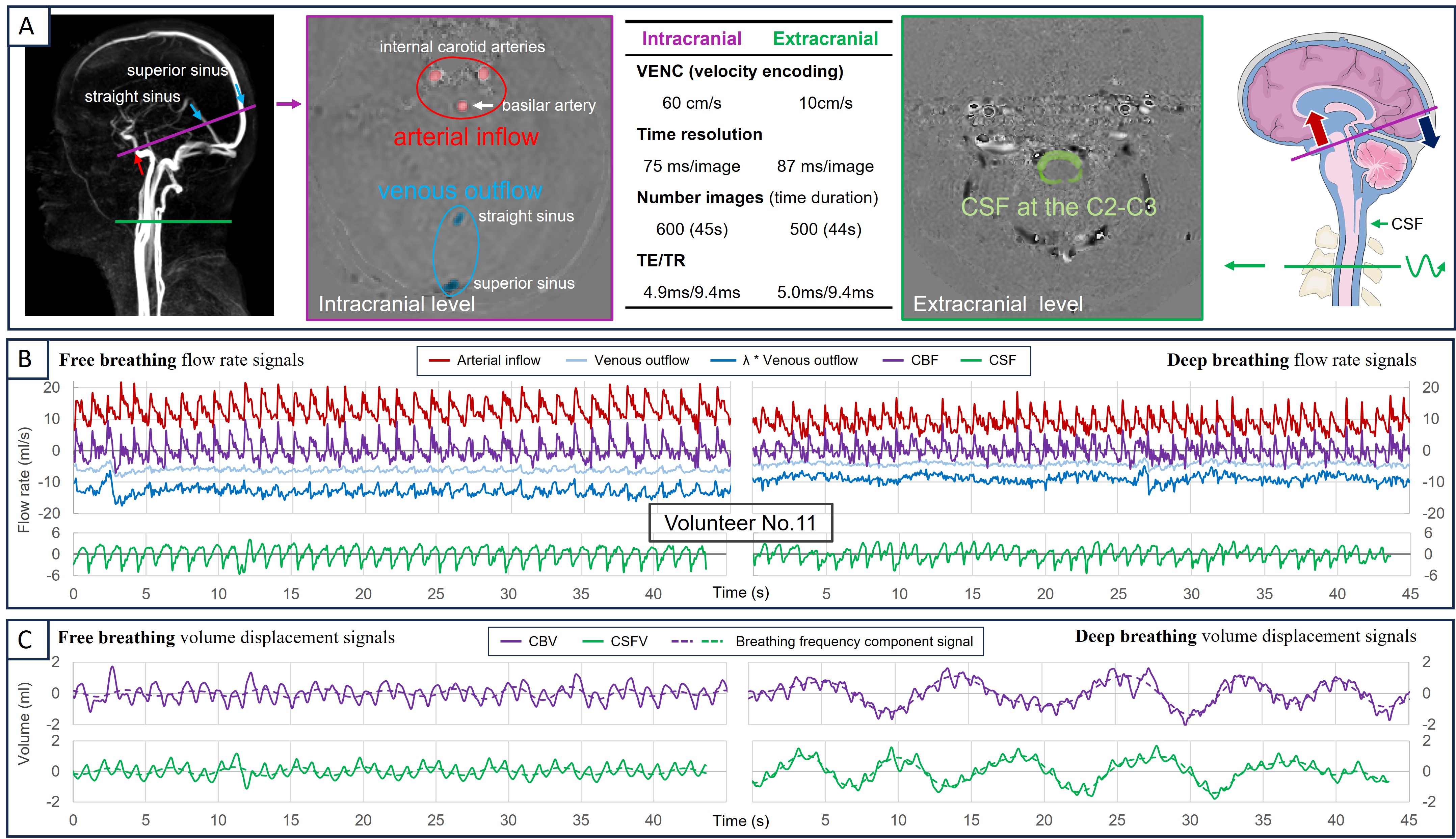
-Image acquisition12 healthy volunteers (age: 20~34) were examined using a clinical 3T scanner and a 32-channel head coil. A finger plethysmograph and a respiratory chest were used to record pulse and breathing signals synchronously during acquisition. Each imaging level was acquired twice: once during free-breathing and once during deep-breathing.
For quantification purposes, the intracranial level was selected for CBV measurements, while the extracranial level, C2-C3, was used for CSF measurements. The RT-PC used in this study was a multi-shot, gradient-recalled echo-planar imaging sequence with parallel acquisition technology. Parameters were: SENSE=2.5, EPI-factor=7, FOV=140*140mm2, matrix acquisition=70*70mm2. Other parameters are shown in Fig.1-A.
-Extraction of CBV and CSFV
All image and signal processing was performed using in-house software – Flow 2.07,8.
Arterial inflow, venous outflow, and CSF oscillations were extracted through post-processing steps, including image segmentation, background field correction, and de-aliasing9.
Venous outflow was adjusted to account for unconsidered peripheral venous drainage and maintain a mean venous flow equal to the mean arterial flow. Subsequently, cerebral blood flow (CBF) was calculated as the sum of the arterial inflow and venous outflow (Fig.1-B).
The CBF and CSF flow signals were then integrated over time to obtain CBV and CSFV signals, respectively. Finally, to preserve the breathing and cardiac frequency components, both volume signals underwent baseline drift correction and high-pass filtering (>0.1 Hz) (Fig.1-C).
-Reconstruction of Cardiac- and Breath-Volume Displacement Signals
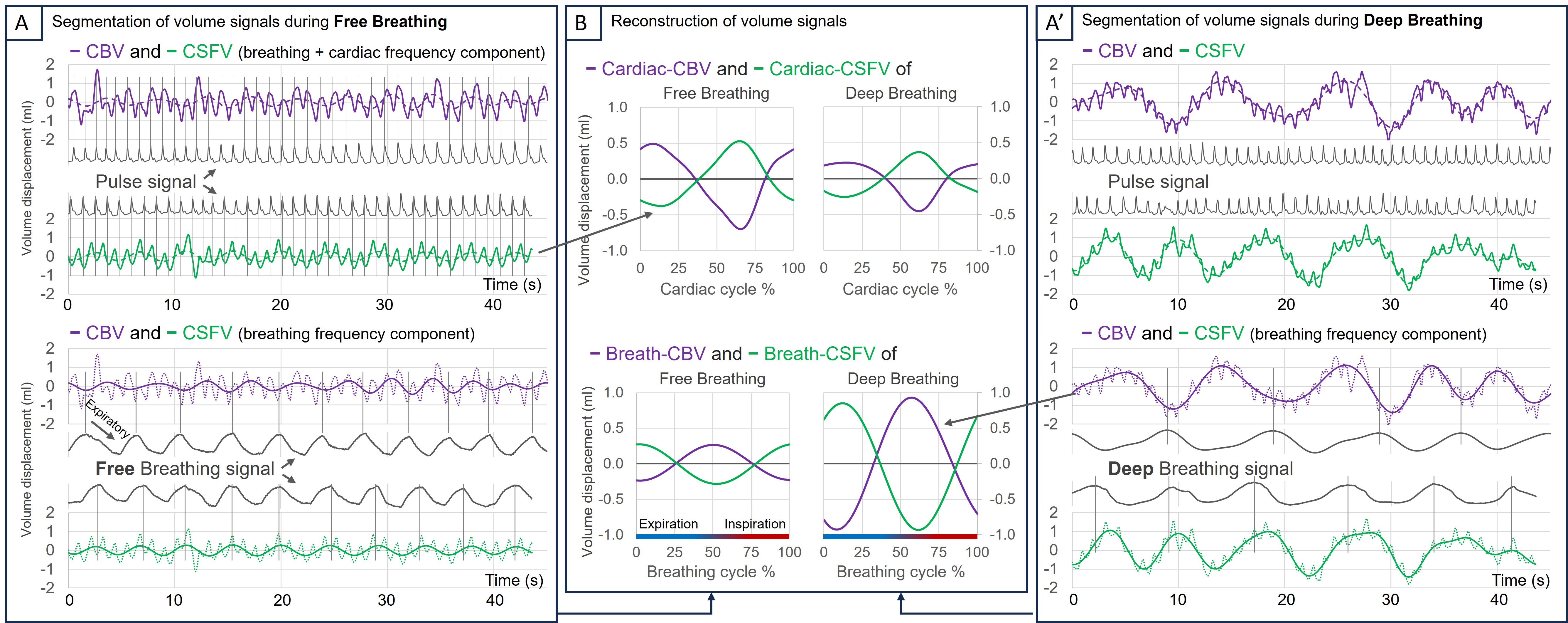
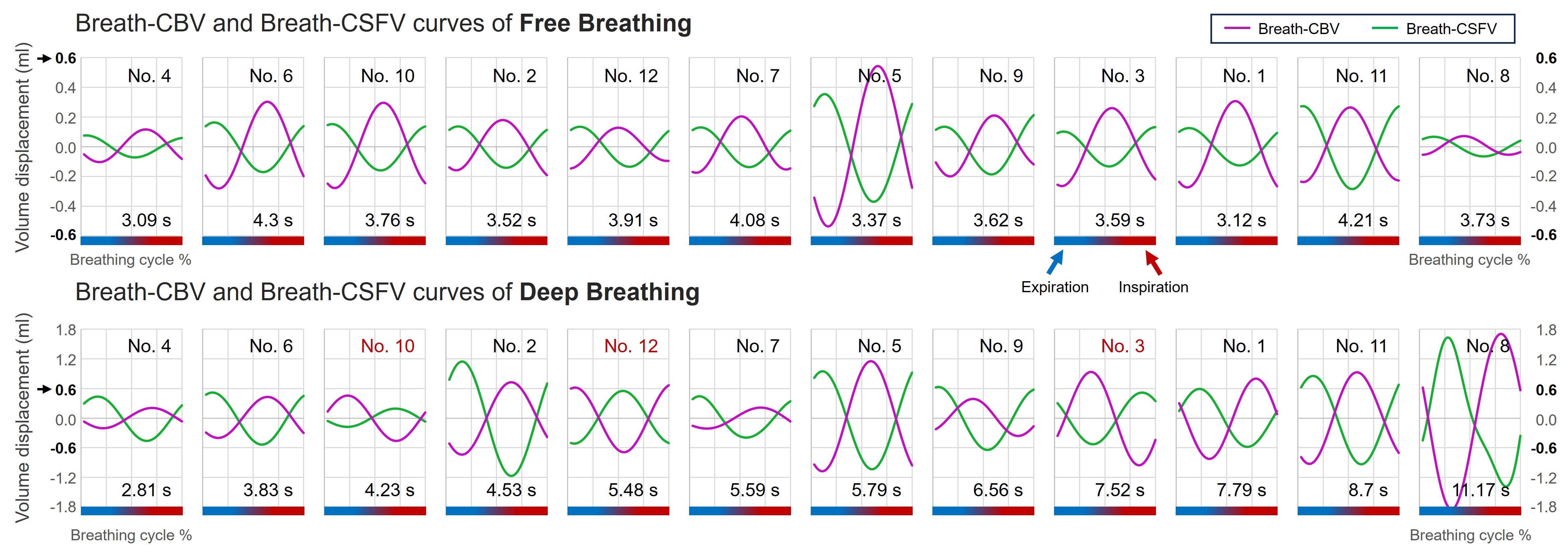
Pulse and breathing signal peaks were identified to segment the volume curves according to the multiple cardiac and respiratory cycles (Fig.2-A&A’). This resulted in reconstructed cardiac-CBV/CSFV and Breath-CBV/CSFV curves, capturing the mean volumetric changes across these physiological cycles (Fig.2-B).
The analysis of the reconstructed volume curves aimed to quantify the breathing effects and to investigate the CBV-CSFV correlation.
Results
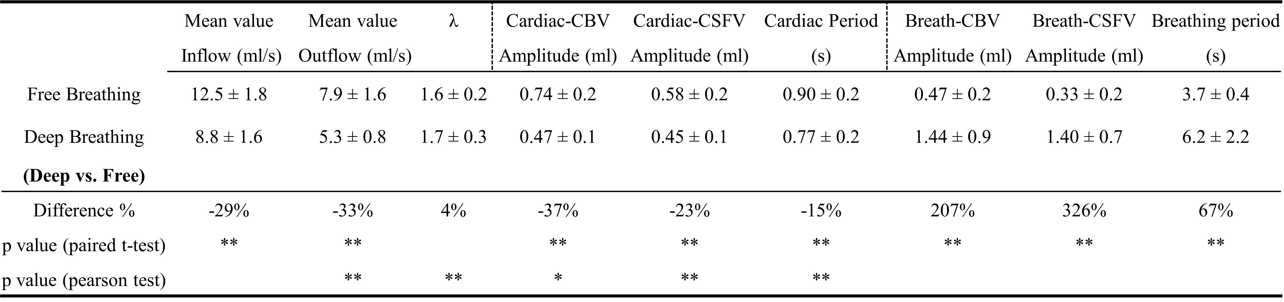
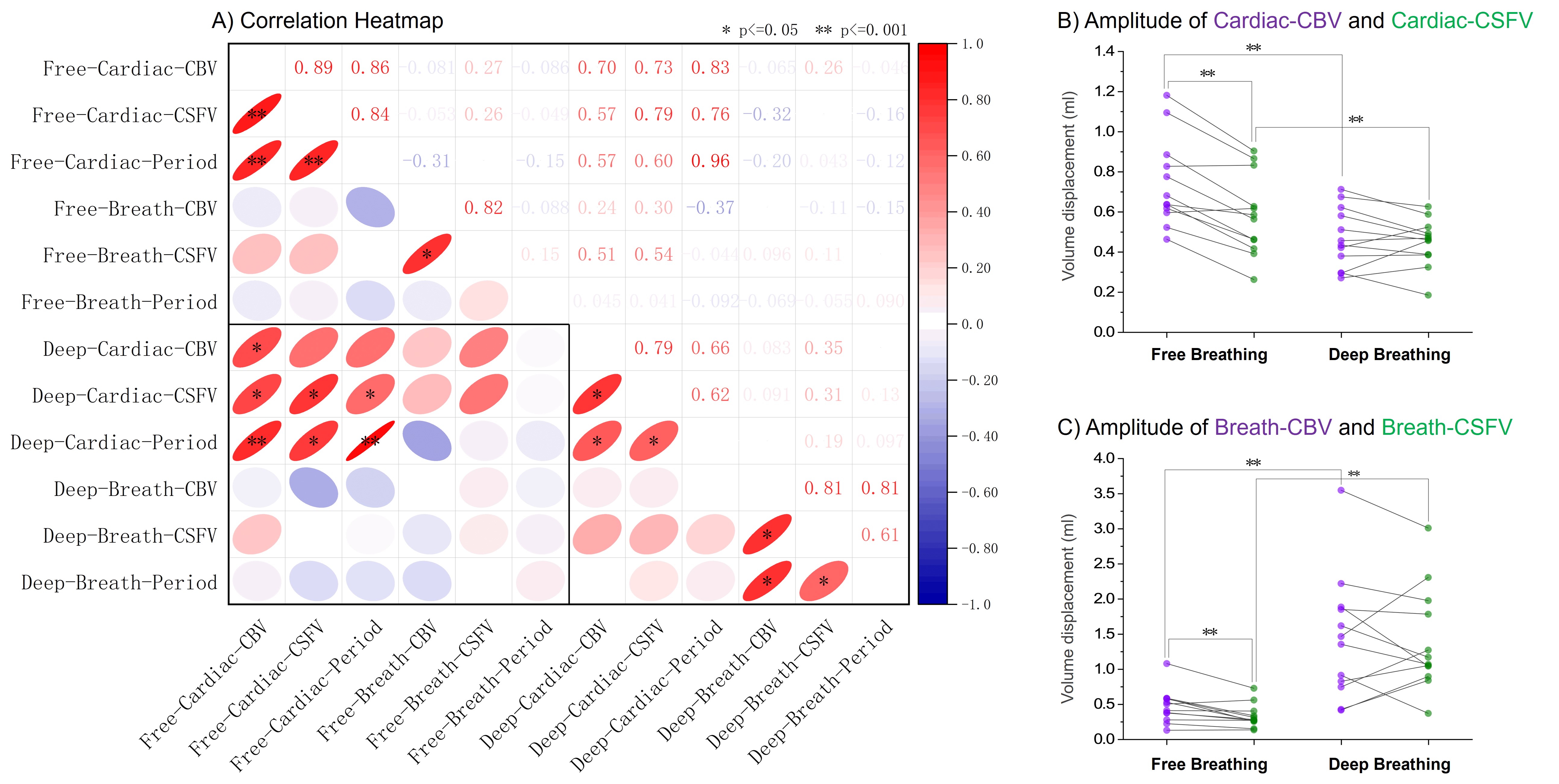
Fig.3 demonstrates that during deep-breathing, compared to free-breathing, inflow decreases by 29%. Cardiac-CBV and Cardiac-CSFV amplitudes decrease by 37% and 23%, respectively, partly due to a 15% shorter cardiac cycle. Conversely, Breath-CBV and Breath-CSFV amplitudes increase by 207% and 326%.Fig.4 illustrates that in both breathing states, a significant correlation was presented between the amplitudes of Cardiac-CBV and Cardiac-CSFV, as well as between the amplitudes of Breath-CBV and Breath-CSFV. No significant amplitude differences were observed between Cardiac/Breath-CBV and Cardiac/Breath-CSFV during deep-breathing. Moreover, Cardiac-CBV/CSFV amplitudes are cardiac period dependent, whereas Breath-CBV/CSFV amplitudes correlate with the breathing period only during deep-breathing.
During free-breathing, CSF flows consistently toward the intracranial compartment during inspiration. However, deep-breathing induces significant phase shifts in three cases, marked in red in Fig.5.
Discussion
Free-breathing can affect CBF and CSF flow10; therefore, CBV changes must be considered in CSF dynamics studies11. This study demonstrates a strong correlation between CBV and CSFV displacements due to cardiac and breathing activities.Using pulse and breathing signals for segmentation and reconstruction of Cardiac-CBV/CSFV and Breath-CBV/CSFV partially overcomes the challenges of simultaneous CBF and CSF measurements. This study demonstrated a clear symmetry between CBV and CSFV. Moreover, this approach facilitates the observation of the volume displacement direction during the different respiratory phases.
The ability of CSF to regulate CBV changes was more pronounced during deep-breathing (Fig.4). This interesting phenomenon is possibly related to alterations in intracranial compliance; however, further investigation is worthwhile.
Although ultra-low frequency components (<0.1 Hz) of CBV and CSFV were observed, they were not analyzed in this study due to frequency resolution limitations. Future studies could further investigate this phenomenon.
Conclusion
This study confirms that changes in CBV resulting from cardiac and breathing activities are the main drivers of CSF dynamics, and the free physiological breathing activity makes a minor contribution to CSF dynamics. This study provides valuable insights into the mechanism of CSF circulation and its potential clinical diagnostic applications in certain diseases.Acknowledgements
This research was supported by EquipEX FIGURES (Facing Faces Institute Guilding Research), Hanuman ANR-18-CE45-0014 and Region Haut de France.Thanks to the staff members at the Facing Faces Institute (Amiens, France) for technical assistance.Thanks to David Chechin from Phillips industry for his scientific support.References
- Balédent O, Idy-peretti I. Cerebrospinal fluid dynamics and relation with blood flow: a magnetic resonance study with semiautomated cerebrospinal fluid segmentation. Investigative radiology. 2001 Jul 1;36(7):368-77.
- Alperin N, Lee SH, Loth F, Raksin P, Lichtor T. MR-Intracranial Pressure (ICP): A method for noninvasive measurement of intracranial pressure and elastance. Baboon and Human Study. Radiology. 2000;217(3):877-85. https://doi.org/10.1148/radiology.217.3.r00dc42877.
- Balédent O, Liu P, Lokossou A, Fall S, Metanbou S, Makki M. Real-time phase contrast magnetic resonance imaging for assessment of cerebral hemodynamics during breathing. In ISMRM 2019-International Society for Magnetic Resonance in Medicine 2019 May 11. https://hal.archives-ouvertes.fr/hal-03736882.
- Yildiz S, Thyagaraj S, Jin N, Zhong X, Heidari Pahlavian S, Martin BA, Loth F, Oshinski J, Sabra KG. Quantifying the influence of respiration and cardiac pulsations on cerebrospinal fluid dynamics using real‐time phase‐contrast MRI. Journal of Magnetic Resonance Imaging. 2017 Aug;46(2):431-9. https://doi.org/10.1002/jmri.25591.
- Gutiérrez-Montes, C., W. Coenen, M. Vidorreta, S. Sincomb, C. Martínez-Bazán, A. L. Sánchez, and V. Haughton. “Effect of Normal Breathing on the Movement of CSF in the Spinal Subarachnoid Space.” American Journal of Neuroradiology 43, no. 9 (September 1, 2022): 1369–74. https://doi.org/10.3174/ajnr.A7603.
- Lloyd, Robert A., Jane E. Butler, Simon C. Gandevia, Iain K. Ball, Barbara Toson, Marcus A. Stoodley, and Lynne E. Bilston. “Respiratory Cerebrospinal Fluid Flow Is Driven by the Thoracic and Lumbar Spinal Pressures.” The Journal of Physiology 598, no. 24 (2020): 5789–5805. https://doi.org/10.1113/JP279458.
- Liu P, Lokossou A, Fall S, Makki M and Bamendent O, 2019. Post Processing Software for Echo Planar Imaging Phase Contrast Sequence. ISMRM 27th, (4823). https://archive.ismrm.org/2019/4823.html
- Liu P, Fall S, Balédent O. Flow 2.0-a flexible, scalable, cross-platform post-processing software for realtime phase contrast sequences. In ISMRM 2022-International Society for Magnetic Resonance in Medicine 2022 May 7. https://archive.ismrm.org/2022/2772.html.
- Liu P, Fall S, Balédent O. Use of real-time phase-contrast MRI to quantify the effect of spontaneous breathing on the cerebral arteries. NeuroImage. 2022 Jun 7:119361. https://doi.org/10.1016/j.neuroimage.2022.119361.
- Liu P, Monnier H, Owashi K, Constant JM, Capel C, Balédent O. “The Effects of Free Breathing on Cerebral Venous Flow: a real-time phase contrast MRI study in healthy adults”. Journal of neuroscience, Accepted, in production.
- Burman, R., & Alperin, N. (2023). CSF‐to‐blood toxins clearance is modulated by breathing through cranio–spinal CSF oscillation. Journal of sleep research, e14029. https://doi.org/10.1111/jsr.14029.
Figures