4919
The relation between sleep indices and indices for brain interstitial fluid dynamics by MRI and the effects of orexin antagonists: a FLUID study.1Department of Innovative Biomedical Visualization (iBMV), Nagoya University, Nagoya, Japan, 2Department of Radiology, Nagoya University, Nagoya, Japan, 3Department of Psychiatry, Nagoya University, Nagoya, Japan, 4Department of Radiological Technology, Nagoya University, Nagoya, Japan, 5Department of Integrated Health Sciences, Nagoya University, Nagoya, Japan, 6Department of Radiology, Juntendo University of Medicine, Tokyo, Japan, 7Medical Headquarters, Eisai Co., Ltd., Tokyo, Japan, 8Department of CT-MR Solution Planning, Canon Medical Systems Corporation, Otawara, Japan, 9Pathophysiology of Mental Disorders Nagoya University Graduate School of Medicine, Nagoya University, Nagoya, Japan
Synopsis
Keywords: Neurofluids, Neurofluids
Motivation: The motivation is to investigate the relationship between sleep and interstitial fluid dynamics influenced by the administration of lemborexant, a dual orexin receptor antagonist.
Goal(s): The goal is to identify changes in indices of interstitial fluid dynamics related to sleep before and after the administration of lemborexant.
Approach: ALPS-index, Ktrans by DSC method, and choroid plexus volume were assessed as indicators of interstitial fluid dynamics.
Results: The sleep indexes on a night were found to correlate with the ALPS-index measured in the evening before that night's sleep. In multiple regression analysis, the ALPS-index was lower when sleep parameters were poor at baseline.
Impact: The sleep indexes on a night were found to correlate with the ALPS-index measured the evening before. The evaluation of the ALPS-index before and after lemborexant administration revealed that the baseline sleep status played a role in improving the index.
Background and purpose
Dual orexin receptor antagonists (DORAs) have been shown to be effective in treating insomnia disorder by blocking wakefulness that interferes with sleep. Lemborexant (LEM) is an orally administered, approved DORA. In the current study, named as the FLUID study (an open-label, single-centre pilot study to investigate the effect of Lemborexant on sleep management in Japanese subjects aged 50 years and older with Insomnia Disorder), we aimed to assess the extent to which the improvement in sleep resulting from lemborexant contributed to the enhancement of interstitial fluid dynamics, as represented by the glymphatic system. The previous reports showed that significant improvements in objective sleep efficiency (SE) and subjective sleep parameters at weeks 4 and 12 following treatment with LEM [1]. Various methods, including diffusion tensor image analysis along the perivascular space (DTI-ALPS) [2], dynamic contrast-enhanced methods to assess tissue vascular permeability (Ktrans), and choroid plexus volume (CPV), were employed to evaluate interstitial fluid dynamics. These imaging indices were correlated with objective sleep recordings, including latency to persistent sleep (LPS), wake after sleep onset (WASO), total sleep time (TST), and sleep efficiency (SE). Furthermore, these indices were examined in relation to clinical findings. MRI and sleep assessments were conducted before (week 0) and 12 weeks after the initiation of lemborexant administration.Materials and Methods
The current study enrolled 29 subjects with insomnia disorder. The inclusion criteria included age 50 years and older, a diagnosis of insomnia disorder based on the Diagnostic and Statistical Manual of Mental Disorders, Fifth Edition, and a score of ≥24 on the Mini-Mental State Examination [3]. All participants underwenrt a home-based sleep test using Zmachine for the night of day 0 at week 0 and 12. MR imaging was conducted in the evening (4 pm; day 0) and in the morning (9 am; day 1) with a sleep examination in between (Figure 1). DTI for the ALPS method was performed using an echo-planar imaging sequence (TR=6600 ms, TE=85 ms, diffusion time = 35.7 ms, MPG=12 axes, b-value=1000 s/mm²). ALPS-indices were calculated using the best target method described in a previous report [4]. The correlation between objective sleep parameters and ALPS-index was examined at baseline and at week 12 after LEM administration in each evening and morning. Whole brain dynamic contrast-enhanced (DCE) imaging was conducted using an axial 3D fast-spoiled gradient echo-recalled T1-weighted sequence with the following settings (TR= 3.8 ms, TE=1.5 ms, FA=10°, FOV=24 cm, slice thickness=4 mm, matrix=192x160) prior to and after gadolinium-based contrast agent administration with a temporal resolution of 18 s for a total scan duration of 10 minutes. Tissue Ktrans was calculated using the Patlak model with custom-made software [5]. To measure the choroid plexus volume, an improved hybrid of reversed images of the positive endolymph signal and the native image of the perilymph signal multiplied with heavily T2-weighted MR cisternography (iHYDROPS-Mi2) method was used. The imaging parameters are described in an existing report [6].Results
In our results, the ALPS-index exhibited an inverse correlation with LPS and a positive correlation with TST and SE on both day 0 and day 1 at 0 week (baseline) (Figure 2). In multiple regression analysis, the ALPS-index was lower when sleep parameters other than LPS were poor at baseline (Figure 3/ Table 1). From week 0 to week 12, the degree of improvement in ALPS-index after LEM administration was greater with worse sleep parameters at baseline (Figure 3/ Table 2). In the evaluation of Ktrans measurements, no statistically significant difference was observed between Ktrans and sleep indices in single measurements. However, in a single regression analysis comparing the differences between measurements at 0 and 12 weeks, a significant correlation was found between the reduction in Ktrans and the shortening of LPS. Regarding CPV measurement, no significant correlation was identified with each sleep index, and no influence of baseline sleep status could be detected.Discussion and conclusion
The investigation of the relationship between changes in sleep indices and changes in cerebral interstitial fluid dynamics indices revealed a significant association between the ALPS-index and sleep indices. The evaluation of each index before and after LEM administration suggested that the sleep status at baseline before administration played a role in the improvement of the ALPS-index after administration. It is anticipated that these cerebral interstitial fluid dynamics indices will be further explored in relation to sleep in future studies.Acknowledgements
No acknowledgement found.References
1. Miyata S, Iwamoto K, Okada I, et al. Long-term efficacy of Lemborexant on objectibe sleep measures in clinical practice. Sleep. 2023;46, abstract ID 411
2. Taoka T, Masutani Y, Kawai H, et al. Evaluation of glymphatic system activity with the diffusion MR technique: diffusion tensor image analysis along the perivascular space (DTI-ALPS) in Alzheimer's disease cases. Jpn J Radiol. Apr 2017;35(4):172-178. doi:10.1007/s11604-017-0617-z
3. Okada I, Iwamoto K, Miyata S, et al. FLUID study: study protocol for an open-label, single-centre pilot study to investigate the efFect of Lemborexant on sleep management in Japanese sUbjects aged 50 years and older with Insomnia Disorder. BMJ Open. Nov 26 2021;11(11):e054885. doi:10.1136/bmjopen-2021-054885
4. Kamagata K, Andica C, Takabayashi K, et al. Association of MRI Indices of Glymphatic System With Amyloid Deposition and Cognition in Mild Cognitive Impairment and Alzheimer Disease. Neurology. Sep 19 2022;99(24):e2648-60. doi:10.1212/WNL.0000000000201300
5. Uchida Y, Kan H, Sakurai K, et al. Iron leakage owing to blood-brain barrier disruption in small vessel disease CADASIL. Neurology. Sep 1 2020;95(9):e1188-e1198. doi:10.1212/WNL.0000000000010148
6. Naganawa S, Nakamichi R, Ichikawa K, et al. MR Imaging of Endolymphatic Hydrops: Utility of
iHYDROPS-Mi2 Combined with Deep Learning Reconstruction Denoising. Magn Reson Med Sci. Sep 1
2021;20(3):272-279. doi:10.2463/mrms.mp.2020-0082
Figures
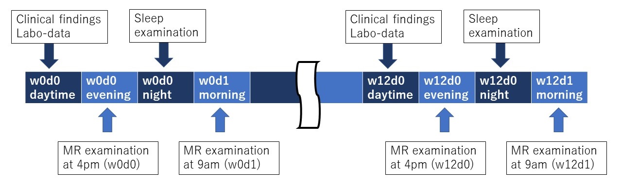
Figure 1
Timetable of measurements Imaging studies were performed in the evening before and the following morning on the night before the night of objective sleep parameter measurements.
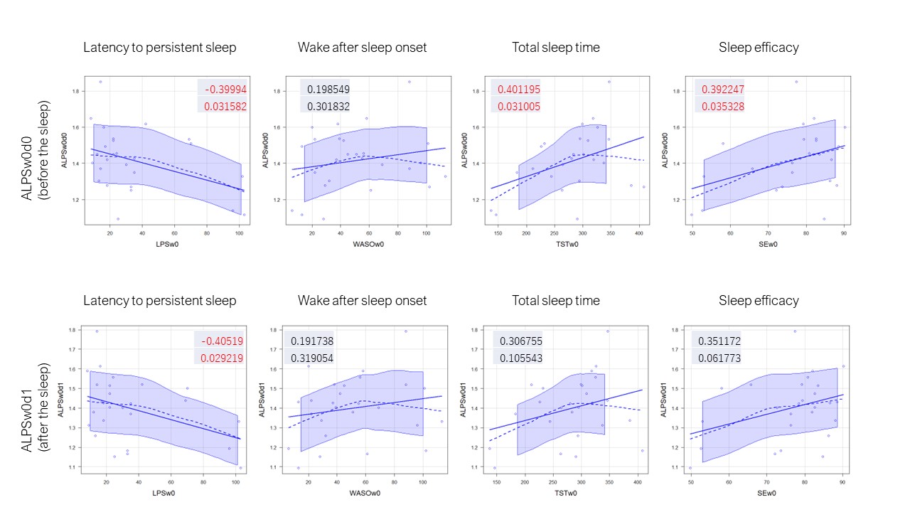
Figure 2
ALPS index and sleep parameters at week0 The ALPS-index in the evening before objective sleep measurement was significantly associated with latency to persistent sleep, total sleep time, and sleep efficacy.
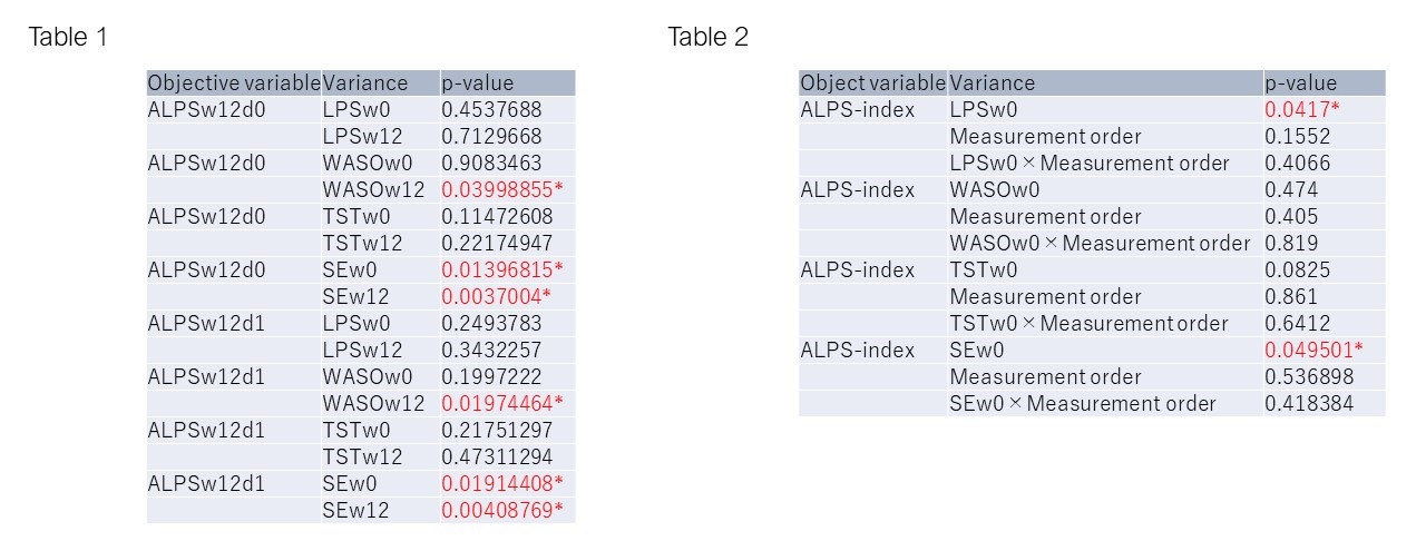
Figure 3
Table 1: Multiple regression analysis with the 0-week and 12-week sleep parameters as variance and the 12-week ALPS-index as objective variable. WASOw12 had a significant effect on the ALPS-index at d0, and SEw0 and SEw12 had significant effects. The ALPS-index in the morning imaging, i.e., d1, also showed a significant effect of WASOw12, as well as SEw0 and SEw12.
Table 2: Linear mixed model analysis of four consecutive ALPS measurements, namely ALPSw0d0, ALPSw0d1, ALPSw12d0, and ALPSw12d1. In this analysis, LPSw0 and SEw0 showed effects with statistical significance.