4912
Predictive value of left atrial and left ventricular strain for sudden cardiac death risk in hypertrophic cardiomyopathy by feature-tracking CMR1the First Affiliated Hospital of Harbin Medical University, Harbin, China, 2Philips Healthcare, Beijing, China
Synopsis
Keywords: Myocardium, Cardiomyopathy
Motivation: The risk stratification for implantable cardioverter-defibrillators (ICD) placement in high-risk patients with hypertrophic cardiomyopathy (HCM) has challenges.
Goal(s): The aim of this study is to investigate the connection between left atrial and left ventricular strain and sudden cardiac death (SCD) risk stratification by feature-tracking CMR.
Approach: All patients were satisfied by the 2020 AHA/ACC HCM Risk-SCD model with feature-tracking CMR for assessing left atrial and left ventricular strain.

Results: Results demonstrated that both left atrial and left ventricular strain show obvious attenuation in high-risk group than low-risk group. LVGRS+LARS combined model exhibited a superior diagnostic value for identifying high-risk SCD.
Impact: The LVGRS+LARS combined model could provide additional predictive value for improving SCD risk stratification in clinical practice, which may provide vital insights into the judicious clinical utilization of ICD to reduce SCD in HCM.
Introduction
Sudden cardiac death (SCD) is the most appalling complication of hypertrophic cardiomyopathy (HCM)[1]. Implantable cardioverter-defibrillator (ICD) implantation is recommended for primary prevention in patients at high risk for SCD, as it has effectively reduced SCD-related mortality[2]. However, the limited positive predictive value of existing clinical markers and deviation in the assessment of SCD risk between guidelines highlight the need for additional indicators for more accurately assessing of SCD risk and guiding ICD implantation while minimizing complications[3,4]. The scattered distribution of late gadolinium enhancement (LGE) or even elevated left ventricular ejection fraction (LVEF) in HCM patients inadequately predicts SCD[5]. Feature-tracking cardiac magnetic resonance (FT-CMR) assessing myocardial strain, as a sensitive and non-invasive measure can provide crucial information on cardiac function and prognostication in clinical practice[6]. However, the relationship between myocardial strain and the risk factors of SCD in HCM patients remains unclear. Therefore, the aim of this study is to investigate the attenuation of left atrial and left ventricular strain in HCM and to assess the predictive value of it for SCD by FT-CMR.Methods
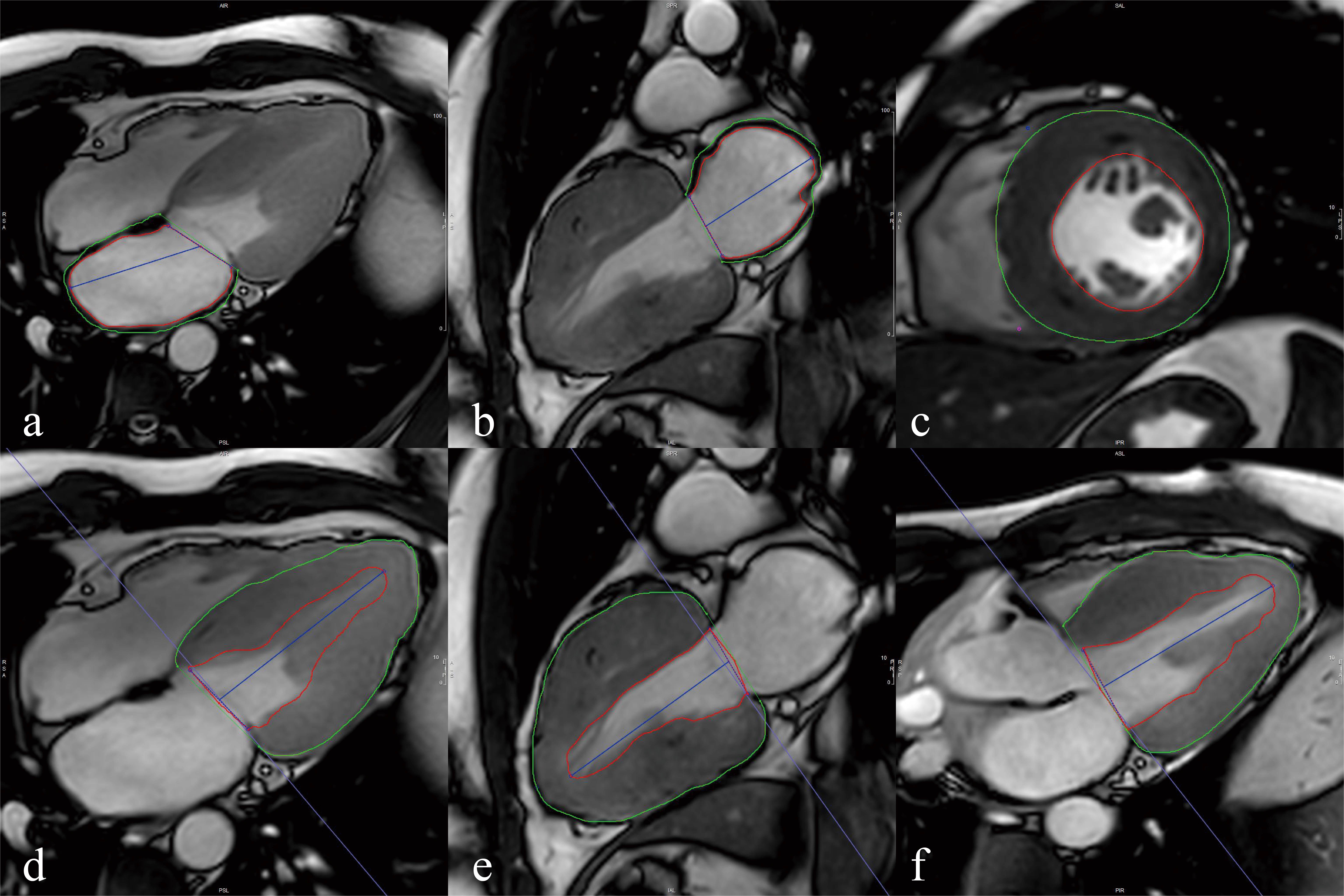
Patients with HCM diagnosed by CMR between February 2021 and July 2023 at the First Hospital of Harbin Medical University were retrospectively enrolled in this study. All patients underwent examination on a 3.0T magnetic resonance scanner (Elition; Philips Healthcare, Best, the Netherlands) and post-processing analysis by CVI42 (Circle Cardiovascular Imaging Inc. Calgary, Alberta, Canada). Left ventricular strain was obtain automatically by feature-tracking, while left atrial strain was measured by manually outlining the left atrial endocardium and epicardium (Figure 1). The included patients were classified into the high and low risk group of SCD according to the 2020 American Heart Association/American College (AHA/ACC) HCM Risk-SCD model. The independent sample t-test was used for comparison between groups of continuous variables conforming to normal distribution. Multivariate logistic regression analysis was performed to further explore the factors influencing SCD risk in HCM. The receiver operating characteristic (ROC) curve was used to determine the area under the curve (AUC). All statistical analyses were performed using SPSS software (version 25.0; IBM, Armonk, NY, USA). p<0.05 was considered statistically significant.Results
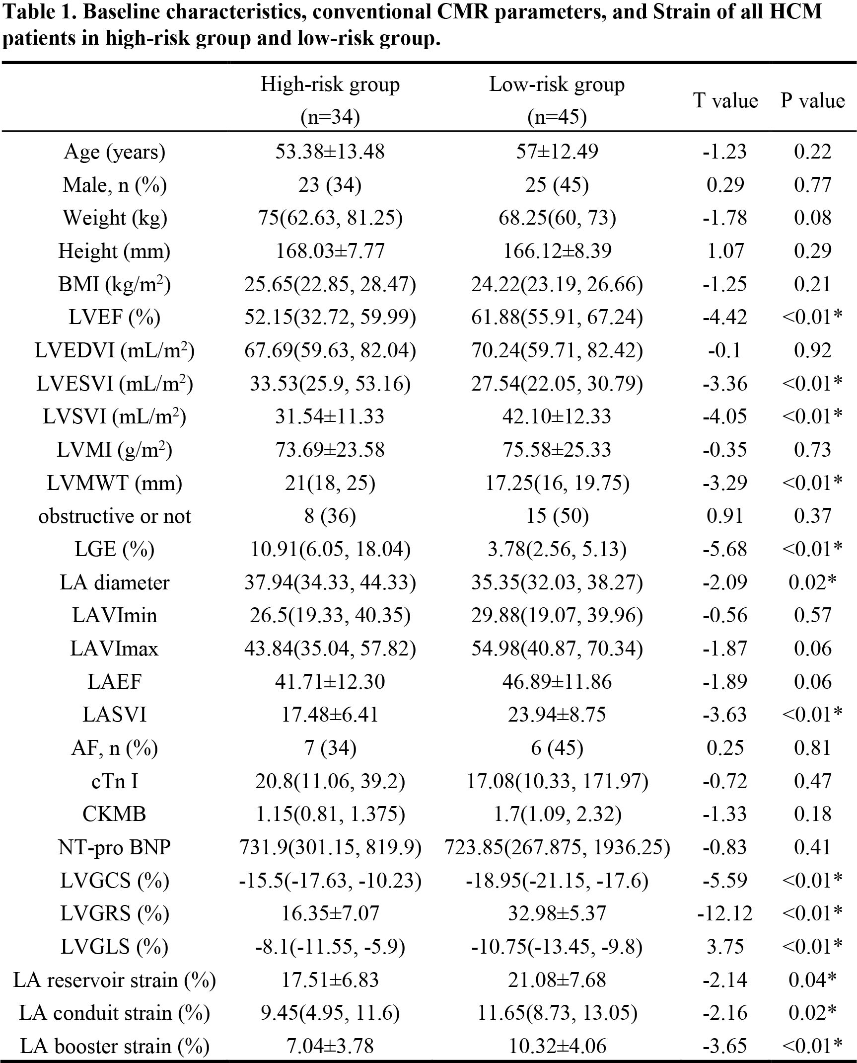

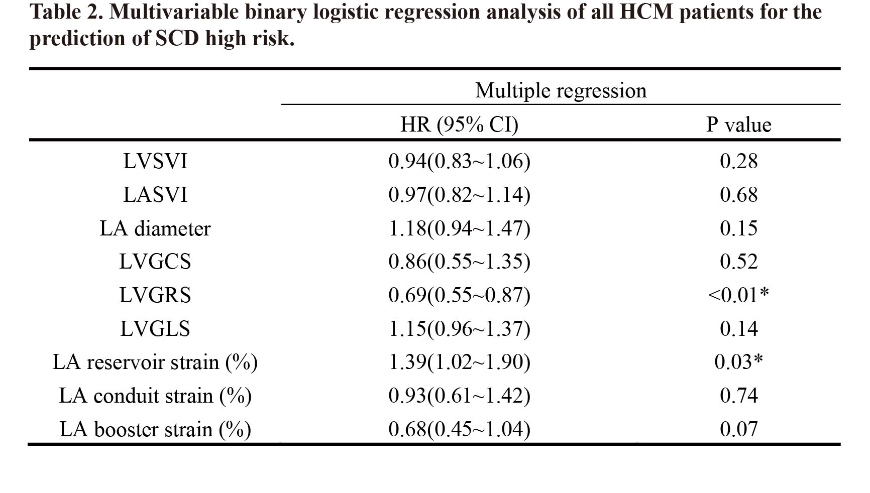
This study analyzed a total of 79 patients with HCM, including 36 patients in high-risk group (mean age 52±14.38) and 50 patients in low-risk group (mean age 55±13.78). Compared with the low-risk group, patients in the high-risk group had lower LVEF, LVSVI, and LASVI, and higher LVESVI, LVMWT, LGE, LA diameter (p<0.01). Patients in the high-risk group were associated with higher GCS, GLS, but lower GRS, LARS, LACS, and LABS compared with the patients in the low-risk group (p<0.05). (Table1 and Figure 2). Multivariate analysis showed that LVGRS and LARS were independent risk factors for predicting the high risk of SCD in HCM patients (LVGRS: OR,0.69; 95% CI: 0.55, 0.87; p<0.01, LARS: OR, 1.39; 95% CI: 1.02, 1.90; p=0.03) (Table 2). LVGRS+LARS combined model exhibited a superior diagnostic value for high-risk SCD (AUC, 0.95; 95%CI: 0.90, 1.00; p<0.01) compared to LARS alone (AUC, 0.63; 95%CI: 0.51, 0.76; p<0.05) (Figure 3).Discussion
Our study revealed that both the left ventricular and left atrial strain showed significant statistical differences between the high-risk and low-risk groups, which was consistent with the study of Lee et al[7]. Left ventricular systolic dysfunction is generally considered an important risk factor for SCD, while the majority of HCM has preserved or elevated LVEF. Global strain is more sensitive to assess LV systolic dysfunction than LVEF and decreased even in HCM with preserved LVEF. In this study, LVGRS and LARS showed excellent predictive value for high risk of SCD. Excessive myocardial hypertrophy can result in increased thickness of the radial ventricular wall and augmented atrial pressure secondary to left ventricular diastolic dysfunction. Xu et al. proposed that changes in radial strain should be closely monitored since radial strain has been declined when early myocardial thickening[8]. Yang et al. demonstrated that HCM patients with impaired LARS are at increased risk for adverse clinical outcomes[9].Our study also exist that LVGRS+LARS combined mode is expected to provide additional predictive value for improving SCD risk stratification in clinical practice.Conclusion
Left atrial and left ventricular strain based on CMR-FT may become a potential risk stratification biomarker for SCD in patients with HCM, and LVGRS+LARS combined model demonstrated a strong predictive value for identifying individuals at high risk of SCD.Acknowledgements
No acknowledgement found.References
1. Reant P, Mirabel M, Lloyd G, et al. Global longitudinal strain is associated with heart failure outcomes in hypertrophic cardiomyopathy[J]. Heart (British Cardiac Society), 2016, 102(10): 741–747.2. Nauffal V, Marstrand P, Han L, et al. Worldwide differences in primary prevention implantable cardioverter defibrillator utilization and outcomes in hypertrophic cardiomyopathy[J]. European Heart Journal, 2021, 42(38): 3932–3944.
3. Limongelli G, Monda E. Implantable cardioverter defibrillator in hypertrophic cardiomyopathy: Time to avoid unnecessary procedure[J]. International Journal of Cardiology, 2022, 355: 30–31.
4. Scatteia A, Baritussio A, Bucciarelli-Ducci C. Strain imaging using cardiac magnetic resonance[J]. Heart Failure Reviews, 2017, 22(4): 465–476.
5. Hou X, Xiong X, Li X, et al. Predictive value of cardiac magnetic resonance mechanical parameters for myocardial fibrosis in hypertrophic cardiomyopathy with preserved left ventricular ejection fraction[J]. Frontiers in Cardiovascular Medicine, 2022, 9: 1062258.
6. Andre F, Steen H, Matheis P, et al. Age- and gender-related normal left ventricular deformation assessed by cardiovascular magnetic resonance feature tracking[J]. Journal of Cardiovascular Magnetic Resonance, 2015, 17(1): 25.
7. Lee H J, Kim H K, Lee S C, et al. Supplementary role of left ventricular global longitudinal strain for predicting sudden cardiac death in hypertrophic cardiomyopathy[J]. European Heart Journal - Cardiovascular Imaging, 2022, 23(8): 1108–1116.
8. Xu H, Chen J, Yang Z, et al. Early marker of regional left ventricular deformation in patients with hypertrophic cardiomyopathy evaluated by MRI tissue tracking: The effects of myocardial hypertrophy and fibrosis[J]. Journal of Magnetic Resonance Imaging, 2017, 46(5): 1368–1376.
9. Yang F, Wang L, Wang J, et al. Prognostic value of fast semi-automated left atrial long-axis strain analysis in hypertrophic cardiomyopathy[J]. Journal of Cardiovascular Magnetic Resonance, 2021, 23: 36.
Figures