4906
DTI/DKI Biomarkers of Chronic mild Traumatic Brain Injury1University of Washington, Seattle, WA, United States, 2VA Puget Sound Health Care System, Seattle, WA, United States
Synopsis
Keywords: Biomarkers, Traumatic brain injury
Motivation: Imaging biomarkers accurately diagnosing and monitoring treatment response for chronic mild Traumatic Brain Injury (mTBI) are needed.
Goal(s): This study aimed to experimentally validate and determine the performance of MRI biomarkers for monitoring chronic repetitive mild traumatic brain injury (mTBI).
Approach: We used diffusion tensor imaging (DTI) and diffusion kurtosis imaging (DKI) to assess white matter injury in mouse brains with chronic mTBI and compared results with those for age-matched control brains.
Results: Reduction in fractional anisotropy and increase of axial kurtosis were observed in the medulla of mTBI brains compared to the parameters in the same region of control brains.
Impact: It is new to monitor DTI and DKI parameters as noninvasive biomarkers in the brain with chronic mTBI.
Introduction
Mild traumatic brain injury (mTBI), or concussion, affects the annual global incidence of 27 to 69 millions(1). This injury can lead to short-term or long-term cognitive, emotional, and behavioral problems. Even though some progress has been made in identifying acute mTBI biomarkers, chronic mTBI remains a substantial unmet need. Identifying noninvasive imaging biomarkers for chronic mTBI will help improve the diagnosis and management of the disease. In this study we used diffusion tensor imaging (DTI) and diffusion kurtosis imaging (DKI) to monitor the mouse brain with chronic mTBI and determine if the DTI/DKI parameters could be noninvasive biomarkers in detecting chronic mTBI.Methods
We measured white matter injury in ex vivo mouse brains using gadollinium-enhanced diffusion tensor MRI. Mild traumatic brain injury was induced by repeated blast overpressure in 5 mice using a custom laboratory shock tube. Five age-matched yoked sham mice, which received equal anesthesia but without blast exposure, were used as control subjects. Animals were terminally anesthetized with an overdose of sodium pentobarbitol. The thoracic cavities were opened and the animals were intracardially perfused. Following perfusion, each animal was decapitated, defleshed, and the lower jaw was removed. Before ex vivo MRI, a brain sample was fixed to a rigid wooden depressor and placed in a 15 mL falcon tube filled with fomblin.Three-dimensional diffusion tensor imaging (3D-DTI) was conducted on a 14 Tesla (T) magnetic resonance (MR) scanner using the echo-planar imaging (EPI) with repetition time TR of 500 ms, echo time of 14.4 ms, number of segmentation of 4, field of view of 12.8 x 12.8 x 17 mm, matrix of 128 x 128 x 34, number of averages of 2, diffusion gradient duration of 2.5 ms, diffusion gradient separation of 7.5 ms, diffusion gradient b values of 1000 and 2000 s/mm2, and number of diffusion directions of 30 and 30.
We used both DSI studio (model free tool: https://dsi-studio.labsolver.org/) and ExploreDTI (for DKI analysis: https://www.exploredti.com/) to analyze the 3D-DTI data. Regions of interest (ROIs) of rostral-to-caudal fibers in green were selected on a slice of 0.5 mm within the medulla. DTI parameters of several ROIs such as the medulla, corpus callosum, and striatum were measured and compared between sham mice and mTBI mice.
Results
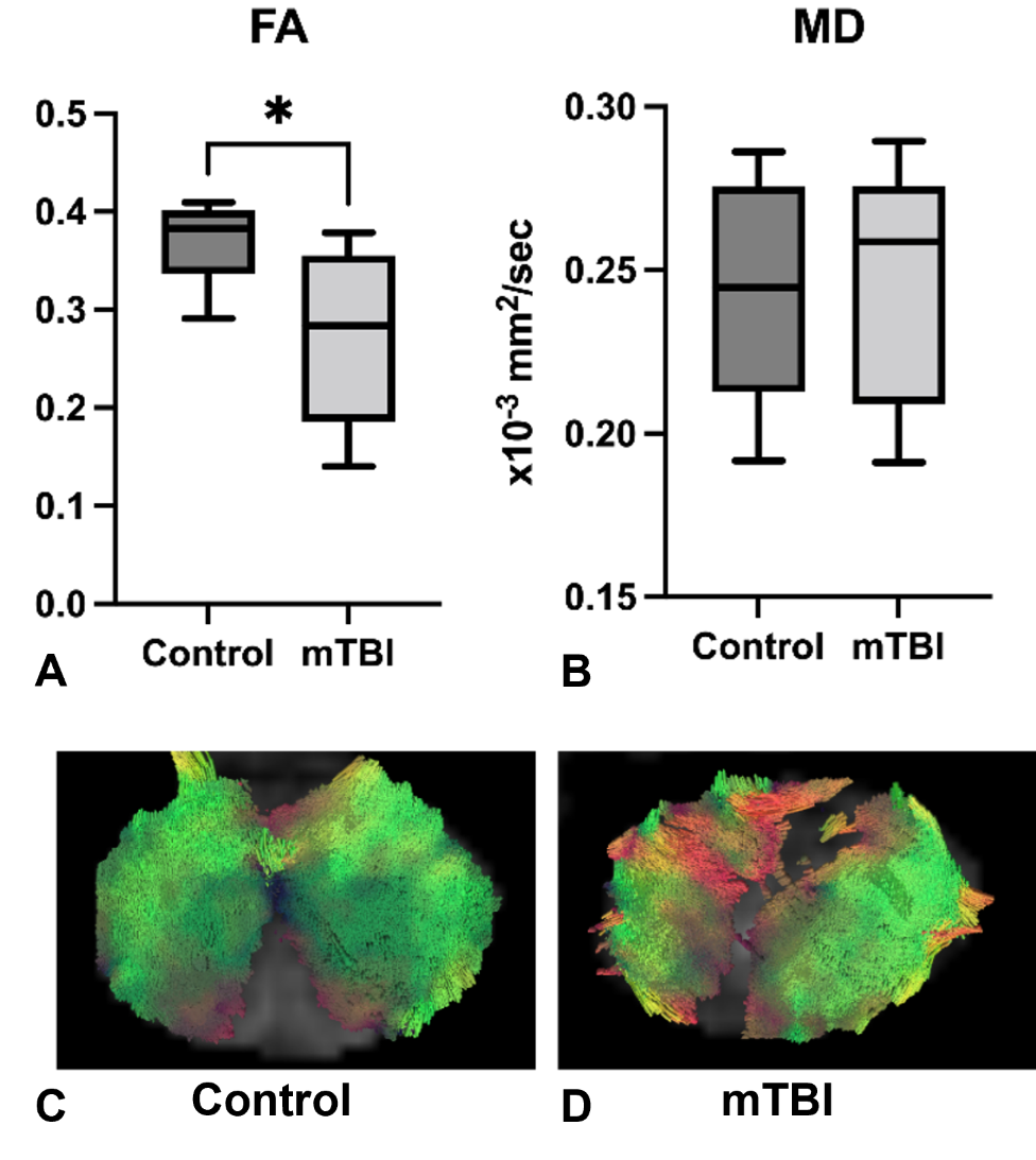
Figure 1 summarizes the comparison results of DTI parameters and fiber tracts for the medulla between control mice and mTBI mice. Fractional anisotropy (FA) values in the medulla were reduced in mTBI mouse brains compared to those in control mouse brains. Slight increases were observed for both mean diffusivity (MD) in mTBI mouse brains. Well-organized fiber tracts were observed in all control mouse brains compared to those in mTBI mouse brains (see Figs 1C & 1D). Only one of five mTBI mouse brains showed normally-organized fiber tracts while the other 4 mTBI mouse brains presented with significant disorganization, as seen in Fig. 1D.DTI parameters were measured in both the corpus callosum and striatum regions. For the corpus callosum, both FA and quantitative anisotropy (QA) values were reduced while MD values were increased in mTBI brains compared to those in control brains (see Fig. 2). However, for the striatum, FA was increased, and MD was decreased in mTBI brains compared to the parameters measured in control brains (see Fig. 3).
DKI parameters of the medulla were further analyzed to show an increase in axial kurtosis (AK) and decrease in axial extra-axonal diffusivity (AxEAD) for mTBI brains compared to AK and AxEAD values measured in the medulla of control brains as shown in Fig. 4.
Discussion
FA and MD values measured in both the medulla and corpus callosum showed the opposite directionality to those measured in the striatum. Medulla and corpus callosum are white matter abundant regions in mice. Hui et al. found that diffusion kurtosis was increased in the acute-to-subacute phase after stroke onset(2). Also, the axonal diffusivity appears to be useful to investigate the white matter. In our study, DKI analysis may be useful to differentiate chronic mTBI from normal brain as compared AK and AxEAD values measured in the medulla as shown in Fig. 4. Conventional DTI uses the assumption that the diffusion follows a Gaussian distribution, which may not be true for complex biological tissues. DKI can address non-Gaussian behavior of biological tissues(3,4).Conclusion
DTI/DKI parameters measured in the medulla demonstrated a good correlation with pathology in the brain and may be effective noninvasive MRI biomarkers to monitor the brain of chronic mTBI. DTI/DKI may be used to non-invasively diagnose and monitor the brain of patients with chronic mTBI.Acknowledgements
We thank Elias Farhan for processing the diffusion parameters of some regions of interest.References
1. Collaborators GTBIaSCI. Global, regional, and national burden of traumatic brain injury and spinal cord injury, 1990-2016: a systematic analysis for the Global Burden of Disease Study 2016. Lancet Neurol 2019;18(1):56-87.
2. Hui ES, Fieremans E, Jensen JH, Tabesh A, Feng W, Bonilha L, Spampinato MV, Adams R, Helpern JA. Stroke assessment with diffusional kurtosis imaging. Stroke 2012;43(11):2968-2973.
3. Jensen JH, Helpern JA. MRI quantification of non-Gaussian water diffusion by kurtosis analysis. NMR Biomed 2010;23(7):698-710.
4. Jensen JH, Helpern JA, Ramani A, Lu H, Kaczynski K. Diffusional kurtosis imaging: the quantification of non-gaussian water diffusion by means of magnetic resonance imaging. Magn Reson Med 2005;53(6):1432-1440.
Figures