4904
Correlation analysis of the behavioral analysis and quantitative T2 map data in the SOD1G93A mouse model1Tokyo Metropolitan University, Tokyo, Japan, 2Central Institute for Experimental Animals, Kawasaki, Japan, 3Hokkaido University, Sapporo, Japan
Synopsis
Keywords: Preclinical Image Analysis, Preclinical, Amyotrophic lateral sclerosis (ALS)
Motivation: Amyotrophic lateral sclerosis is still an understudied disease, and early detection is important.
Goal(s): This study aimed to clarify the transition of the pathophysiology and evaluate the relationship between the abnormalities detected on magnetic resonance imaging and the development of clinical symptoms by combining the T2 map data obtained in the previous study with behavioral analysis.
Approach: Behavioral analysis was performed on four parameters, namely, bodyweight, general condition, hind-foot reflex test, and landing foot-splay test. Correlation analysis of the behavioral analysis data and T2 map data was performed in each voxel.
Results: A correlation was detected in the trigeminal motor nucleus.
Impact: Changes in the trigeminal motor nucleus are indicated to be closely related to early changes in amyotrophic lateral sclerosis (ALS). This finding is suggested to be useful for the early diagnosis of ALS in humans and research on novel treatments.
Introduction
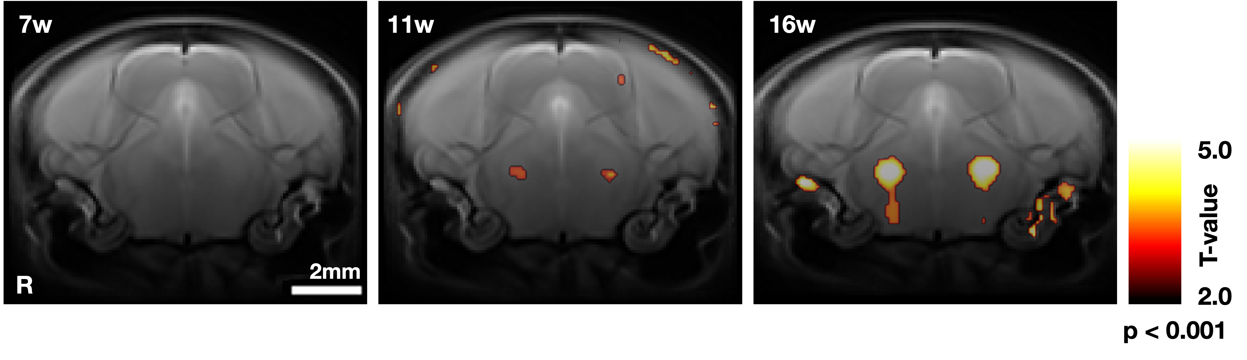
Amyotrophic lateral sclerosis (ALS) is a degenerative neurological disease that affects motor neurons, resulting in muscle paralysis, including the muscles necessary for breathing, ultimately leading to fatality. The exact cause remains unclear, and there is no cure. Early ALS detection and understanding of a more accurate disease progression are vital for patient treatment. Magnetic resonance imaging (MRI), a noninvasive imaging technique, can detect early changes in ALS1,2. Previous studies have suggested that voxel-based relaxometry (VBR) and region of interest analysis with T2 values can detect early changes in the trigeminal motor nucleus before symptom onset (Figure1). In this study, in addition to the T2 value data, noninvasive behavioral analysis was used to observe symptom onset. A correlation analysis was performed between T2 map data and behavioral analysis data to better understand ALS progression and explore the relationship between abnormalities detected on MRI and the development of clinical symptoms.Methods
ALS model mice with the human mutant SOD1 gene and normal mice (wild type [wt]) were used. SOD1 is one of the causative genes of ALS and accounts for 2% of all ALS cases. Behavioral assessments were conducted on both groups at 7, 11, 13, 14, and 15 weeks of age. The evaluation parameters included bodyweight, general condition, hind-foot reflex test, and landing foot-splay test. For the assessment of general condition, the behavioral score system developed by Vercelli was used3,4. For the hind-foot reflex test, limb extension was observed when the mice were lifted by the tail and held suspended downward5,6. For the landing foot-splay test, the mice were then dropped from a height of approximately 30 cm, and the landing foot-splay distance was evaluated5. T2 maps were acquired using a 7-T MRI scanner (Biospec 70/16, Bruker BioSpin, Germany) and cryoprobe with the following parameters (echo time, 10–200 ms [20 steps]; repetition time, 4000 ms; field of view, 19.2 × 19.2 mm; image matrix, 128 × 128; slice thickness, 0.5 mm; slices, 18; scan time, 6 min 24 s). T2 map data were normalized using the same parameters used to normalize T2-weighted image, and smoothed for image distortion, position correction and noise removal. The correlation between the preprocessed T2 map and behavioral analysis was analyzed using SPM12 by performing multiple regressions and calculated for each voxel.Results
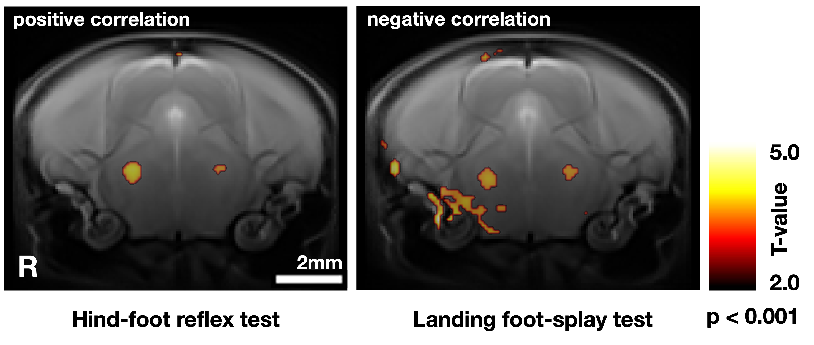
Behavioral analysis is summarized in Figure2. Bodyweight was gradually increased in wt mice but gradually decreased from 11 weeks in the ALS model mice. At 16 weeks, it was significantly lower in the ALS model mice than in the wt mice. In the assessment of general condition, a score in the ALS model mice was lower starting from 13 weeks, with scores significantly lower than in the wt mice at 14 and 15 weeks. In the hind-foot reflex test, a score in the ALS model mice was significantly higher than that in the wt mice starting from 11 weels. In the landing foot-splay test, the landing foot-splay distance in the ALS model mice was significantly shorter than that in the wt mice at 13, 14, and 15 weeks. A positive correlation was found in the hind-foot reflex test and a negative correlation in the landing foot-splay test in the trigeminal motor nucleus (Figure3).Discussion
In the behavioral analysis, all four parameters showed significant differences between wt and ALS model mice, indicating ALS-induced motor neuron damage leading to muscle atrophy, loss of muscle strength and motor control, and age-related behavioral changes. Significant changes in bodyweight and assessment of general condition occurred later in ALS onset, possibly because muscles take time to actually lose weight after the loss of motor control in neurodegeneration. The correlation analysis showed a correlation in the trigeminal motor nucleus for the hind-foot reflex test and the landing foot-splay test. This nucleus, in which VBR detected early changes due to neurodegeneration in ALS, shows significantly increased T2 values in ALS (Figure1). As it controls muscles involved in chewing, trigeminal motor nucleus impairment in ALS may result in reduced food intake, contributing to weight loss. The tests showing correlation in this study, reflecting neurodegenerative effects directly, suggest the potential of the trigeminal motor nucleus as an early indicator of ALS.Conclusion
The study indicated that the trigeminal motor nucleus is closely associated with the early changes in ALS by combining MRI with behavioral analysis and may be a crucial region to monitor for early diagnostic purposes. This result suggests its potential to aid in the early diagnosis of ALS in humans. More studies in this direction could contribute to the development of early intervention strategies for ALS.Acknowledgements
This research was supported by AMED under Grant Number JP21ek0109455h0002, “MRI platform” as a program for advanced research equipment platforms for Promoting public Utilization of Advanced Research Infrastructure of the MEXT and JSPS KAKENHI Grant Number 20K08095.References
1. Dai Z, Chen Y, Yan G, Xiao G, Shen Z, Wu R (2019). Progress of magnetic resonance imaging in amyotrophic lateral sclerosis. Radiology of Infectious Diseases;6(1), 1-7.
2. Evans, MC et al. (2014). T2-weighted MRI detects presymptomatic pathology in the SOD1 mouse model of ALS. Journal of Cerebral Blood Flow and Metabolism;34(5), 785-793.
3. Knippenberg S, Thau N, Dengler R, Petri S (2010). Significance of behavioural tests in a transgenic mouse model of amyotrophic lateral sclerosis (ALS). Behavioral Brain Research;213(1), 82-87. doi: 10.1016/j.bbr.2010.04.042.
4. Vercelli A et al. (2008). Human mesenchymal stem cell transplantation extends survival, improves motor performance and decreases neuroinflammation in mouse model of amyotrophic lateral sclerosis. Neurobiology of Disease;31(3), 395-405.
5. Aoki M, Warita H, Mizuno H, Suzuki N, Yuki S, Itoyama Y (2011). Feasibility study for functional test battery of SOD transgenic rat (H46R) and evaluation of edaravone, a free radical scavenger. Brain Research;1382, 321-325. doi: 10.1016/j.brainres.2011.01.058.
6. Peng Y et al. (2014). Deficient import of acetyl-CoA into the ER lumen causes neurodegeneration and propensity to infections, inflammation, and cancer. Journal of Neuroscience;34(20), 6772-6789. doi: 10.1523/JNEUROSCI.0077-14.2014.
Figures
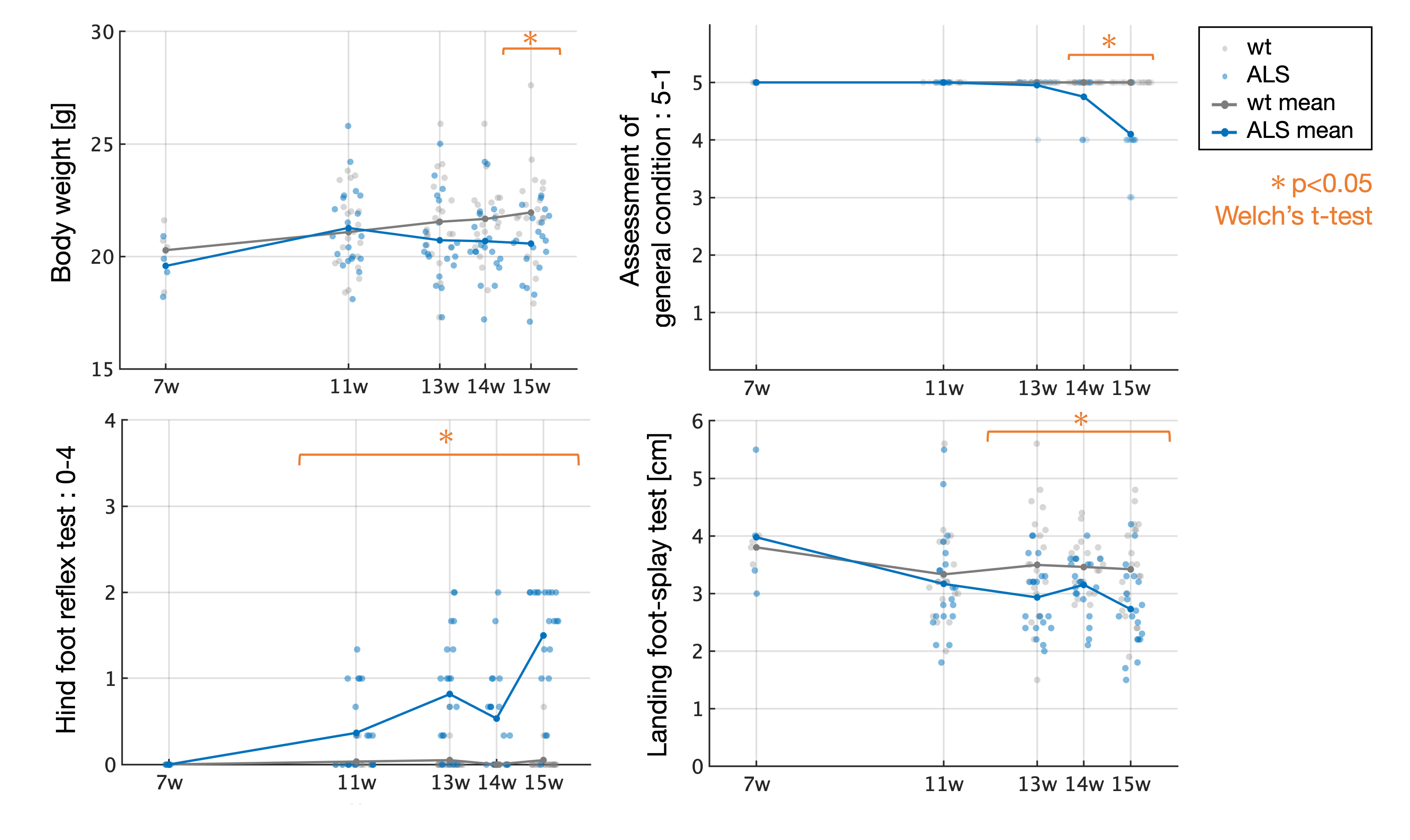
Scatter plot of the result of the behavioral analysis. The straight lines show the mean change at each week of age. The vertical axis shows the score value of each parameter, and the horizontal axis shows the age of mice in weeks. General condition is assessed from 1 to 5 by gait instability and degree of paralysis3. For the hind-foot reflex test, a score of 0–4 was given based on how many limbs were bent, and all limbs fully extended is normal5,6.