4901
Identifying potential therapeutic targets for focal transcranial magnetic stimulation (TMS) in a rat model of cocaine dependence1Neuroimaging Research Branch, National Instititute on Drug Abuse, NIH, Baltimore, MD, United States
Synopsis
Keywords: Small Animals, Brain, TMS, CBV, basal metabolism
Motivation: Transcranial magnetic stimulation (TMS) shows promise as a therapeutic intervention for many neuropsychiatric conditions. Yet, pinpointing the appropriate stimulation target remains elusive.
Goal(s): To identify brain regions affected by prolonged cocaine exposure, and to identify potential targets for focal TMS.
Approach: Basal CBV were mapped by injecting iron-oxide contrast. Multi-echo gradient echo and spin echo sequences were employed to generate maps of 05732972-a323-4027-8676-ccee7744913e">R2.
Results: Many brain regions, including the prelimbic cortex, exhibited a significant decrease in basal CBV following prolonged cocaine exposure. The prelimbic cortex is situated relatively close to the cortical surface and presents a promising candidate for TMS.
Impact: Likely the first attempt to identify TMS targets using the CBV mapping approach.
INTRODUCTION
There is no FDA-approved medication for treating cocaine use disorder. TMS has been approved by FDA for treatment-resistant major depression and more recently as an add-on treatment for nicotine use disorder. Our lab recently developed focal TMS technologies, achieving a focality of 2 mm1. The goal of this study is to use fMRI to identify potential targets for focal TMS, and to explore the therapeutic potentials of TMS treatment in a rat model of cocaine dependence. In this regard, basal CBV, a physiological parameter closely related to basal CBF and cerebral metabolism, was mapped to identify brain regions whose basal metabolism were impacted by prolonged cocaine exposure. We hypothesize that potential TMS targets can be derived from basal CBV mapping.METHODS
Adult SD rats (n=12) underwent jugular vein catheterization, and were trained to lever-press for cocaine infusion (0.5 mg/kg/infusion) for 3 weeks. MRI scans took place 1 day before (pre-cocaine baseline) and 1 day post the 3-week cocaine training. Basal CBV were mapped using 1) multi-echo gradient echo (MGE) and 2) multi-echo spin echo (MSME) sequence with the aid of iron oxide contrast agent (iron dose 15 mg/kg IV for MGE and 20 mg/kg for MSME). Data were acquired on a Bruker 9.4T scanner. Scan parameters for MGE: TR=700ms, 10 echoes, TE1=2ms, echo space=2ms, FOV=35 35mm2, matrix size=128 128, 19 slices with a slice thickness of 1mm; for MSME: TR=1500ms, TE1=6ms, echo space=6ms, 10 echoes. Prior to MRI contrast injection, resting state BOLD fMRI data were also acquired. Rats were anesthetized under a combination of low-dose dexmedetomidine and isoflurane during the scans2.For MGE data, voxel-wise R2* value was calculated by fitting the multi-echo data with a single exponential decay model. Differences in values pre- and post-contrast agent injection reflected basal CBV. Voxel-wise was normalized to mean value of the individual rat brain. Alteration in CBV caused by cocaine exposure was assessed by comparing values between pre-cocaine baseline and post-cocaine SA, corrected for multiple comparisons. MSME data were analyzed in the same fashion as described above except that instead of was the subject of interest.RESULTS
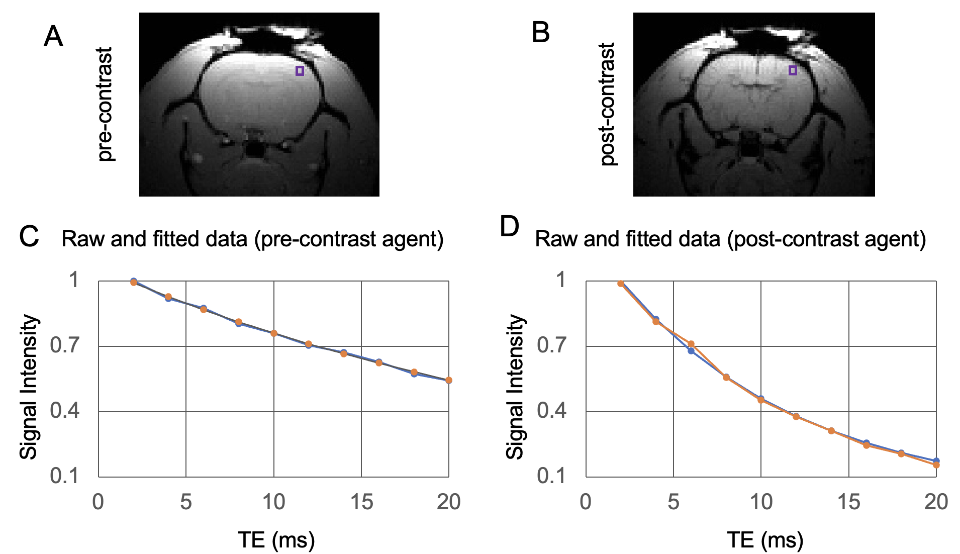
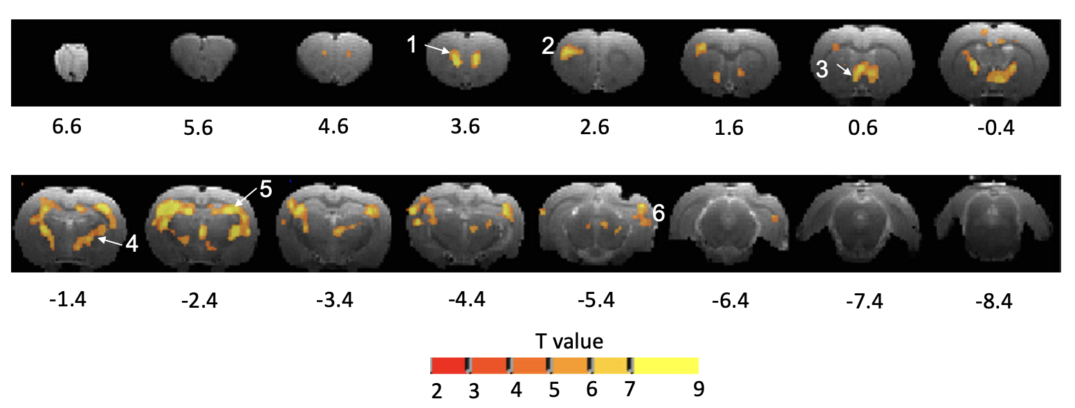
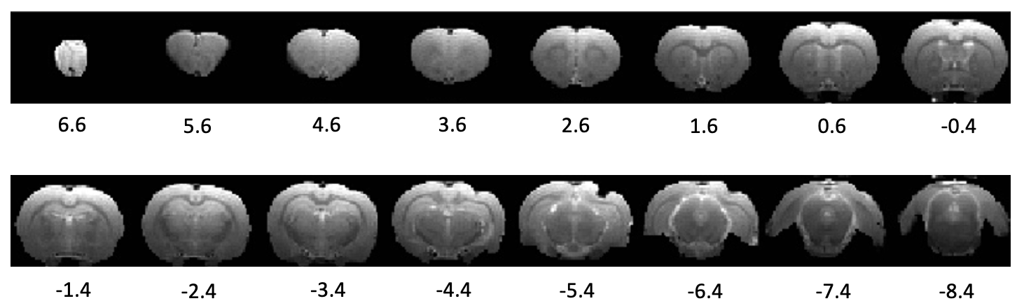
Figure 1 depicts representative MGE images (TE=TE1) both prior to and following the injection of a contrast agent, accompanied by the fitting of the multi-echo data utilizing a single exponential decay model. As anticipated, the iron-oxide contrast agent substantially increased the relaxation rate, evident in the contrast between images (A-B) and the signal intensity plots across different TE values (C-D). Figures 2 and 3 show T-statistical maps comparing and values between pre-cocaine baseline and after 3 weeks of cocaine SA, respectively. Data acquired with the MGE sequence (Fig. 2) reveals many regions showing significant alterations in basal CBV induced by prolonged cocaine exposure, but not a single region in data acquired with the MSME sequence (Fig. 3).CONCLUSION and DISCUSSION
The electric field induced by a TMS coil is always strongest on brain surface and diminishes from superficial to deep brain regions3. Thus, superficial cortical regions are favored for TMS targeting. Of the regions showing significant basal CBV changes in Fig. 2, the prelimbic cortex (region 1 in Fig. 2) is superficial and is a preferred candidate site for TMS administration. Furthermore, while TMS cannot directly access some of the deep brain regions shown in Fig. 2, it is possible to modulate activity in these brain regions through functional connectivity4. Resting state fMRI data acquired in the same cohort of animals should shed light on this.Figures 2-3 illustrate a notable dissimilarity in the sensitivity of MGE and MSME techniques in detecting CBV changes resulting from prolonged cocaine exposure. Our initial hypothesis posited that cocaine exposure would induce alterations in basal cerebral metabolism, likely manifesting in the microvasculature. It is well-documented that gradient echo and spin echo methods exhibit distinct sensitivities to relaxation rate changes in the presence of intravascular contrast agents, with spin echo being more specific in capturing susceptibility changes in the microvasculature5,6. In addition to acquiring gradient echo data, we also obtained spin echo data using a higher dose of the contrast agent (20 mg/kg vs. 15 mg/kg of iron7). Somewhat unexpectedly, all the regions that exhibited significant differences in the MGE acquisition did not meet the statistical threshold in the MSME acquisition. These data suggest that MGE is the preferred sequence in future studies like the one reported here.Acknowledgements
This work was supported by the NIDA Intramural Research Program, NIHReferences
1. Meng Q, Jing L, Badjo JP, et al. A novel transcranial magnetic stimulator for focal stimulation of rodent brain. Brain Stimul. 2018;11(3):663-665. doi: 10.1016/j.brs.2018.02.018. Epub 2018 Mar 1.2.
2. Brynildsen JK, Hsu LM, Ross TJ, Stein EA, Yang Y, Lu H. Physiological characterization of a robust survival rodent fMRI method. Magn Reson Imaging. August 2016. doi:10.1016/j.mri.2016.08.0103.
3. Heller L, Van Hulsteyn DB. Brain stimulation using electromagnetic sources: theoretical aspects. Biophysical Journal. 1992;63(1):129-138. doi:10.1016/S0006-3495(92)81587-44.
4. Fox MD, Buckner RL, Liu H, Chakravarty MM, Lozano AM, Pascual-Leone A. Resting-state networks link invasive and noninvasive brain stimulation across diverse psychiatric and neurological diseases. Proc Natl Acad Sci U S A. 2014;111(41):E4367-75. doi:10.1073/pnas.14050031115.
5. Boxerman JL, Hamberg LM, Rosen BR, Weisskoff RM. Mr contrast due to intravascular magnetic susceptibility perturbations. Magnetic Resonance in Medicine. 1995;34(4):555-566. doi:10.1002/mrm.19103404126.
6. Schmainda KM, Rand SD, Joseph AM, et al. Characterization of a First-Pass Gradient-Echo Spin-Echo Method to Predict Brain Tumor Grade and Angiogenesis. American Journal of Neuroradiology. 2004;25(9):1524-1532.7.
7. Lu H, Scholl CA, Zuo Y, Stein EA, Yang Y. Quantifying the blood oxygenation level dependent effect in cerebral blood volume-weighted functional MRI at 9.4T. Magn Reson Med. 2007;58(3):616-621. doi:10.1002/mrm.21354
Figures