4899
Brain Tissue Displacement and Strain Measures in an Alzheimer’s Disease Cohort using DENSE1Department of Medical Physics, University of Wisconsin, Madison, Madison, WI, United States, 2Department of Medicine, Department of Medical Physics, University of Wisconsin, Madison, Madison, WI, United States, 3Department of Radiology, University of Wisconsin. Madison, Madison, WI, United States, 4Department of Medicine, University of Wisconsin, Madison, Madison, WI, United States, 5Department of Medical Physics, Department of Biomechanical Engineering, Department of Radiology, University of Wisconsin, Madison, Madison, WI, United States
Synopsis
Keywords: Neurofluids, Alzheimer's Disease, Aging, Brain, Blood Vessels, Cardiovascular, Dementia, Flow, Neuro, Neurodegeneration, Neurofluids, Velocity & Flow
Motivation: Cardiac driven motions may affect Alzheimer's Disease.
Goal(s): The goal was to provide preliminary results on the success of measuring the displacement and strain of brain tissues due to Cardiac Arterial Pulsations (CAPs) using DENSE (Displacement ENcoding with Stimulated Echoes) in patients with AD and age-comparable controls.
Approach: DENSE scan data from a sample of 133 volunteers was processed and evaluated for trends in the derived displacement and strain information.
Results: High variability was found in the displacement measures, and future work is needed to determine the confounding factors behind the variability and to what degree those factors can be minimized going forward
Impact: DENSE MR was used to measure displacement of brain tissue from cardiac effects in Alzheimer’s disease (AD) patients and age-comparable controls. Preliminary results are inconclusive and future work is needed to determine what confounding factors are affecting the displacement measures.
Introduction
Prior research has provided evidence for arterial stiffening1-3, increased pulsatile pressure2-3, and a loss of brain tissue biomechanical integrity with increased age4-6 and also with the onset of Alzheimer’s disease (AD)5,7-9. Higher pulsatile pressure of the blood entering the capillaries may result in increased displacement of the brain tissue over the cardiac cycle and increase mechanical forces to brain tissue leading to neuronal damage10. Unfortunately, quantifying brain biomechanics is challenging. MR Elastography provides measures of brain stiffness; however, it only provides brain stiffness and does not provide measures relating to cardiac driven motions. DENSE (Displacement Encoding with Stimulated Echoes) MRI has been used in prior research to measure brain tissue displacement due to Cardiac Arterial Pulsations (CAPs) in small cohorts of young, healthy individuals11-15, and these methods have yet to have demonstrated utility in aging and AD populations. DENSE16 uses strong gradients to encode displacement information into the phase of a stimulated echo sequence and is thus sensitive to confounding factors. The goal of this research is to gauge the ability of DENSE to measure displacement due to CAPs in a cohort of AD patients and age-comparable controls.Methods
Subjects undergoing AD studies were non-specifically recruited to undergo an additional DENSE scan during a research imaging visit. Scans of 133 of volunteers (44 males, 84 females, 5 undisclosed) between ages 49 and 93 (mean age of 65.75) were included. Using 3.0T MR scanners and photo-plethysmography gating (PPG), a 2D, single-shot, spiral readout DENSE sequence was used to acquire 10 frames over the cardiac cycle, using a 1.8ms echo time, fat saturation, 10 degree flip angle, and 5.72 cycles/mm displacement encoding frequency, producing a 240x240mm FOV, 10mm thick slice with 4x4mm in-plane resolution. A single 2D slice was collected with a plane placed just superior to the lateral ventricles.Scans were reconstructed using a NUFFT. Images were normalized over the cardiac cycle and underwent background field subtraction, phase unwrapping, and linear detrending to remove bulk motion. Displacement maps were generated using the pseudo-inverse of the encoding matrix to estimate displacement fields and quantifiable metrics (e.g. maximum displacement). Strain maps were created using the derivatives of the displacement maps and an estimate of the maximum strain was computed using the magnitude of a pseudo 3D strain tensor. Each set of images was categorized using a 4 point Likert scale according to image quality. ROIs were drawn for the left and right hemispheres, excluding ventricles when present. In pixel-wise analyses, pixel measures which fell outside a 90% percentile range when compared to other pixels within the ROI were excluded.
Results
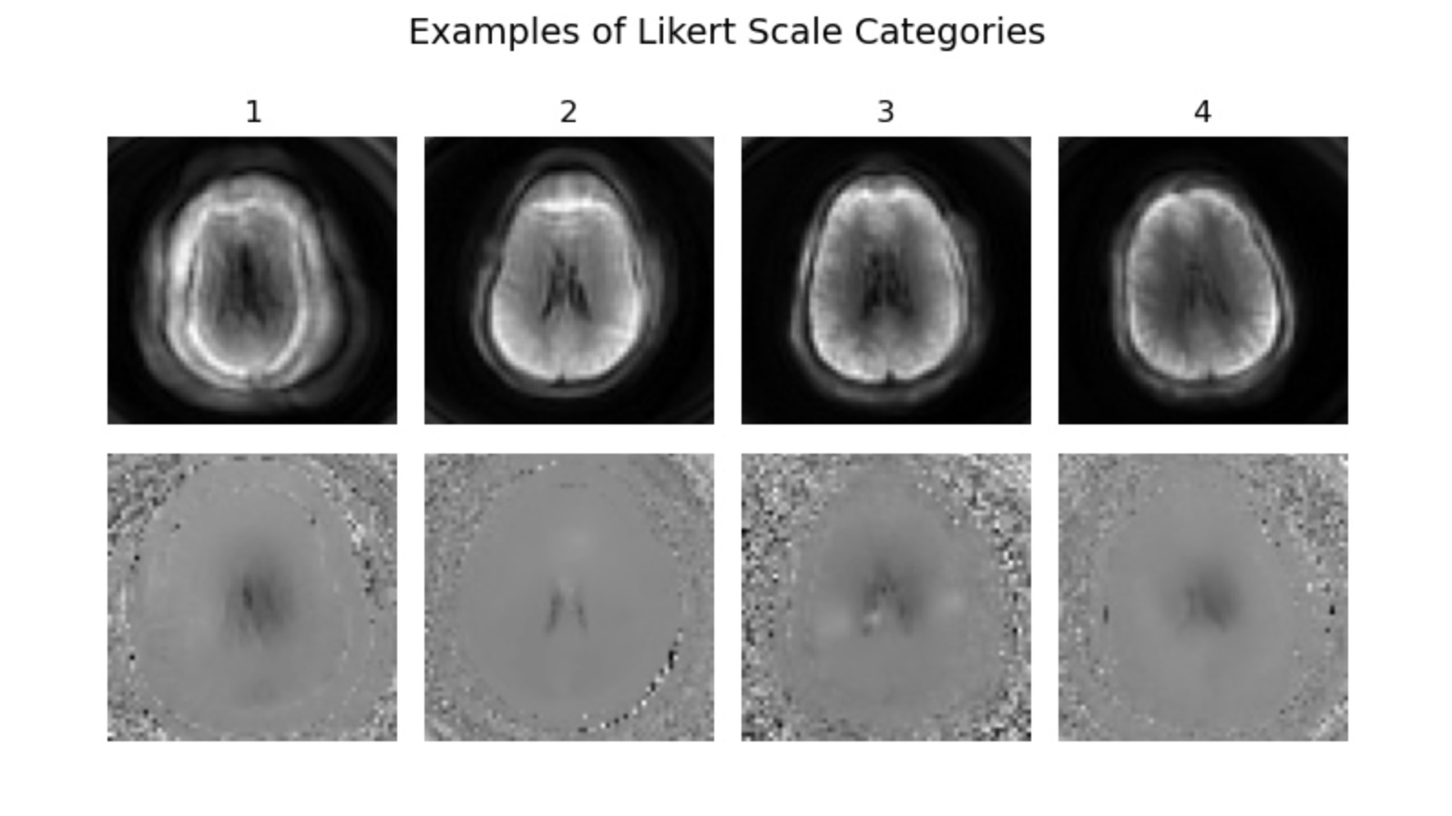
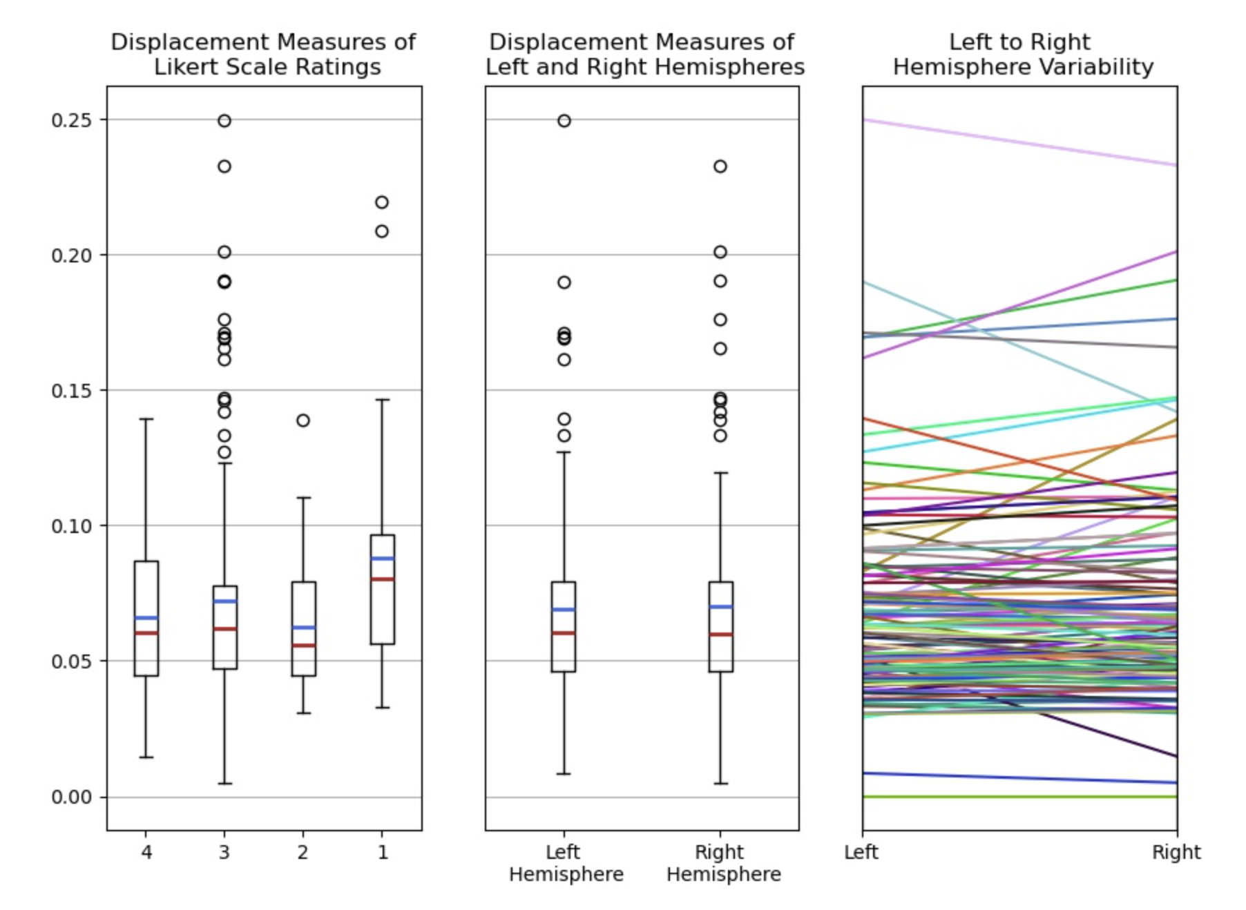
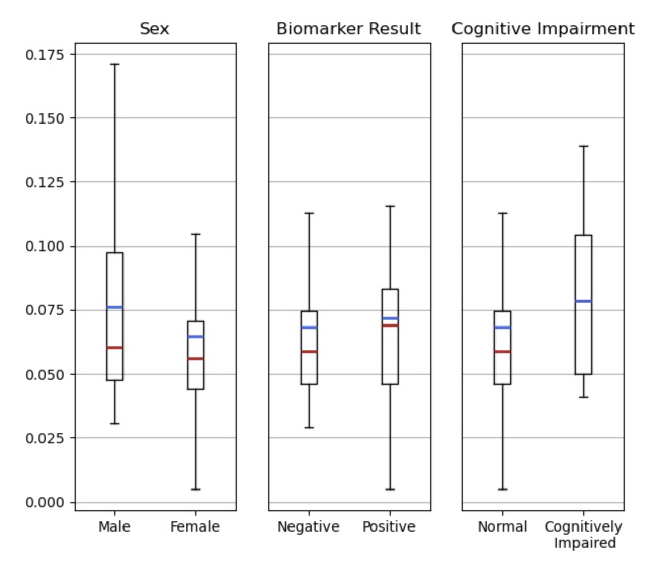
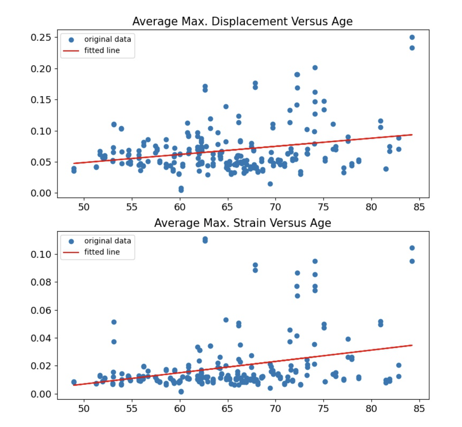
120 of 133 scans were deemed of sufficient quality [2-4 on Likert scale] to be included in the analysis. Figure 1 provides magnitude images and displacement maps representative of the image quality within each Likert scale rating. Plot 1 of Figure 2 shows the variance in maximum displacement within each Likert scale rating. 10% of the data was categorized in the lowest rating, 13.5% in the second rating, 57.9% in the third rating, and 18.7% in the fourth rating. Plots 2 and 3 of Figure 2 show a high degree of consistency in maximum displacement measures between the left and right hemispheres. Figure 3 compares average maximum displacement measures between various patient variables and found no significant correlation. Figure 4 shows both average maximum displacement and strain measures display a small positive correlation with age, as well as the high degree of variability in both. The strain measures were deemed too “noisy” for substantial interpretation.Discussion and Conclusions
Our study suggests further work is needed to understand the contributing and confounding factors to DENSE measures in aging cohorts. There was a high degree of consistency in displacement measures between the left and right hemispheres; however, few variables were found to explain the substantial variability across the patient cohort (e.g. sex, amyloid status, image quality). The resolution of the data limited our ability to exclude CSF contamination from ROIs, as well as inconsistencies in slice placement and difficulties related to scanning older populations, such as a greater potential for bulk motion or less accurate PPG readings, were likely contributors to the high variability observed in the data. Prior research demonstrating the successful use of DENSE within the brain has been done on smaller cohorts, younger demographics and at higher resolutions than what was done here. Future research is needed to determine what biological and technical factors may be causing the variability in the data and to what extent technical variability can be mitigated in future sequence design and post-processing.Acknowledgements
We gratefully acknowledge funding support from NIH grants R01AG075788 and R21NS125094 and research support from GE Healthcare.References
1. Fonck, Edouard, et al. Effect of Aging on Elastin Functionality in Human Cerebral Arteries. Stroke, vol. 40, no. 7, 28 May 2009, pp. 2552–2556, https://doi.org/10.1161/strokeaha.108.528091.
2. Thorin-Trescases, Nathalie, and Eric Thorin. Lifelong Cyclic Mechanical Strain Promotes Large Elastic Artery Stiffening: Increased Pulse Pressure and Old Age-Related Organ Failure. Canadian Journal of Cardiology, vol. 32, no. 5, 1 May 2016, pp. 624–633, https://doi.org/10.1016/j.cjca.2015.12.022.
3. Vaitkevicius, P V, et al. Effects of age and aerobic capacity on arterial stiffness in healthy adults. Circulation, vol. 88, no. 4, Oct. 1993, pp. 1456–1462, https://doi.org/10.1161/01.cir.88.4.1456.
4. Takamura, Tomohiro, et al. Influence of Age on Global and Regional Brain Stiffness in Young and Middle‐Aged Adults. Journal of Magnetic Resonance Imaging, vol. 51, no. 3, 1 Aug. 2019, pp. 727–733, https://doi.org/10.1002/jmri.26881.
5. Hiscox, Lucy V., et al. Aging brain mechanics: Progress and promise of magnetic resonance elastography. NeuroImage, vol. 232, 15 May 2021, p. 117889, https://doi.org/10.1016/j.neuroimage.2021.117889.
6. Hiscox, Lucy V., Curtis L. Johnson, et al. High-resolution magnetic resonance elastography reveals differences in subcortical gray matter viscoelasticity between young and healthy older adults. Neurobiology of Aging, vol. 65, 1 May 2018, pp. 158–167, https://doi.org/10.1016/j.neurobiolaging.2018.01.010.
7. Murphy, Matthew C., et al. Decreased brain stiffness in Alzheimer's disease determined by magnetic resonance elastography. Journal of Magnetic Resonance Imaging, vol. 34, no. 3, 12 July 2011, pp. 494–498, https://doi.org/10.1002/jmri.22707.
8. ElSheikh, Mona, et al. MR Elastography Demonstrates Unique Regional Brain Stiffness Patterns in Dementias. American Journal of Roentgenology, vol. 209, no. 2, Aug. 2017, pp. 403–408, https://doi.org/10.2214/ajr.16.17455.
9. Calik, Nazmi, et al. Altered diastolic function and aortic stiffness in Alzheimer’s disease. Clinical Interventions in Aging, 16 July 2014, p. 1115, https://doi.org/10.2147/cia.s63337.
10. Meaney, David F., et al. Biomechanical Basis of Traumatic Brain Injury. Youmans Neurological Surgery, 2011, pp. 3277–3287, https://doi.org/10.1016/b978-1-4160-5316-3.00328-2.
11. Soellinger, Michaela, et al. 3D cine displacement‐encoded MRI of pulsatile brain motion. Magnetic Resonance in Medicine, vol. 61, no. 1, 2008, pp. 153–162, https://doi.org/10.1002/mrm.21802.
12. Adams, Ayodeji L., et al. Quantifying cardiac‐induced brain tissue expansion using DENSE. NMR in Biomedicine, vol. 32, no. 2, 2018, https://doi.org/10.1002/nbm.4050.
13. Pahlavian, Soroush Heidari, et al. Regional Quantification of Brain Tissue Strain Using Displacement-Encoding With Stimulated Echoes Magnetic Resonance Imaging. Journal of Biomechanical Engineering, vol. 140, no. 8, 2018, https://doi.org/10.1115/1.4040227.
14. Sloots, Jacob Jan, et al. Strain Tensor Imaging: Cardiac-induced brain tissue deformation in humans quantified with high-field MRI. NeuroImage, vol. 236, 2021, p. 118078, https://doi.org/10.1016/j.neuroimage.2021.118078.
15. Zhong, Xiaodong, et al. Tracking brain motion during the cardiac cycle using spiral cine-DENSE MRI. Medical Physics, vol. 36, no. 8, 1 July 2009, pp. 3413–3419, https://doi.org/10.1118/1.3157109.
16. Aletras, Anthony H., et al. DENSE: Displacement Encoding with Stimulated Echoes in Cardiac Functional MRI. Journal of Magnetic Resonance, vol. 137, no. 1, 1999, pp. 247–252, https://doi.org/10.1006/jmre.1998.1676.
Figures