4897
Inhomogeneity-informed Field-fitting for Quantitative Susceptibility Mapping (if-QSM)1Electrical and Computer Engineering, Cornell University, New York, NY, United States, 2Radiology, Weill Cornell Medicine, New York, NY, United States, 3Biomedical Engineering, Cornell University, New York, NY, United States
Synopsis
Keywords: Bioeffects & Magnetic Fields, Artifacts
Motivation: Inhomogeneity in measured multi-gradient echo (mGRE) field data corrupts reconstructions of quantitative susceptibility maps by obscuring the tissue field of interest with strong background field.
Goal(s): To extend the voxel spread function (VSF) library implementation to a nonzero phase offset and demonstrate improvements on QSM.
Approach: The measured field was estimated and inhomogeneity contributions computed using the extended library implementation. The inhomogeneity field was then estimated and subtracted from the total field to reduce the influence of strong background fields in the QSM reconstruction
Results: Inhomogeneity-informed field-fitting for QSM is shown to improve total field reconstructions of the brain, carotid, and cervical spine.
Impact: The voxel spread function (VSF) library implementation is extended to include initial phase offset contributions and reduce the effect of field inhomogeneity in quantitative susceptibility map (QSM) reconstruction.
Introduction
The ill-posed problem of dipole deconvolution used in quantitative susceptibility mapping (QSMs)1 relies on a robust estimate of the field map. Dipole-incompatible errors in the estimated field map generate nonlocal artifacts in QSM due to the wave propagator solution to the dipole deconvolution.2 One source of such errors is field inhomogeneity, induced by the anatomical interfaces where susceptibility changes abruptly (e.g., air-tissue), and causing non-local image distortions. The effects of this distortion are typically ignored in in the background removal methods3-12 In the present work, we propose the extension of the voxel spread function (VSF) correction method to address this shortcoming. We demonstrated the improvement in QSM reconstruction using use QSM Challenge 2.0 data.13 Additionally, performance of this method in is assessed in cervical spine and carotid QSM reconstructions.Theory
The measured mGRE signal $$$S_n(TE)$$$ can be decomposed as a product of the true signal $$$\sigma_n(TE)$$$ and field inhomogeneity contribution $$$F_n(TE)$$$, such that $$$S_n(TE)=\sigma_n(TE)F_n(TE)$$$.14 Here$$F_n(TE)=\frac{1}{|S_n(0)|}\sum_m \Psi_{nm}(TE)|S_m(0)|$$ Where $$$n$$$ and $$$m$$$ are the neighbor indices, is the complex mGRE signal at voxel $$$S_n(TE)$$$ and echo time $$$TE$$$, $$$|S_k(0)|$$$ is the signal magnitude at voxel $$$k$$$, $$$\Psi_{nm}$$$ is the voxel-wise kernel and phase of signal $$$S_m(TE)$$$ at neighboring voxel $$$m$$$. From this, inhomogeneity correction factor $$$F_n(TE)$$$ can be computed to correct field inhomogeneity as described in the original voxel spread function (VSF) implementation14 and the accelerated library implementation.15 The voxel-wise kernel is a function of the VSF $$$\eta(n,m;q_m(TE))$$$, which describes the effect of phase dispersion on the measured signal: $$\Psi_{nm}(TE)=\eta(n,m;q_m(TE))e^{i\phi_{0,m}+i\gamma b_m TE}$$ Here, $$$q_m(TE)$$$ is the phase dispersion, $$$\phi_{0,m}$$$ is the phase offset, $$$\gamma$$$ is the gyromagnetic ration and $$$b_m$$$ is the field map. Previously, this method has been shown to improve $$$R_2^*$$$ maps. However, the correction factor $$$F_n(TE)$$$ is complex and can therefore also be used to correct the signal phase, $$$\angle S_n(TE)$$$. The original library implementation15 consisting of matching each voxel of the field gradient with a precomputed kernel value to accelerate the calculation of $$$\eta(n,m;q_m(TE)$$$, is hereby extended by allowing $$$\phi_0 \neq 0$$$.Methods
Complex multi-gradient echo (mGRE) data was simulated via dipole convolution with the QSM challenge 2.0 data13 in the presence of decay and complex Gaussian noise ($$$SNR=50$$$) at $$$7T$$$. To avoid introducing further phase wrapping from complex division of the measured signal by the inhomogeneity factor,14,15 the phase correction was applied after fitting both the measured field and the field inhomogeneity maps. The simulated data was processed using the nonlinear field fitting algorithm16 followed by ROMEO17 phase unwrapping and VSF correction. ROMEO phase unwrapping and field fitting was applied for both the field map estimation and the VSF estimation for the in vivo brain and spine and carotid. The corrected field map and VSF were computed iteratively for $$$K$$$ ($$$1$$$ for brain, $$$10$$$ for carotid, $$$15$$$ for brain and spine) iterations. The original and VSF-corrected field maps were used in nonlinear preconditioned total field inversion (npTFI)18 with preconditioner $$$P=20$$$ and $$$\lambda=100$$$. The cervical spine images were acquired using 3D mGRE at $$$3T$$$ with $$$6$$$ echo times and repetition time $$$TR=38ms$$$, flip angle $$$15°$$$, matrix size $$$256\times256\times72$$$ and voxel size $$$0.9375\times0.9375\times2mm^3$$$. The carotid images were acquired using 3D mGRE19 with $$$4$$$ echo times and $$$TR=20ms$$$, first echo time $$$TE_1=\Delta TE=4.7ms$$$, voxel size $$$0.63\times0.63\times2mm^3$$$, flip angle $$$30°$$$, scan time = $$$4$$$ minutes.Results
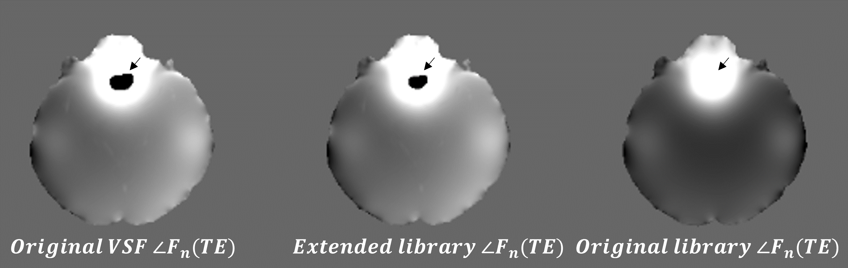
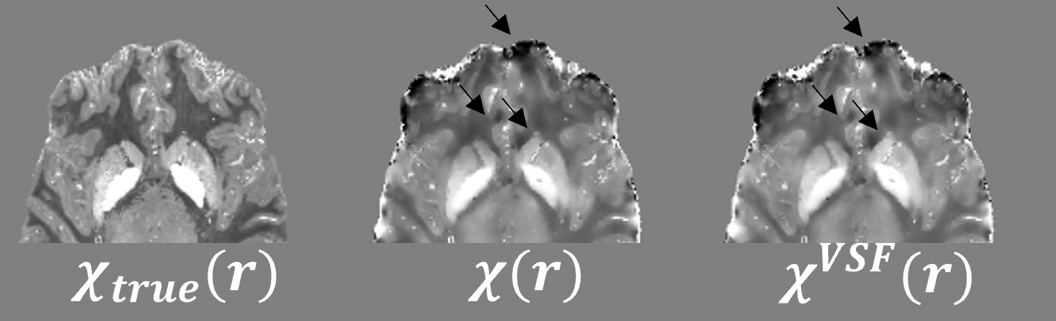
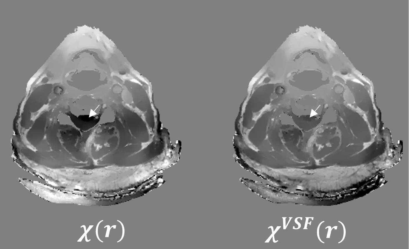
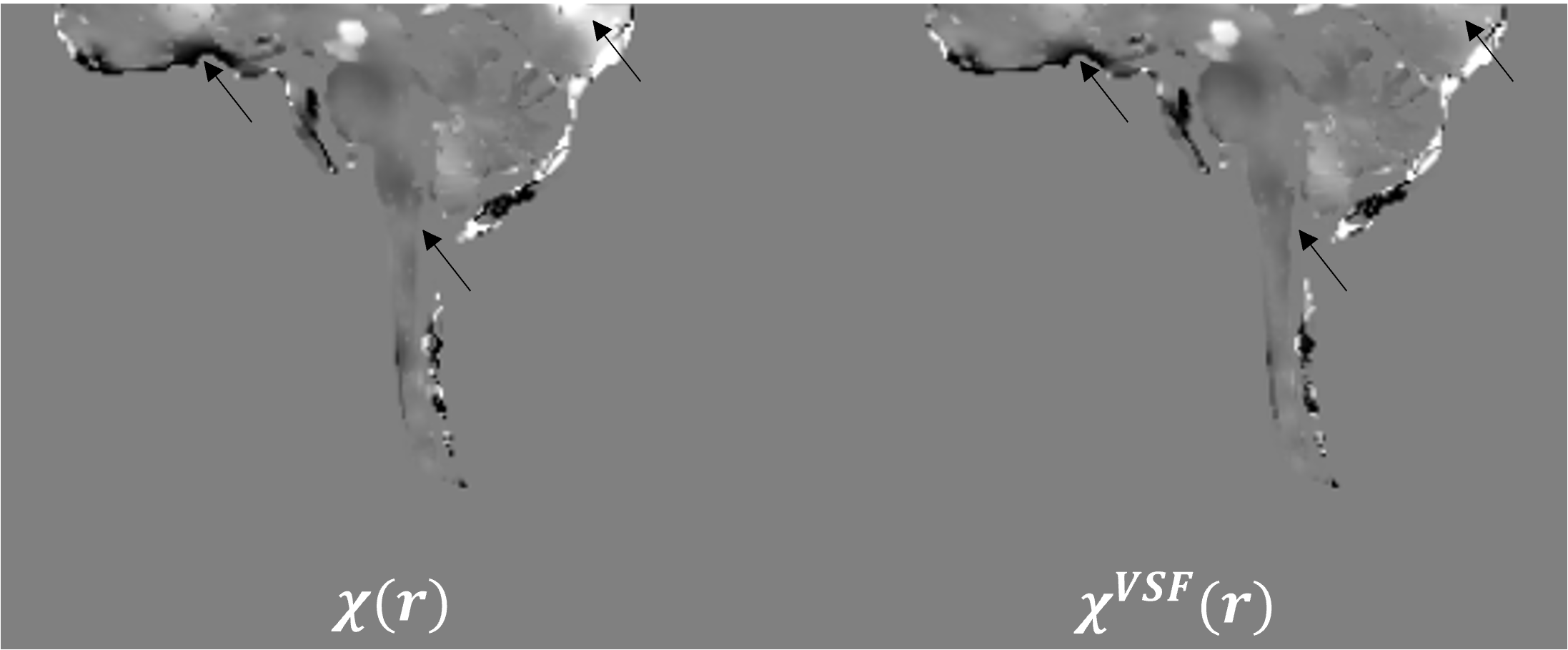
The phase offset extension reduces the mean-squared phase error between the original implementation and library implementation by several orders of magnitude ($$$0.0024$$$ and $$$0.2866$$$, respectively) as shown in Figure 1. The inhomogeneity correction in the nonlinear field fitting reduces ground truth error ($$$0.43$$$ versus $$$0.42$$$) and the presence of nonlocal artifacts in the simulation (Figure 2). This method extends to improve inhomogeneity artifacts in the carotid (Figure 3) and cervical spine (Figure 4).Conclusion
The proposed phase offset extension of the VSF library implementation reduces artifacts across multiple field strengths and improves accuracy of reconstructed QSM.Acknowledgements
No acknowledgement found.References
1. Wang Y, Liu T. Quantitative susceptibility mapping (QSM): Decoding MRI data for a tissue magnetic biomarker. Magnetic Resonance in Medicine. 2015;73(1):82-101. doi:10.1002/mrm.25358 2. Youngwook Kee ZL, Liangdong Zhou, Alexey Dimov, Junghun Cho, Ludovic de Rochefort, Jin Keun Seo, and Yi Wang. Quantitative Susceptibility Mapping (QSM) Algorithms: Mathematical Rationale and Computational Implementations. IEEE Transactions on Biomedical Engineering. 2017;64(11):2531-2545. doi:10.1109/tbme.2017.2749298
3. Li W, Wu B, Liu C. Quantitative susceptibility mapping of human brain reflects spatial variation in tissue composition. NeuroImage. 2011;55(4):1645-1656. doi:10.1016/j.neuroimage.2010.11.088
4. Liu Z, Spincemaille P, Yao Y, Zhang Y, Wang Y. MEDI+0: Morphology enabled dipole inversion with automatic uniform cerebrospinal fluid zero reference for quantitative susceptibility mapping. Magnetic Resonance in Medicine. 2018;79(5):2795-2803. doi:10.1002/mrm.26946
5. Milovic C, Bilgic B, Zhao B, Langkammer C, Tejos C, Acosta‐Cabronero J. Weak‐harmonic regularization for quantitative susceptibility mapping. Magnetic Resonance in Medicine. 2019;81(2):1399-1411. doi:10.1002/mrm.27483
6. Roberts A, Spincemaille P, Nguyen T, Wang Y. MEDI-d: Downsampled Morphological Priors for Shadow Reduction in Quantitative Susceptibility Mapping. presented at: International Society for Magnetic Resonance in Medicine; 2021; Vancouver, Canada. https://cds.ismrm.org/protected/21MPresentations/abstracts/2599.html
7. Roberts A, Spincemaille P, Nguyen T, Wang Y. MEDI-FM: Field Map Error Guided Regularization for Shadow Reduction in Quantitative Susceptibility Mapping. presented at: International Society for Magnetic Resonance in Medicine; 2022; London, England. https://archive.ismrm.org/2022/2359.html 8. Roberts A, Spincemaille P, Nguyen T, Wang Y. Whole Brain Spherical Mean Value Filtering for Shadow Reduction in Quantitative Susceptibility Mapping. Paper No. 2172. presented at: International Society for Magnetic Resonance in Medicine; 2023; Toronto, Canada.
9. Roberts AG, Romano DJ, Sisman M, et al. Maximum Spherical Mean Value (mSMV) Filtering for Whole Brain Quantitative Susceptibility Mapping. arXiv pre-print server. 2023-04-22 2023;doi:None arxiv:2304.11476
10. Schweser F, Deistung A, Lehr BW, Reichenbach JR. Quantitative imaging of intrinsic magnetic tissue properties using MRI signal phase: An approach to in vivo brain iron metabolism? NeuroImage. 2011;54(4):2789-2807. doi:10.1016/j.neuroimage.2010.10.070
11. Wei H, Dibb R, Zhou Y, et al. Streaking artifact reduction for quantitative susceptibility mapping of sources with large dynamic range. NMR in Biomedicine. 2015;28(10):1294-1303. doi:10.1002/nbm.3383 12. Schweser F, Robinson SD, De Rochefort L, Li W, Bredies K. An illustrated comparison of processing methods for phase MRI and QSM: removal of background field contributions from sources outside the region of interest. NMR in Biomedicine. 2017;30(4):e3604. doi:10.1002/nbm.3604
13. Marques JP, Meineke J, Milovic C, et al. QSM reconstruction challenge 2.0: A realistic in silico head phantom for MRI data simulation and evaluation of susceptibility mapping procedures. Magnetic Resonance in Medicine. 2021;86(1):526-542. doi:10.1002/mrm.28716
14. Yablonskiy DA, Sukstanskii AL, Luo J, Wang X. Voxel spread function method for correction of magnetic field inhomogeneity effects in quantitative gradient-echo-based MRI. Magnetic Resonance in Medicine. 2013;70(5):1283-1292. doi:10.1002/mrm.24585
15. Liu Y, Ye Q, Zeng F, et al. Library‐driven approach for fast implementation of the voxel spread function to correct magnetic field inhomogeneity artifacts for gradient‐echo sequences. Medical Physics. 2021;48(7):3714-3720. doi:10.1002/mp.14904
16. Liu T, Wisnieff C, Lou M, Chen W, Spincemaille P, Wang Y. Nonlinear formulation of the magnetic field to source relationship for robust quantitative susceptibility mapping. Magnetic Resonance in Medicine. 2013;69(2):467-476. doi:10.1002/mrm.24272
17. Dymerska B, Eckstein K, Bachrata B, et al. Phase unwrapping with a rapid opensource minimum spanning tree algorithm (ROMEO). Magnetic Resonance in Medicine. 2021;85(4):2294-2308. doi:10.1002/mrm.28563
18. Liu Z, Kee Y, Zhou D, Wang Y, Spincemaille P. Preconditioned total field inversion (TFI) method for quantitative susceptibility mapping. Magnetic Resonance in Medicine. 2017;78(1):303-315. doi:10.1002/mrm.26331
19. Nguyen TD, Wen Y, Du J, et al. Quantitative susceptibility mapping of carotid plaques using nonlinear total field inversion: Initial experience in patients with significant carotid stenosis. Magnetic Resonance in Medicine. 2020;84(3):1501-1509. doi:10.1002/mrm.28227
Figures