4895
Pushing the image quality by integrating FatNav and pTx Universal Pulses in MPRAGE and MP2RAGE sequences at 7T1University Paris-Saclay, CEA, CNRS, BAOBAB, NeuroSpin, Gif-sur-yvette, France, 2Siemens Healthcare SAS, Courbevoie, France, 3CUBRIC, Cardiff, United Kingdom
Synopsis
Keywords: High-Field MRI, High-Field MRI
Motivation: Ultra-high field MRI of the brain suffers from an increased B1+ inhomogeneity as well as involuntary motion artifacts when very high spatial resolution is targeted.
Goal(s): Integration of a FatNav technique into pTx Universal Pulse sequences would be beneficial to reach the best image quality at very high resolution.
Approach: An MP(2)RAGE sequence using GRAPE pTx universal pulses was modified to integrate a FatNav module. High-resolution protocols were acquired in vivo on the brain.
Results: Very high-quality images were obtained throughout the brain and cerebellum thanks to FatNav motion correction and pTx Universal Pulses.
Impact: The ultra-high field community targetting high-resolution protocols on the whole brain would benefit from a FatNav-enabled PASTEUR package to bring robust protocols against B1+ inhomogeneities and involuntary motion of the head.
Introduction
Ultra-high field MRI has demonstrated its ability to provide very high-resolution images to study the fine structures of the brain. Unfortunately, scan time lengthens rapidly with increased resolution, thereby leading to image blurring in the presence of motion. Technical solutions have been proposed recently by the research community to limit the effects of motion-induced artifacts. For instance, 3D fat navigator (FatNav) is a method very well suited for 3D segmented sequences at very high resolutions1,2. In addition to motion, UHF MRI suffers from B1+ inhomogeneities that have successfully been tackled using parallel transmission (pTx) technology. To spare the users additional B0 and B1+ measurements, and lengthy calculations, the Universal Pulse concept3 was introduced as a plug-and-play pTx solution needing no extra expertise. For the same scanner and RF coil types, it was also demonstrated to be a portable solution4. Thus, a package of anatomical sequences containing UPs, called PASTEUR5 package, has been shared with the UHF research Siemens 7T community. The goal of this work is to demonstrate the feasibility of associating two key technologies, FatNav and UPs, and provide a FatNav-enabled MP(2)RAGE solution using Universal Pulses.Methods
Acquisitions were performed on a healthy volunteer on an investigational Magnetom 7T scanner (Siemens Healthineers, Erlangen, Germany) equipped with the Step 2.3 pTX hardware and an 8Tx/32Rx pTx coil (Nova Medical, Wilmington, MA, USA). The MP(2)RAGE pulse sequence from the PASTEUR package4,5 was modified to insert a FatNav1,2 module on every TR. Thus, inversion and excitation pulses were Universal GRAPE6 pTx pulses designed on a database of 20 subjects composed of B0 and B1+ field maps. The FatNav module used fat-selective binomial RF pulses which were played in circularly polarized mode. The resolution of the FatNav volumes was 2mm isotropic. Additional parameters for the module were TE/TR 1.47/3.2ms, a 4x4 GRAPPA acceleration, 6/8 partial Fourier undersampling, and a total acquisition time for one FatNav volume of 1.24s. The scanning session was composed of two MPRAGE protocols of 0.65 and 0.45mm isotropic resolutions and two MP2RAGE protocols of 0.65 and 0.5mm isotropic resolutions, with relatively long acquisition times. Detailed parameters are given in table 1. The whole setup was compatible with Siemens protected mode step 2.3 and the protected mode of the Nova coil, i.e. with peak amplitude limits of 165V per channel and average power limits of 1.5W per channel and 8W total defined at coil plug. The volunteer was instructed not to move during the scans. Motion correction and image reconstruction were performed offline using the open-source RetroMocoBox7 toolbox in Matlab (The MathWorks, Inc., Natick, MA) during which reconstructed 3D FatNavs were co-registered using the ‘realign’ tool in SPM (Statistical Parametric Mapping) to estimate the motion parameters.Results
Figure 2 reports the results obtained with the 0.65mm MPRAGE protocol. Estimated rigid body motion parameters, expressed in root-mean-square (RMS) displacement and rotation, obtained from 3D FatNav volumes were significantly lower than the image resolution. Thus, improvement in image quality of the motion-corrected images was found to be insignificant for this acquisition. Figures 3 and 4 show results obtained with 0.45mm MPRAGE and 0.5mm MP2RAGE protocols respectively. In both cases, estimated rigid body motion parameters were comparable to the image resolution, and improvement in image sharpness was visible between non-corrected and motion-correction reconstructions, which resulted in an increase in the conspicuity of small vessels and sharpness in transition between tissues such as gray and white matter. Figure 5 presents the results of MP2RAGE acquisition at 0.65mm resolution. Estimated rigid body motion parameters were larger than image resolution in this case, leading to a significant deterioration of the non-corrected image that was well recovered in the motion-corrected image. In all four protocols, the use of Universal GRAPE pTx pulses led to a smooth signal and contrast throughout the brain and cerebellum thanks to an improved homogeneity of the inversion and excitation flip angles. Receive sensitivity profiles were not corrected in MPRAGE images.Discussion/Conclusion
As expected with the use of Universal GRAPE pulses available in the PASTEUR package, B1+ inhomogeneities in MPRAGE and MP2RAGE acquisitions were well mitigated in the brain and the cerebellum. Yet, the relatively moderate motion estimates obtained with the involuntary motion of the head were large enough to degrade image sharpness. The FavNav acquisition and its retrospective motion-corrected reconstruction helped improving image quality for very high-resolution protocols. The FatNav-enabled MP(2)RAGE UP sequence is readily available for dissemination. Future work includes incorporating the module in 3D-SPACE sequences to foster further very high-resolution acquisitions in the growing UHF community.Acknowledgements
This work received financial support from the Leducq Foundation (large equipment ERPT program, NEUROVASC7T project) and from FET-Open H2020 (AROMA project, grant agreement n°885876).
References
1. Gallichan D. et al. (2016). Retrospective correction of involuntary microscopic head movement using highly accelerated fat image navigators (3D FatNavs) at 7T. Magn Reson Med. 75(3):1030-9. doi: 10.1002/mrm.25670.
2. Federau C. and Gallichan D. (2016). Motion-Correction Enabled Ultra-High Resolution In-Vivo 7T-MRI of the Brain, PloS one vol. 11,5 e0154974. 9 May. doi:10.1371/journal.pone.0154974
3. Gras V. et al. (2017). Universal pulses: A new concept for calibration-free parallel transmission. Magn Reson Med. 77(2): 635-643. doi:10.1002/mrm.26148
4. Mauconduit F. et al. (2022). Traveling Pulses Visit 7T Terra Sites: Getting Ready For Parallel Transmission In Routine Use. In Proceedings of the 31th Annual Meeting of ISMRM, London, UK, p. 2091.
5. Gras V. et al. (2019). PASTeUR: Package of Anatomical Sequences using parallel Transmission UniveRsal kT-point pulses. In Proceedings of the 28th Annual Meeting of ISMRM, Montréal, Canada, p. 4626.
6. Van Damme, L. et al. (2021). Universal nonselective excitation and refocusing pulses with improved robustness to off-resonance for Magnetic Resonance Imaging at 7 Tesla with parallel transmission. Magn Reson Med. 85(2): 678–693. doi:10.1002/mrm.28441
7. https://github.com/dgallichan/retroMoCoBox
Figures
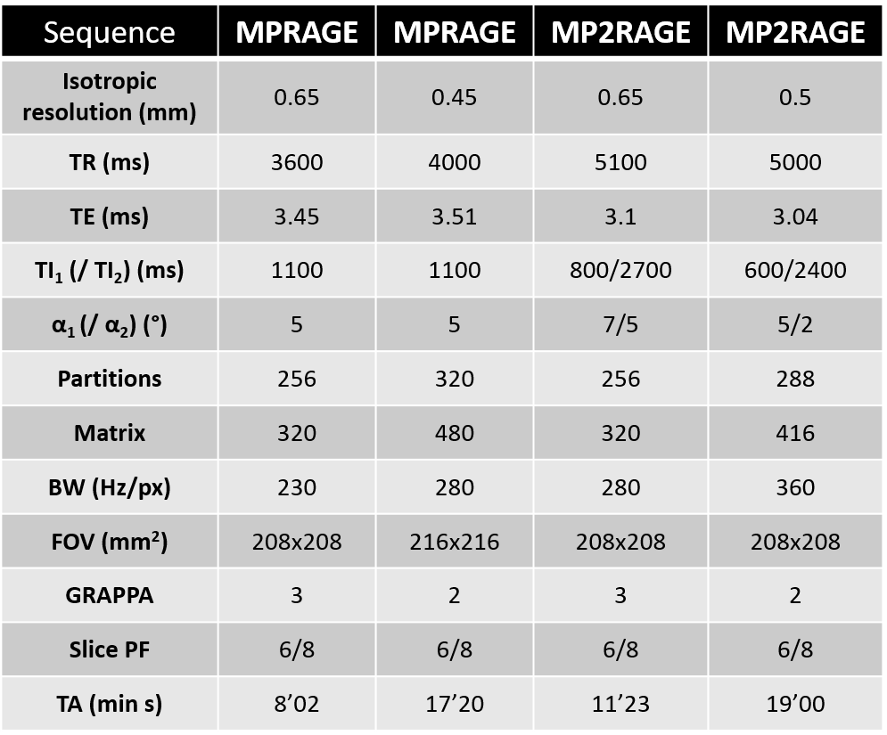
Table 1. Table of sequence parameters for each of the MPRAGE and MP2RAGE protocols. The protocols include inversion and excitation GRAPE UPs as well as a FatNav module of 1.24s long within every repetition time.
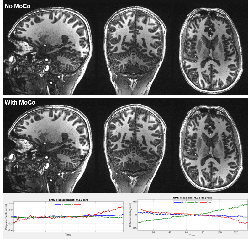
Figure 2. Sagittal, coronal, and axial views of a T1-weighted 3D MPRAGE acquisition at 0.65mm resolution (8min02 scan time) without (top row) and with motion correction (bottom row). Estimated rigid body motion parameters are depicted. RMS displacement and rotation were estimated as 0.12mm and 0.23° respectively, explaining the similarity of both reconstructions. No clear improvement was observed due to the low level of motion. In terms of B1+ inhomogeneities, UPs helped preserving the brain and cerebellum contrasts.
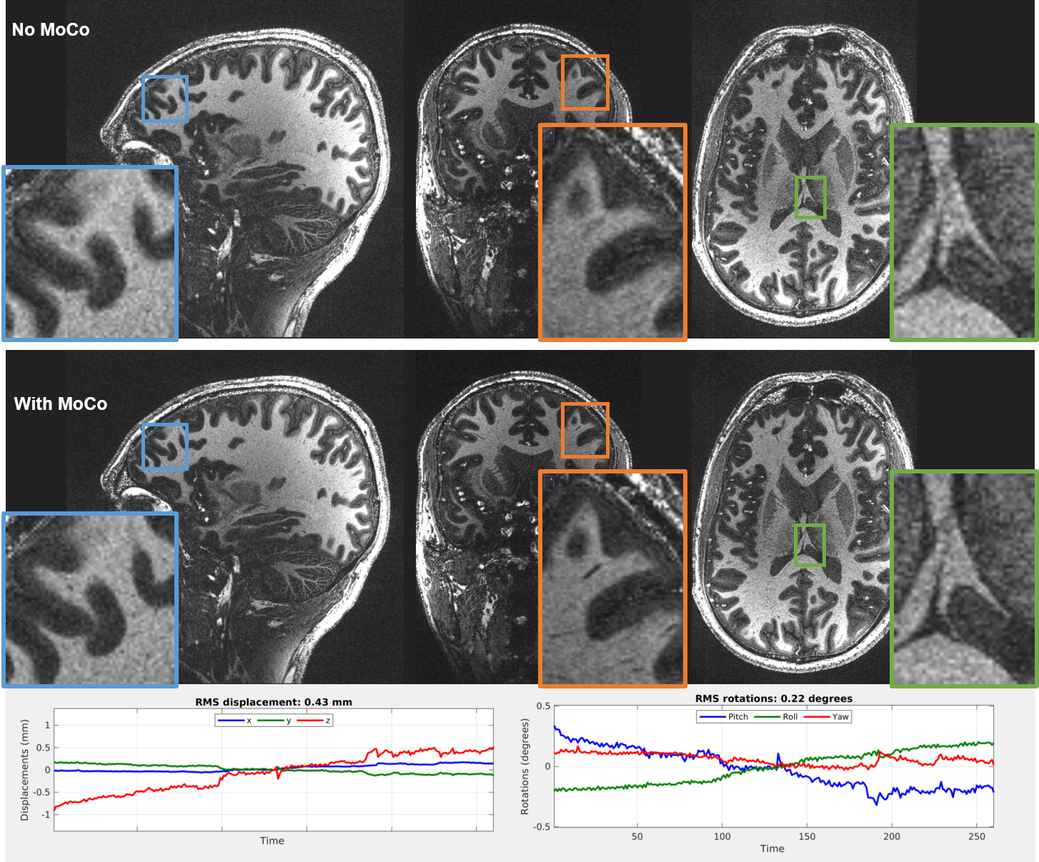
Figure 3. Sagittal, coronal, and axial views of a T1-weighted 3D MPRAGE acquisition at 0.45mm isotropic resolution (17min20 scan time) without motion correction (top row) and with FatNav motion correction (bottom row). Zooms are depicted to reveal the improvement in sharpness of the motion-corrected reconstruction. RMS displacement and rotation were estimated as 0.43mm and 0.22° respectively.
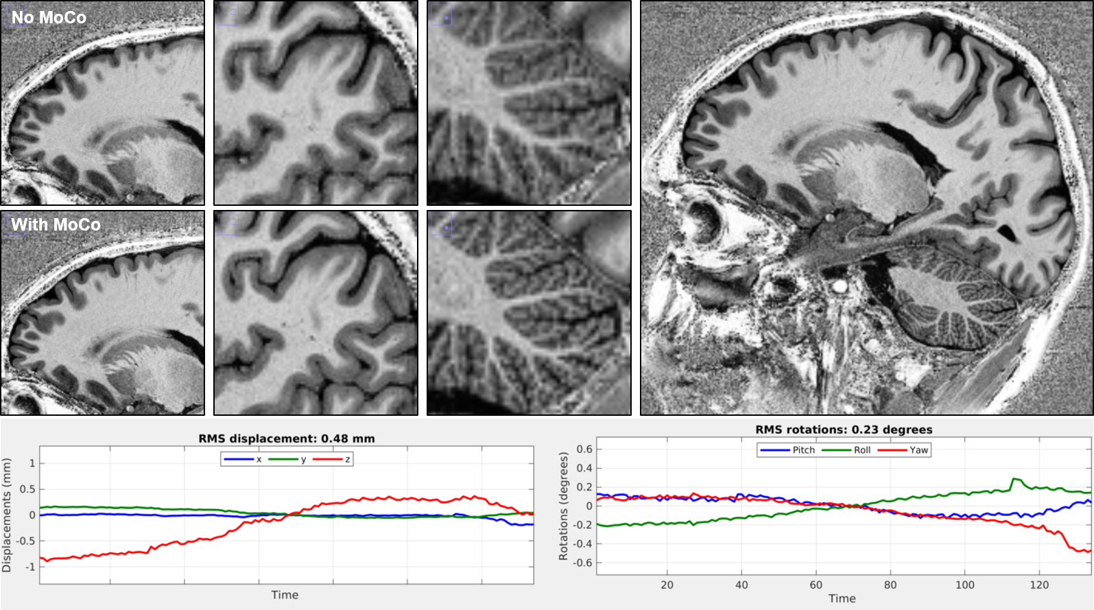
Figure 4. Resulting T1-weighted images were obtained from a 3D MP2RAGE acquisition at 0.50mm isotropic resolution (19min00 scan time). In Zoom captions, without motion correction (top row) and with motion correction (bottom row). The image on the right is a sagittal view of the motion-corrected reconstruction. RMS displacement and rotation were estimated as 0.48mm and 0.23° respectively, leading to a significant improvement of the motion-corrected images.
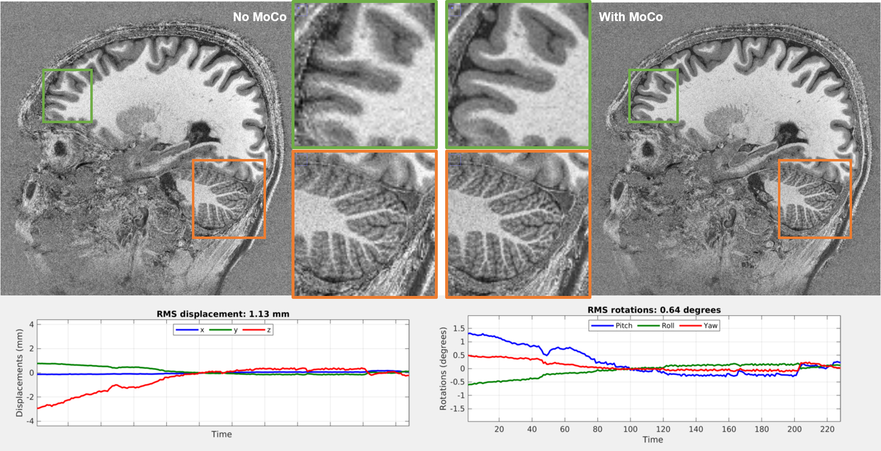
Figure 5. Sagittal view of UNI image reconstructed from a 3D MP2RAGE acquisition at 0.65mm isotropic resolution (11min23 scan time) without motion correction (left image and zooms) and with motion correction (right image and zooms). RMS displacement and rotation were estimated as 1.13mm and 0.64° respectively, leading to a deteriorated native image that is well recovered by the FatNav motion-corrected reconstruction.