4894
Automated Vendor-Independent Open-Source Quality Assurance Protocol using Pulseq1Division of Medical Physics, Department of Radiology, University Medical Center Freiburg, Faculty of Medicine, University of Freiburg, Freiburg, Germany, 2Department of Radiology and Nuclear Medicine, St. Olav's University Hospital, Trondheim, Norway, 3Department of Circulation and Medical Imaging, NTNU - Norwegian University of Science and Technology, Trondheim, Norway, 4Functional MRI Laboratory, University Of Michigan, Ann Arbor, MI, United States
Synopsis
Keywords: System Imperfections, System Imperfections: Measurement & Correction, Vendor-Neutral Automated Quality Assurance
Motivation: Ensuring comparable performance of MR protocols across vendors and over time duration.
Goal(s): To implement an easy-to-use vendor-independent quality control pulse sequences and data analysis routines.
Approach: Relying on Pulseq as a vendor-independent MR pulse sequence environment, we implemented the established quality assurance protocols (ACR/fBIRN). Following the image reconstruction from the acquired data, performed either on the scanner or off-line in Gadgetron, images are analyzed by the open-source Matlab pipeline.
Results: The proposed protocol has been tested on three 3T Siemens scanners with over a decade difference in the manufacturing date and has been successfully executed on a 3T GE scanner.
Impact: The proposed protocol and post-processing scripts allow for easy and streamlined quality assurance, contributing an essential component for Pulseq to become usable in large scale multicenter imaging studies.
Introduction
Neuroimaging research relies heavily on the consistent image quality. This concerns both structural anatomic imaging and functional MR imaging (fMRI). Especially the fMRI, that associates blood oxygenation level dependent (BOLD) signal changes to brain function, depends critically on the system stability. Furthermore, intricate details of data acquisition, image reconstruction and integrated post-processing also have a great effect on the results of fMRI studies. Quality assurance (QA) protocols can test the stability of MR scanners and help researches to detect errors during longitudinal MRI studies, thereby greatly enhancing their success rate. In multicenter settings, QA protocols help establishing consistent data standards across scanners and ensuring data comparability, which is essential for data pooling. However, heterogeneity of the imaging hardware may present substantial challenges for cross-scanner and cross-center quality comparisons due to the necessity of implementing consistent protocols in varying vendor-specific environments.In this work we use Pulseq to implement an open-source transparent quality assessment MR protocol based on recommendations of the American Congress of Radiology (ACR) and the Function Biomedical Informatics Research Network (fBIRN), that is accompanied by an open-source data processing pipeline. The proposed protocol has been thoroughly tested on three 3T Siemens scanners with the manufacturing date separated by over 17 years and also successfully executed on one from 3T GE scanner.
Methods
Pulse sequencesA multi-slice spin echo (SE) sequence with two Shinnar-Le Roux radiofrequency pulses for excitation and refocusing was implemented with the Pulseq framework to measure the structural quality. The sequence parameters are based on those of the ACR Axial T1 series [1]: TR/TE = 500/20; field of view (FOV) = 250 * 250 mm2; number of slices = 11; slice thickness/gap = 5/5 mm; repetition = 2; matrix size = 256 * 256; total scan time = 2:16. A 2D slice-selective echo-planar imaging (EPI) sequence was also implemented using Pulseq for fMRI scan. The fMRI acquisition parameters follow Friedman et al [2]: TR/TE=2000/22 ms; FOV=220*220 mm2; number of slices = 27; slice thickness/gap = 4/1 mm; flip angle=90°; number of volumes = 200; scan time = 6:40 min. Siemens product SE and EPI protocols were configured to closely match the corresponding Pulseq protocols.
Automatic processing
An iterative circle detection algorithm was used to automatically detect the position of phantom. This position information was used to automatically calculate the structural quality and functional quality. The structural quality metrics are[1]: signal-to-noise ratio (SNR), percent signal ghosting (PSG), Percent Image Uniformity (PIU). The functional quality metrics are [3]: signal-to-fluctuation-noise ratio (SFNR), temporal SNR (tSNR), temporal signal-to-background noise ratio (tSBNR), and functional signal-to-noise ratio (SNR-functional).
Measurement
A water-filled phantom doped with a NiCl/NaCl mixture was used as an alternative of the fBIRN phantom. A first fMRI acquisition was performed to let the system reach a temperature equilibrium. And the second fMRI scan and the SE sequence from Siemens and Pulseq were executed to produce data for QA metrics calculation.
Results and Discussion
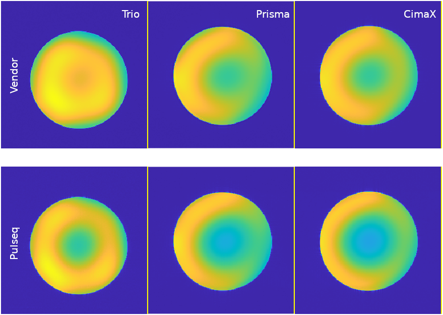
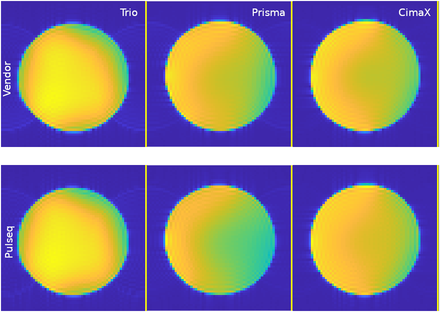
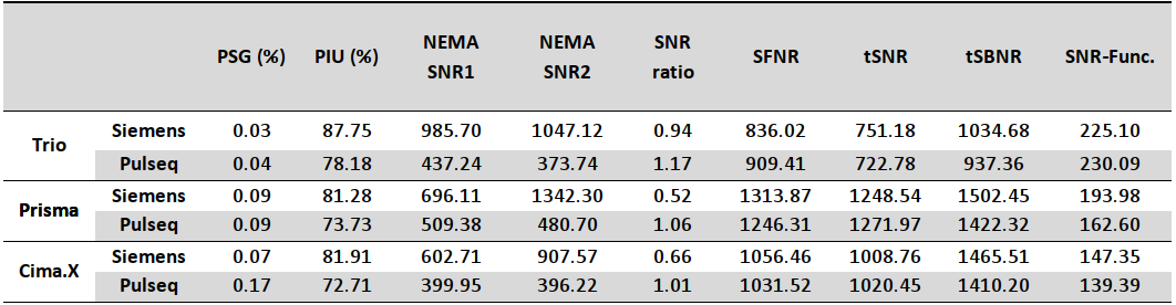
Figure 1 presents example phantom images acquired with the spin-echo protocol. As seen, a consistent image quality can be observed both for vendor sequences on three scanners and the Pulseq counterparts, although image homogeneity varies, which can be attributed to differences in receiver coil sensitivities, which were not normalized here. Figure 2 shows EPI results for both vendor and Pulseq sequences.Detailed numerical analysis can be seen in Table 1, which shows a general tendency for the image quality to improve with new scanner generations, with a slight exception for EPI performance on CimaX, which needs to be investigated further.
The same protocol has been tested on MR750 3T GE scanner, but the automated data processing pipeline was not finalized at the time of this writing. The cross-platform capability of the framework has however been verified.
Acknowledgements
This work was supported by research grants NIH R01 EB032378 and NIH U24 NS120056.References
1. Albus K. Large and Medium Phantom Test Guidance for the MRI Accreditation Program. Am Coll Radiol. Published online 2022. 2.
2. Friedman L, Glover GH. Report on a multicenter fMRI quality assurance protocol. J Magn Reson Imaging. 2006;23(6):827-839. doi:10.1002/jmri.20583
3. Layden, E. A. (2020). MRIqual: MRIqual: A Matlab toolbox for examining the quality of structural (SNR) and functional (tSNR, SFNR) MRI. Zenodo. http://doi.org/10.5281/zenodo.3735471
Figures