4893
Bias and Variability of Mean Diffusivity from DTI Across Two Head Coils: a Traceable Phantom Study1University College London, London, United Kingdom, 2National Physical Laboratory, Teddington, United Kingdom, 3Great Ormond Street Hospital for Children NHS Foundation Trust, London, United Kingdom
Synopsis
Keywords: Phantoms, Quantitative Imaging, Reproducibility
Motivation: Variability and bias of diffusion parameters need to be assessed to separate the measurement-induced variability from biological effects.
Goal(s): Our goal was to assess the bias and variability of mean diffusivity (MD) from the Diffusion Tensor Model using a traceable phantom.
Approach: The NIST/QIBA phantom was scanned multiple times using two different head coils on the same system. The differences between MD measured using different head coils and bias to NIST-reported values were investigated.
Results: A significant difference in measured MD with two coils can be found across multiple vials. Both coils introduce non-negligible bias to NIST-reported values for lower MD values.
Impact: Using traceable phantoms is essential in the development of Diffusion MRI-based Quantitative Imaging Biomarkers (QIBs). Different head coils can introduce significant differences across MD values and should be treated as confounding factors in QIBs studies.
Introduction
Rotationally-invariant diffusion parameters have the potential to become Quantitative Imaging Biomarkers (QIB) for various diseases. Therefore, it is important that the effect of measurement variability on the parameters is understood. This variability can be quantified using a well-characterised traceable phantom.The effects of different multi-channel head coils on measurements of diffusion parameters have been studied in the brain1-3 and by using phantoms4,5. However, these studies do not account for Region-Of-Interest (ROI) variability, or physiological noise, or do not separate inter-scanner effects from the effects of different coils. The assumption that different multi-channel head coils have a marginal effect on the results needs to be investigated.
This study aims to assess the bias and variability of mean diffusivity (MD) from the Diffusion Tensor Imaging (DTI) model using a traceable phantom, two multi-channel head coils, and a single 3T MRI scanner.
Methods
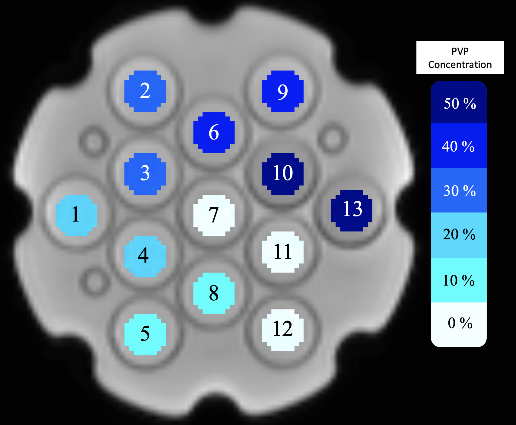
A NIST/QIBA Diffusion Phantom6 was scanned on a 3T scanner 20 times during 3 months, using 20- and 64-channel head coils, to acquire a set of diffusion-weighted images. Each scan was pre-processed using the pipeline routinely applied to in-vivo data at our centre. MD maps were obtained using DIPY7. Semi-automated binarize and erode procedure, and the scipy.ndimage.label function separated the 13 vials into distinct regions (Fig.1).The temperature was recorded during scanning using a built-in Liquid Crystal Thermometer8. The mean was 19.25 °C (± 1 °C uncertainty). The NIST-reported values were linearly interpolated for this temperature. To account for uncertainty in temperature, the uncertainties of NIST values were expanded.
The variability of MD measured with different head coils was assessed with the coefficient of variation (CoV). The bias of MD measured with different head coils to NIST values was assessed and considered non-negligible if it was higher than NIST uncertainty.
To assess the significance of the difference in MD measured with both coils, Welch’s t-test was performed. Statistical significance was set for p-value <0.05.
Results
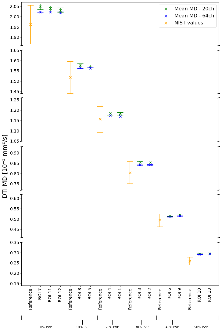
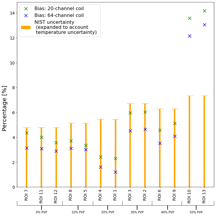
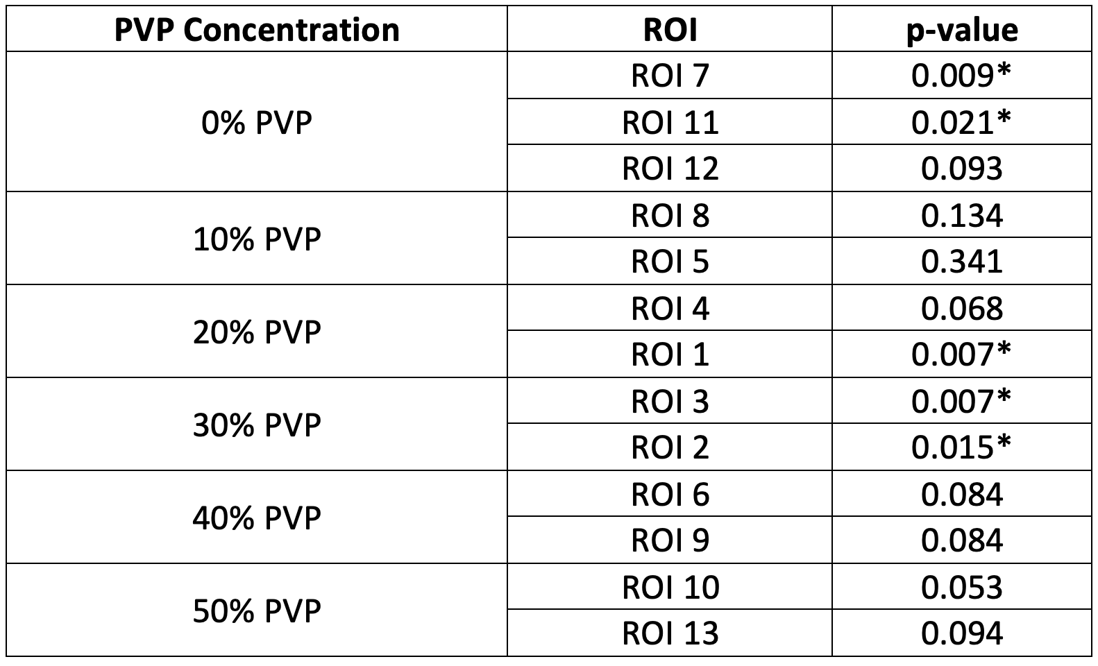
Mean CoV of MD measured with the 20-channel coil was 1.19% (95% CI: 1.06%, 1.33%) and 0.42% with the 64-channel coil (95% CI: 0.39%, 0.46%). Fig.2 shows the mean MD values in each ROI. Fig.3 shows the bias of acquired values to NIST values. The bias ranges 2.31-14.17% and 1.2-13.06% for 20- and 64-channel coils, respectively, and is non-negligible for vials with 50% PVP (ROI 10, 13). MD measured by different coils is significantly different (Table 1) in 0% PVP vial (ROI 7, 11), 20% PVP vial (ROI 1), and 30% PVP (ROI 3, 2). Other ROIs have no significant difference in mean MD between head coils.Discussion
Since diffusivity increases by 3%/°C9, we would expect the MD variation to be less than 3% if the coils have a marginal effect on the variability. The CoV was consistently below 3% for both head coils, suggesting that the variability for each coil is not significant.We found a non-negligible bias in measured MD compared to the NIST values in 50% PVP vials for both coils. In other vials, the bias falls within the limits of expanded NIST uncertainty. This is largely due to the imprecision of the temperature measurement. A measurement with reduced temperature uncertainty could potentially reveal non-negligible bias in other ROIs.
The significant difference between two coils in measured MD is found in 5 ROIs in vials with 0%, 20% and 30% PVP concentration. This suggests that across some values of MD, different multi-channel coils may add to the non-biological variability. If results are coming from two different coils, then this should be taken into consideration as confounding factor.
Conclusions
We assessed the bias and variability of MD from DTI acquired using different multi-channel head coils. We found a significant difference in measured MD between the two head coils. Additionally, both coils introduce non-negligible bias to NIST-reported values for lower MD values (NIST reference values: 0.259 ± 0.019 (×10-3 mm2/s). Significance of bias in other vials was reduced by imprecision of the temperature measurement. Reduced uncertainty in temperature could potentially reveal non-negligible bias in other vials, suggesting that the effects of head coils need to be considered if diffusion parameters are studied to become QIBs for various diseases. One factor that has not been comprehensively studied here is bias from positioning. Future studies could investigate whether distance from the isocentre has any effect on the results.Acknowledgements
This project is funded by UCL and the National Physical Laboratory as part of the EPSRC iCASE 2021-22 programme. The NPL contribution was funded by the Department for Science, Innovation and Technology (formerly known as Business, Engineering and Industrial Strategy) through the National Measurement Strategy under the Data Science Modelling & Analytics Applications theme.References
1. Saski et al. Variability in Absolute Apparent Diffusion Coefficient Values across Different Platforms May be substantial: a multivendor, multi-institutional comparison study, Radiology 2008
2. Giannelli et al. Technical note: DTI measurements of fractional anisotropy and mean diffusivity at 1.5 T: comparison of two radiofrequency head coils with different functional designs and sensitivities, Med. Phys. 2011
3. Panman et al. Bias Introduced by Multiple Head Coils in MRI Research: An 8 channel and 32 channel coil comparison, Front. Neurosci. 2019
4. Crop et al. Efficiency and accuracy evaluation of multiple diffusion-weighted MRI techniques across different scanners, J. Magn. Reson. Med. 2023
5. Timmermans et al. Potential of a Statistical Approach for the Standardization of Multicenter Diffusion Tensor Data: A Phantom Study, J. Magn. Reson. Imaging 2019
6. CaliberMRI, Model 128 Diffusion Phantom Spec Sheet: Revision B. ULR: https://qmri.com/wp-content/uploads/2023/05/Model-128-Diffusion-Phantom-specs-Rev-B.pdf Accessed online: 02/11/2023
7. Garyfallidis et al. Dipy, a library for the analysis of diffusion MRI data, Front. Neuroinform. 2014
8. Keenan et al. MRI-visible liquid crystal thermometer, Magn. Reson. Med. 2020
9. Tofts et al. Test Liquids for Quantitative MRI Measurements of Self-Diffusion Coefficient In Vivo, Magn. Reson. Med. 2000
Figures