4891
Characterisation of 3D Printed Materials for MRI Applications1MRI Physics, NHS Greater Glasgow and Clyde, Glasgow, United Kingdom
Synopsis
Keywords: Phantoms, Precision & Accuracy, 3D Printing, Phantoms, Quantification, Material Characterisation
Motivation: 3D printed materials offer the capability of manufacturing custom phantoms or coil prototypes. While materials for other modalities (such as CT) are widely available, the same cannot be said for MRI.
Goal(s): The goal of this project was to characterise a wide range of 3D printed materials for use in MRI.
Approach: Using standardised sample sizes, and relaxation property mapping sequences.
Results: Some materials, such as Nylon, proved invisible to MRI. Other materials, such as OrganLike, showed relaxation properties similar to those in the brain at 3T.
Impact: 3D printing offers the potential to rapidly create low cost, reproducible prototypes for MRI. This work provides an extensive list of the properties of materials, aiding others in narrowing down a material of choice.
Introduction
In recent years, 3D printing has become widely used in medicine, from the printing of anatomically accurate surgical planning models, to prosthetics and imaging phantoms. While 3D printing technology is rapidly evolving, there has been little work done on the use of MRI visible materials. Work done by He et al 2019 [1] suggests that this manufacturing method affords the possibility of creating an anthropomorphic phantom for MRI, with a design which can be used to reproduce an identical phantom anywhere. This is supported by work done by Talawala and Beer et al (2020) [2], who did work with some experimental materials included in this study.While finding MR visible materials is important for the production of imaging phantoms, it is also important to understand which materials produce minimal signal for MRI, for prototyping and producing of MR Safe MRI accessories, as well as a 'negative' for MRI Phantoms.
Methods
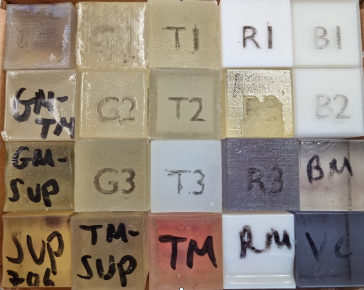
Over 45 different 2cm x 2cm x 2cm blocks of 3D printed materials were tested to evaluate their T1 and T2 properties. These materials were printed using a variety of different additive manufacturing (3D Printing) techniques.Due to the large number of samples to be tested, it was important to use a rapid method to broadly characterise these materials before a subset could be evaluated more closely. To do this, Siemens MyoMaps T1 and T2 mapping sequences were used.
To ensure both traceability and reliability of these rapid methods, they were also performed on the NIST Premium System phantom, alongside the protocols recommended by CaliberMRI.
Results
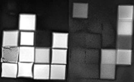
Testing of all 3D printed materials was done using a Siemens Magnetom Prisma 3T MRI scanner. A pointwise encoding time reduction with radial acquisition (PETRA) (256 x 256, TR = 5ms, TE = 0.07ms, TA = 9s) sequence was used to visualise the blocks. The sequence (Fig 3) of the first 45 blocks of materials showed that even with an ultra-short TE, only 19 materials were visible.Siemens MyoMaps T1 mapping sequence implements a Modified Look Locker Inversion Recovery (MOLLI) in this case with TI = 100,180,350,430,600,680,850,1100ms, TR = 250ms TE = 1.27ms. The MyoMaps T2 mapping sequence used was a Fast Low Angle Shot (FLASH) with TR = 330ms and 18 TE's between 1.27ms and 18ms. Both sets of maps were produced using a non-linear curve fitting within the scanner.
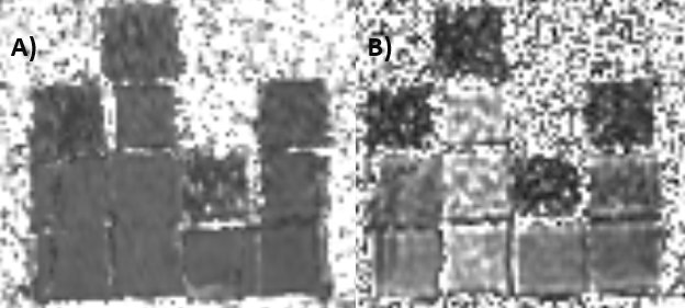
The 3D printed materials had a range of T1 values from 641+-21ms to 87 +- 30ms. The T1 maps produced using this method showed some materials as imperceptible above noise. The T2 values range from 21ms +- 8ms to 126ms +- 19ms. The maps from one row of these materials can be seen in Figure 3.
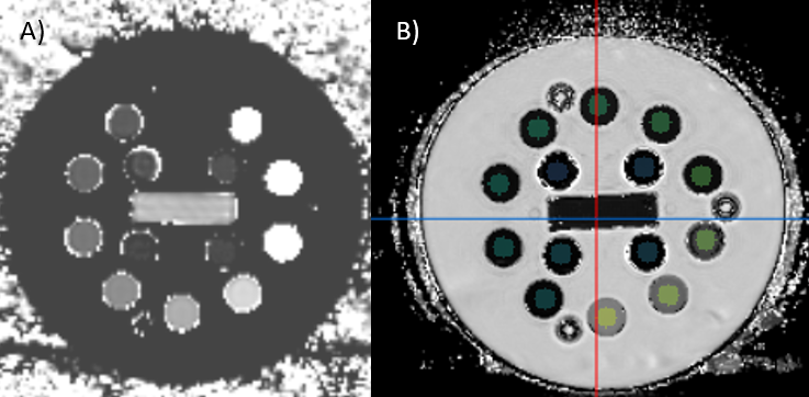
The NIST Premium System Phantom was also scanned using the same MyoMaps sequences, and agreement was found to their reference values of within 15% across the targeted ranges. Excellent agreement was found with the Caliber sequences and the reference values provided for this phantom, with the mean difference being 4.85%.
Discussion
The results thus far show that the range of relaxation properties of these materials is wide. The MyoMaps sequences provided a reliable method of rapidly characterising these materials. That said, in future, it will be useful to use more comprehensive methods to obtain detailed values for the most promising materials for a given application. Many materials show little to no signal, which would be useful for MR Safe accessories. While many of the approximated T1 and T2 values are not close to being representative of human tissue, some materials are coming close. Further investigation of these materials, using the sequences provided by CALIBER, will be commencing soon. Other further work around this project will involve investigating the dielectric and mechanical properties of the most promising materials.Acknowledgements
My thanks to the NHS GGC MRI Physics department for their ongoing support throughout this project, in particular my supervisors, Pauline HallBarrientos and David Brennan. Thanks also to those at the National Physics Laboratory who loaned the NIST Premium System Phantom for use in this project, alongside their support and guidance on appropriate use of the phantom. Finally, thank you to all the other NHS GGC departments and other organisations who provided me with sample materials free of charge, without whom this would not have been possible.References
[1] He, Y et al (2019). Characterizing mechanical and medical imaging properties of polyvinyl chloride-based tissue-mimicking materials. Journal of applied clinical medical physics, 20(7), 176-183 .
[2] Talalwa, Lotfi, et al. "T 1 -mapping and dielectric properties evaluation of a 3D printable rubber-elastomeric polymer as tissue mimi cking materials for MRI phantoms." Materials Research Express 7.1 1 (2020): 1 1 5306.13] Bullitt E, Zeng D, Gerig G, Aylward S, Joshi S, Smith JK, Lin W, Ewend MG (2005) Vessel tortuosity and brain tumor malignancy: A blinded study. Academic Radiology
Figures