4887
Comparing Bowsher’s Method to MRgBSREM in PET Recon with MR-priors in the Presence of Mismatch Between MRI and PET Images1Stanford University, Stanford, CA, United States, 2GE HealthCare, Waukesha, WI, United States
Synopsis
Keywords: PET/MR, PET/MR, MR Priors, Motion Correction
Motivation: Using MR-priors in PET reconstruction has been performed in numerous studies. While MR-priors provide more SNR and better resolution ti PET-images, without considering motion there may be misalignment between PET and MR images which leads to crosstalk artifacts. Another concern is mismatch between MR and PET images which can potentially affect the final PET image.
Goal(s): To compare the two most-widely used PET recon with MR-priors methods: the Bowsher's method and MRgBSREM.
Approach: We created an MR-series with severe line artifacts and used in as MR-priors in both methods.
Results: We have shown that MRgBSREM handles these mismatches better than the Bowsher’s method.
Impact: MRgBSREM is more robust to mismatches between PET and MR. This is achieved by adding a PET-seed image to identify similar-voxels which avoids the situation in which voxels with significantly different PET-uptake would be considered similar based only on MR-images.
Objectives
More studies are using anatomical priors to reconstruct PET images with higher signal-to-noise (SNR) ratio and improve spatial resolution. A recent study1 has shown that motion is prevalent in most PET/MR brain studies by analyzing more than 500 exams. Having good alignment between PET and MR images is vital for the accurate application of MR priors, and since motion can cause misalignment between the PET and MR images, accurate motion correction is very important. Among PET reconstruction with MR priors, two methods have been used widely: Bowsher’s method2 and MRgBSREM3, both of which can be combined with motion correction. One of the main differences between these 2 methods is the way they identify similar voxels in order to construct the penalty function. While in Bowsher’s method, only MR images are used to identify similar voxels, for the MRgBSREM method, an initial PET reconstruction is done (called the PET seed) and is used along with MR images to identify similar voxels. The addition of this PET seed to MRgBSREM method makes it more robust to mismatches between MR and PET images. In other words, in the areas where PET uptake is significantly different between otherwise similar voxels based on their MR images (i.e., in Bowsher’s method), MRgBSREM will not consider them as similar voxels and does not allow the mismatch between MR and PET to affect the reconstruction of those voxels. We have tested this property of MRgBSREM by manipulating the MR priors.Methods
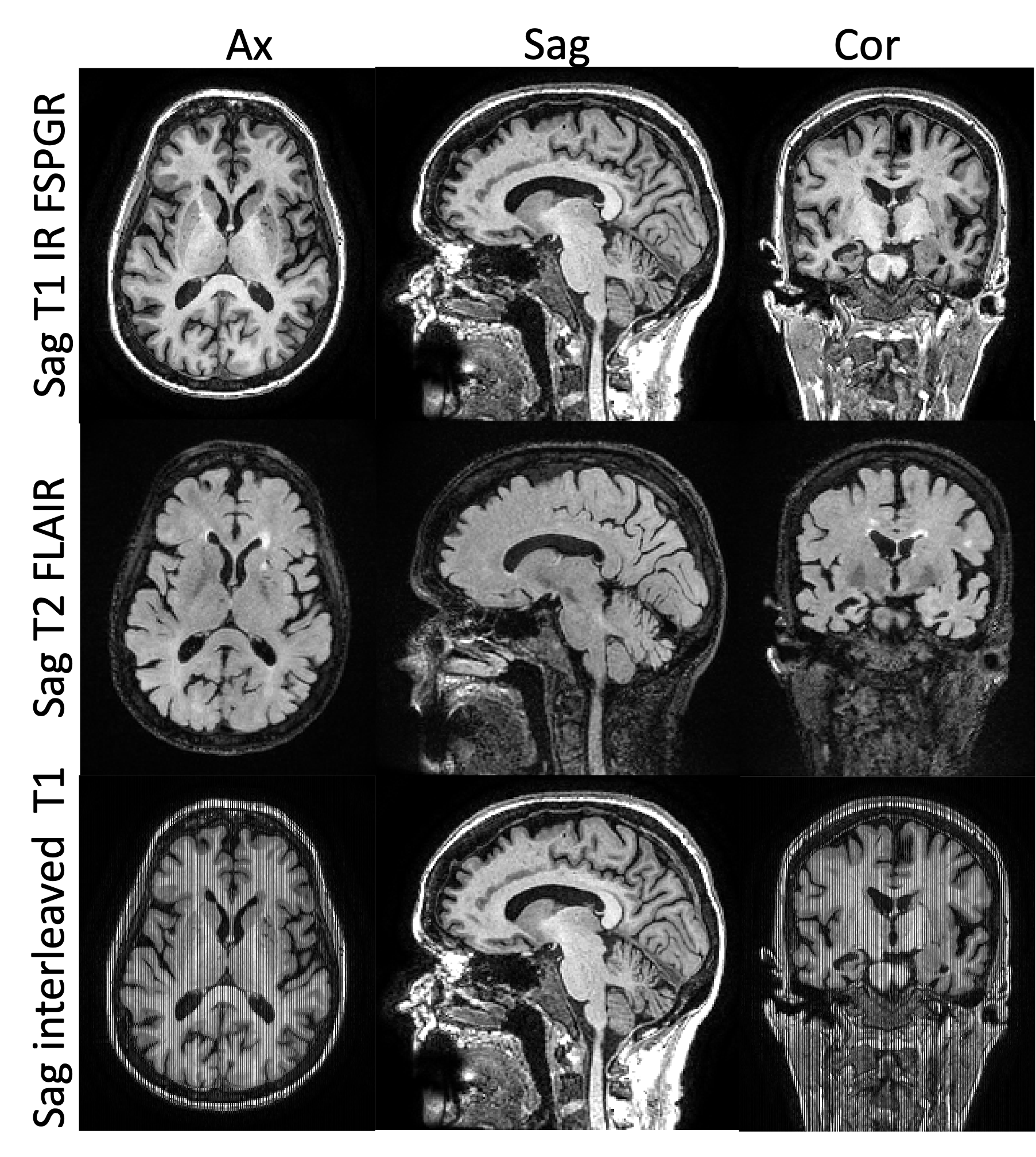
One subject was injected with 8 mCi of 18F-florbetaben (FBB, amyloid tracer) and after 70 min uptake time, underwent a 40-minute brain scan on a SIGNA PET/MR (GE HealthCare, Waukesha, WI). The study was approved by Stanford's Institutional Review Board and the subject provided written consent. 3D T1 IR FSPGR and 3D T2 FLAIR CUBE images were acquired simultaneously with PET. The PET list file was used to estimate motion over the entire exam4. A 20 min frame between 90-110 min after injection was reconstructed with TOF-BSREM (b = 75) with 1 mm isotropic resolution and motion correction. The anatomical images were used to reconstruct the PET images with MRgTOF-BSREM with b=15 and bm=100 over the same matrix and field of view (i.e., 1 mm isotropic) using motion correction. Then the 3D T1 and T2 weighted images were interleaved to generate a mixed set of anatomical images which showed severe line artifacts in the A/P direction. This new mixed MR images were used to reconstruct the same PET images with both Bowsher’s and MRgBSREM method and the results were compared.Results
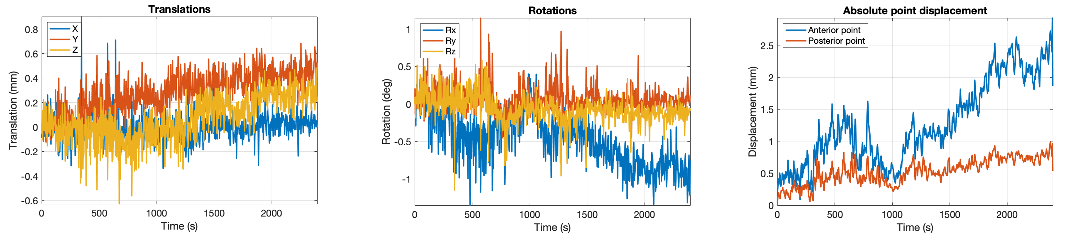
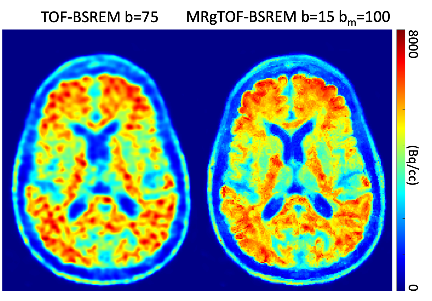
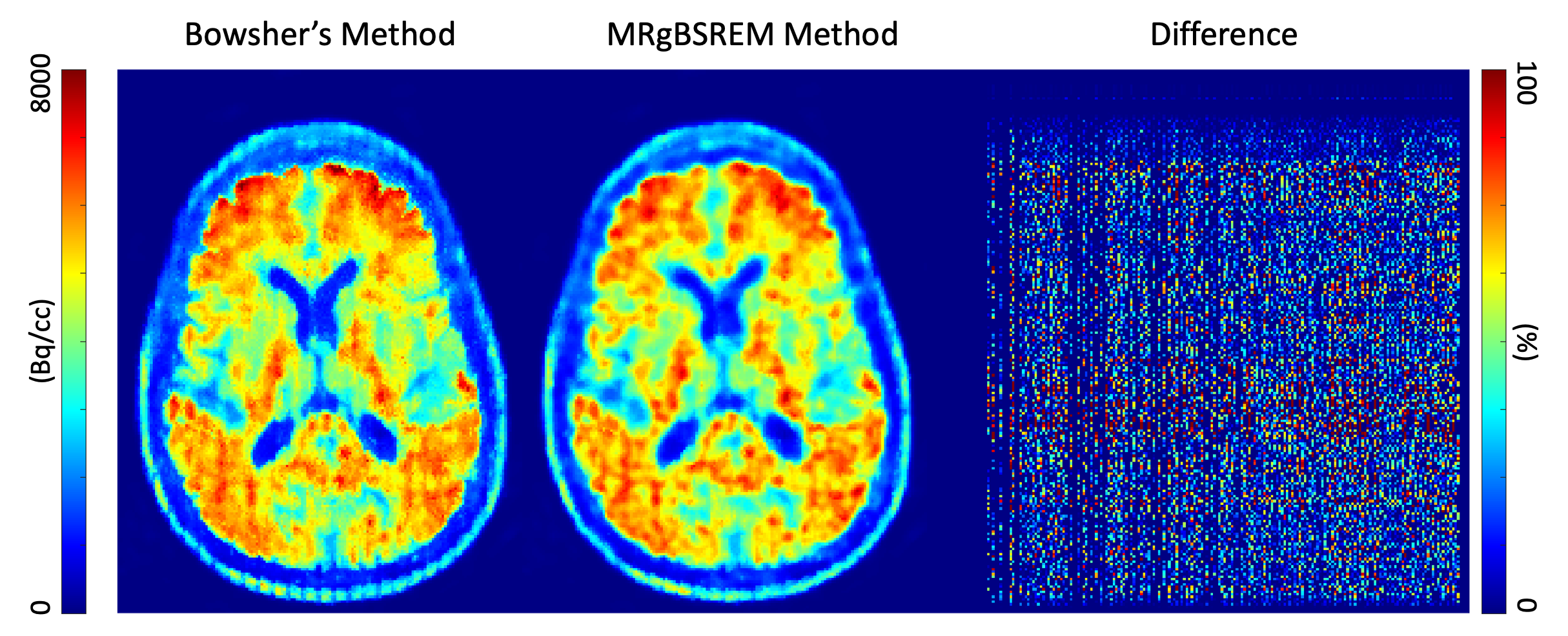
Figure 1 shows the anatomical images used as MR priors. The top rows are 3D T1 and T2 FLAIR images. The bottom row is a set of artificially made MR images by interleaving between T1 and T2 sagittal images and shows severe line artifacts in A/P direction. Figure 2 shows the measured motion of the subject during the 40 min exam. The tracker points (70 mm anterior and posterior to the center of the brain) show a motion of about 2.5 mm. Figure 3 shows the PET reconstructions with TOF-BSREM (b=75) and MRgTOF-BSREM (b=15 and bm=100). The PET reconstruction with MR priors shows a higher SNR and increased spatial resolution compared to TOF-BSREM method. Figure 4 shows a comparison between the Bowsher’s method and MRgBSREM method when the manipulated MR images with line artifacts (Figure 1, bottom row) are used as priors. The line artifacts are visible in the reconstruction with the Bowsher’s method and can easily be seen in the difference image between the two methods.Discussion
The MRgBSREM method is more robust to mismatches between PET and MR images. This is achieved by adding a PET seed image when similar voxels are to be identified. This will avoid the situation in which voxels with significantly different PET uptake would be considered similar based only on their similarity on MR images. The weight of the PET seed during the regularization can be adjusted with the bm parameter.Acknowledgements
No acknowledgement found.References
1. Khalighi MM, Spangler-Bickell M., Jansen F., et al. “Studying Rigid Head Motion on PET/MR Brain Exams,” ISMRM-SNMMI workshop on PET/MRI, October 2023, Los Angeles, CA.
2. Bowsher JE, Yuan H, Hedlung LW, et al., “Utilizing MRI information to estimate F18-FDG distributions in rat flank tumors.” Nucl Sci Symp Conf Rec 2004 IEEE. 2004;4(C):2488–2492.
3. Khalighi MM, Deller T., et al. “High Quality Isotropic Whole-body PET Imaging Using MR Priors,” J Nucl Med May 1, 2020 vol. 61 no. supplement 1 1477.
4. Spangler-Bickell M., Hurley S., Pirasteh A., Perlman S., Deller T., McMillan A., “Evaluation of Data-Driven Rigid Motion Correction in Clinical Brain PET Imaging,” Journal of Nuclear Medicine, October 2022, 63 (10) 1604-1610.
Figures