4882
Development and in-vivo testing of an MR-compatible biofeedback device for MR-guided radiotherapy simulation and treatment1MD Anderson Cancer Center, Houston, TX, United States
Synopsis
Keywords: New Devices, Motion Correction, Biofeedback
Motivation: Biofeedback during radiotherapy simulation and treatment on conventional linacs improves the accuracy and reproducibility of patient alignment, especially those using breath-hold. However, commercial devices cannot operate in MR environments.
Goal(s): The goal of this study was to develop an MR-compatible biofeedback device and test its utility in volunteer studies.
Approach: The device consists of a single-board computer and addressable LED strip, which was automatically controlled by tracking anatomic motion in CINE MR images.
Results: Initial volunteer studies using the device in an MR linac demonstrated that biofeedback-guided breath-hold resulted in more accurate and consistent target alignment compared to self-guided breath-hold.
Impact: MR-compatible biofeedback devices can facilitate and improve breath-hold techniques for MR-guided radiotherapy simulation and treatment. Furthermore, incorporation of CINE MR images to measure changes in internal anatomy improves target alignment relative to existing devices that use surface imaging.
Introduction
For tumors in the thoracic and abdominal cavities, respiratory motion can greatly impact radiotherapy efficacy and side effects due to the reduced dose to the target and increased dose to nearby normal tissue. To mitigate the effects of motion, breath-hold techniques are routinely implemented, where the patient holds their breath and expands their diaphragm to a reproducible level while the linac beam is on. Biofeedback devices, which are used to indicate the accuracy of the breath-hold to the patient, have proven successful at improving accuracy and reproducibility of breath-hold techniques1-3. However, commercial biofeedback devices cannot be used in MR-guided simulations and treatments due to the high magnetic field and enclosure of the patient in the MRI bore. The goal of this study was to develop a biofeedback device that could operate in the MR environment and guide a patient’s breath-hold based on anatomic motion measured in CINE MR images.Methods
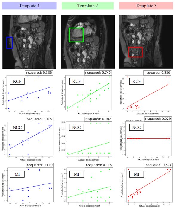
The development of this device comprised of both software and hardware components. For the software component, several open-source motion tracking algorithms were adapted and tested for both computational speed and tracking accuracy. These included Normalized Cross Correlation (NCC), Mutual Information (MI), and Kernalized Correlation Filter (KCF)4. For the hardware component, several configurations of a video grabber, single-board computer, strip of addressable light-emitting diodes (LED), and shielded fiber optic cables were tested for operability in the MR-Linac vault and operation latency.With an operational prototype, volunteer studies were conducted, following institutional review board policy, to test the device’s ability to guide breath-holds. Consenting volunteers were imaged with CINE MRI and asked to perform a series of breath-holds. The resultant anatomic displacement over time was measured for self-guided and biofeedback-guided breath-holds.
Results
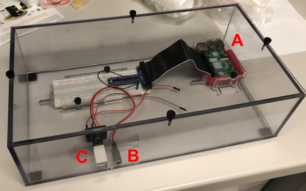
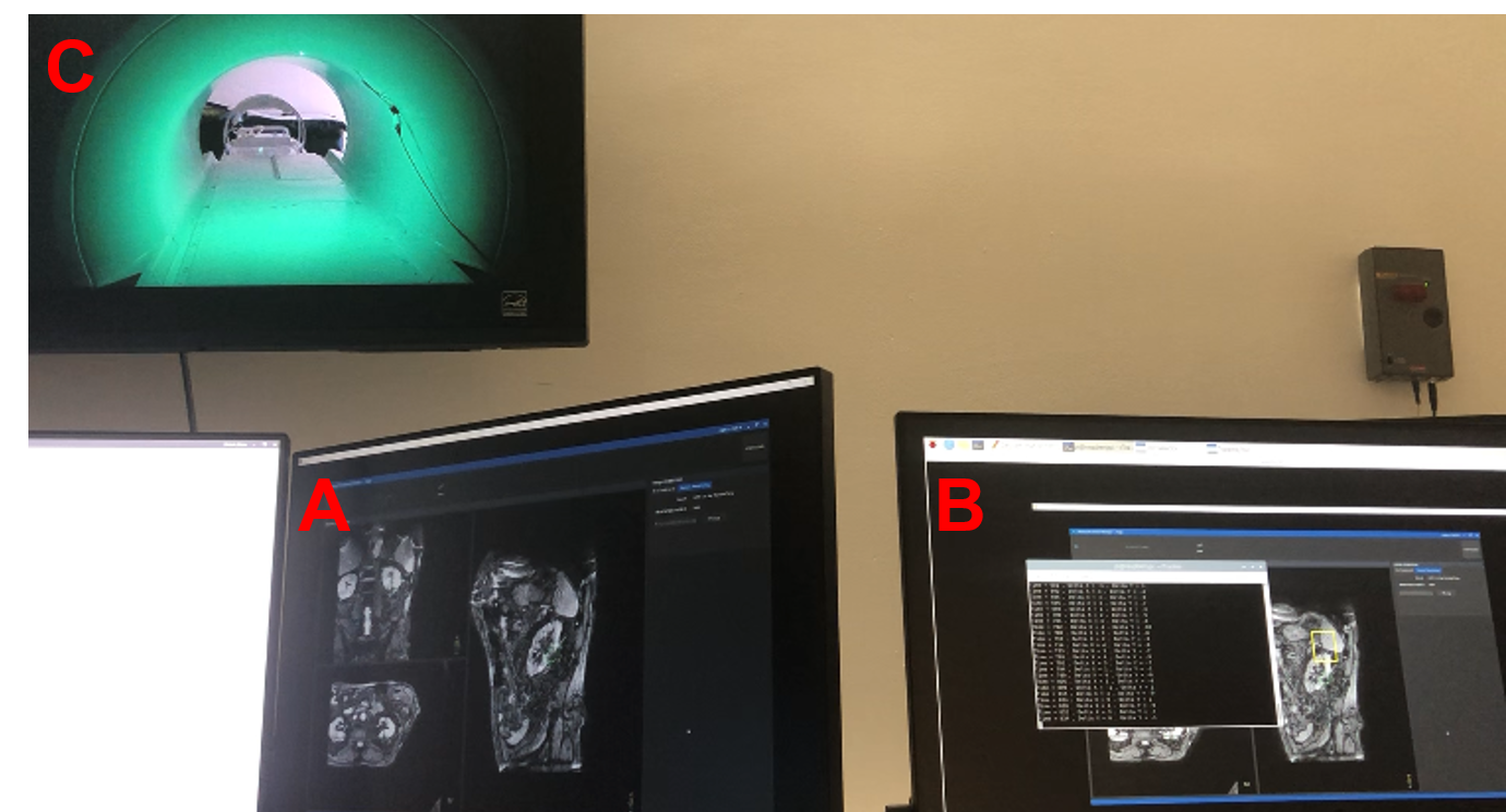
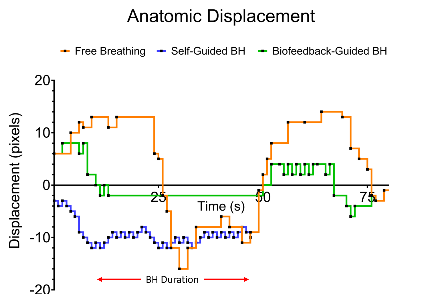
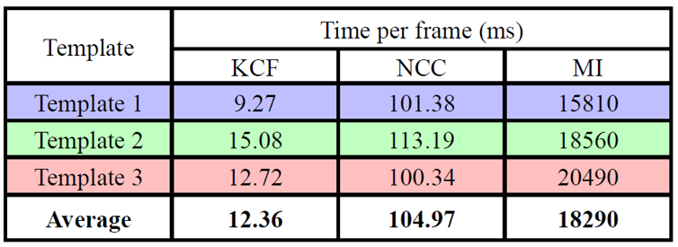
Among the three tracking algorithms, KCF had both the lowest computation time (12.36 ms) and highest average correlation coefficient (0.65) between calculated displacement and actual displacement. Thus, this algorithm was incorporated into python script, whose output was used in a modified open-source script to control the LED strip. The hardware was made operational by running the script on a single-board computer. CINE MR images were inputted via HDMI cable and processed, and the resultant voltage and data signal was outputted via general-purpose input/output pins on the computer. These signals were transmitted via shielded fiber optic cables into the MRI vault and integrated with the LED strip to control individual unit illumination and color. The LED strip was attached to the superior side of the inside of the MR linac bore. No impact on MR signal acquisition or image quality was observed during the operation of the LED strip. The measured end-to-end latency between CINE frame update and LED strip illumination was approximately 800 ms.Volunteers are still being recruited for the in vivo analysis, but a sample of one volunteer’s self-guided and biofeedback-guided breath-hold is illustrated in the associated figure. The biofeedback-guided breath-hold resulted in a smaller median anatomic displacement (2 vs. 10 pixels), which was more stable (0-2 pixels vs. 8-12 pixels) compared to the self-guided breath-hold.
DIscussion
The developed biofeedback device can reliably operate in a high magnetic field within the enclosed MRI bore. Because the device extracts frames from CINE MR, the motion of the actual target can be directly measured, rather than a surrogate such as the patient surface. This is achieved with no additional radiation dose unlike fluoroscopy-based real-time imaging. One drawback of the device is the 800 ms latency, which is higher than many biofeedback devices. However, for the purposes of assessing breath-hold accuracy, this is acceptable.Another feature of this device is that it is highly customizable. The tracking algorithm can be easily replaced since only the tracking output is used as the input for the LED illumination. The displacement tolerance resulting in a change in LED illumination can be any number of pixels and in any direction, and the color and number of illuminated LED units can be customized. The CINE MR images were extracted from the console via screen grabber, so this device can be installed at any MR console that displays CINE MR images in real-time. Thus, in addition to testing the device at an MR linac, it will also be deployed at an MR simulator to help guide breath-holds during simulation imaging for treatment planning.
Conclusion
A prototype MR-compatible biofeedback device was successfully constructed and tested in the MR linac environment. Initial volunteer testing demonstrates promising results that the device can effectively guide patient breath-holds to improve target alignment during MR-guided radiotherapy simulation and treatment.Acknowledgements
No acknowledgement found.References
1. Cerviño LI, Gupta S, Rose MA, et al. Using surface imaging and visual coaching to improve the reproducibility and stability of deep-inspiration breath hold for left-breast-cancer radiotherapy. Phys Med Biol. 2009;54(22):6853-6865. doi:10.1088/0031-9155/54/22/007
2. Stock M, Kontrisova K, Dieckmann K, et al. Development and application of a real-time monitoring and feedback system for deep inspiration breath hold based on external marker tracking. Med Phys. 2006;33(8):2868-2877. doi:10.1118/1.2219775
3. Lee D, Greer PB, Lapuz C, et al. Audiovisual biofeedback guided breath-hold improves lung tumor position reproducibility and volume consistency. Adv Radiat Oncol. 2017;2(3):354-362. doi:10.1016/j.adro.2017.03.002
4. Singh, SP, Mittal, A, Gupta, M, et al. Comparing Various Tracking Algorithms In OpenCV. Turkish J. Comput. Math. Educ. 2021;12(6): 5193–5198
Figures