4878
Direct Revascularization Improves Vascular Hemodynamics in Pediatric Moyamoya Patients1Department of Neurosurgery, Stanford University, Stanford, CA, United States, 2Department of Radiology, Stanford University, Stanford, CA, United States
Synopsis
Keywords: Arterial Spin Labelling, Perfusion, Pediatrics
Motivation: Moyamoya is a progressive cerebrovascular disorder affecting hemodynamics and the disease often begins during childhood. However, the non-invasive assessments of hemodynamics in pediatric Moyamoya patients remain an open question.
Goal(s): Investigate the CBF change before and after revascularization in pediatric Moyamoya patients.
Approach: CBF in 42 patients was measured using arterial spin labeling on a 3T MRI 1 week before and within 2 weeks after revascularization. CBF in regions affected by vasculopathy was compared before and after revascularization.
Results: CBF in regions affected by vasculopathy increased significantly after revascularization.
Impact: ASL can be utilized to evaluate CBF in pediatric Moyamoya patients before and after direct revascularization.
Introduction
Cerebral blood flow (CBF) is an important metric to determine the health of vascular hemodynamics. Studies suggest that decreased CBF may lead to a higher risk for neurological disorders and cerebrovascular diseases, such as acute strokes1. Moyamoya is a profound cerebrovascular disease that causes cerebral vasculopathies associated with decreased CBF2. Studies have found that revascularization procedures can significantly improve CBF in adult Moyamoya patients3. Since Moyamoya is a progressive disease that often begins during childhood4, evaluating CBF in pediatric Moyamoya patients may offer important evidence about disease prognosis. However, measuring CBF in the pediatric population has been challenging due to a lack of non-invasive techniques and the heterogeneity of the developing brain. Arterial spin labeling (ASL) is a non-invasive and contrast-free MRI technique that allows quantitative assessments of CBF. Several prior studies have demonstrated the feasibility of ASL in measuring pediatric CBF.Here, we investigate the hemodynamic change in pediatric Moyamoya patients before and after their direct revascularization procedures. Specifically, we present the results of CBF measured by ASL in regions affected by vasculopathy.
Methods
A cohort of 46 pediatric Moyamoya patients (28 females, 11 months to 18 years) was included in this work. Each patient underwent direct revascularization surgery at our institution and completed MRI exams 2 weeks before and 1 week after their surgeries. In short, a direct revascularization connects the frontal or temporal branch of the superficial temporal artery with the M4 segment of the middle cerebral artery under mild hypothermia (33–35°C)5. All patients were asked not to consume caffeine at least 6 hours before their imaging exam. MRI scans were conducted using 3-T MRI systems (Discovery MR 750 and SIGNA Premier, GE Healthcare, Waukesha, WI, USA). The MRI protocol includes single-delay PCASL (labeling duration = 1450 msec; PLD = 1525 msec, 3 repeats, scanning time = 4:13 min), DWI, MRA, FLAIR, and T1-weighted. CBF from single-delay PCASL was measured using the FSL BASIL software6. The mean CBF within regions affected by Moyamoya vasculopathy was computed for each patient. Paired t-tests were performed to compare the mean CBF before and after surgery in regions affected by vasculopathy. A p-value <0.05 was considered statistically significant.Results
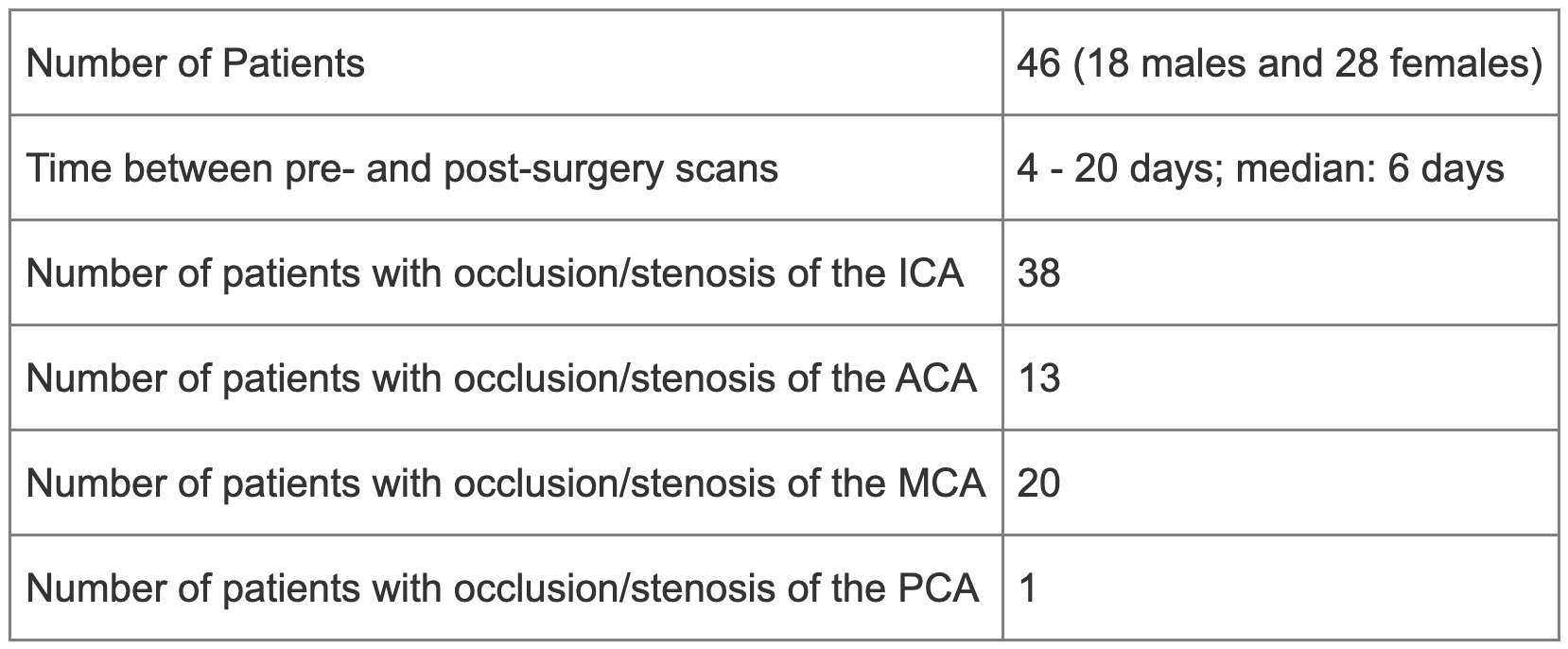
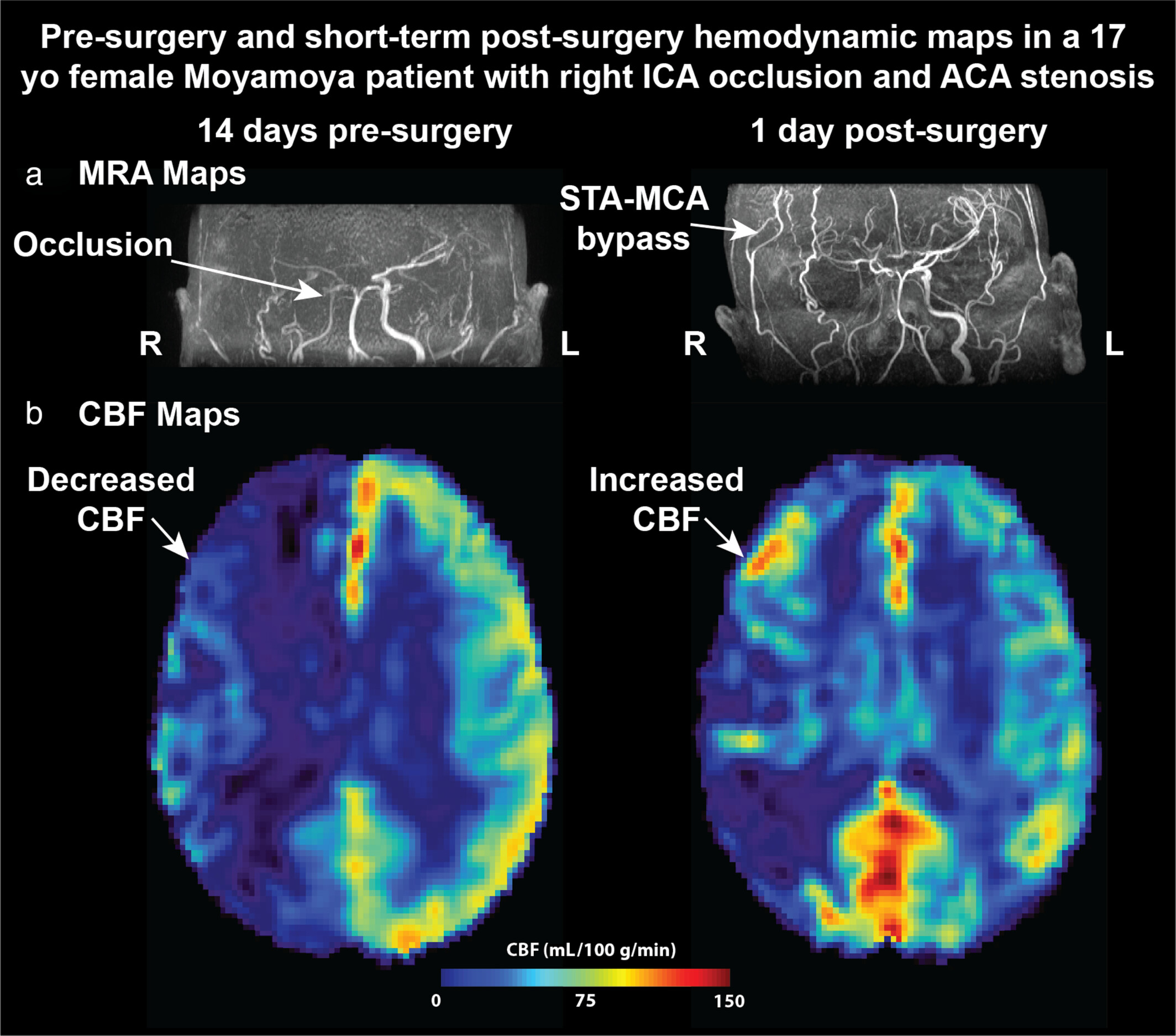
Table 1 shows the clinical characteristics of this cohort. All patients had their pre- and post-surgical scans within 3 weeks. The majority of vasculopathy occurred at the supraclinoid ICA, ACA, and MCA.Figure 1 shows the pre- and post-surgical MRA and CBF maps of a patient with Moyamoya affecting the right hemisphere. MRA images show occlusion at the supraclinoid segment of the right ICA and moderate stenosis of the A1 segment of the right ACA. Post-surgery MRA shows a successful bypass. Before revascularization, CBF of the right hemisphere was significantly lower than the left. After surgery, CBF of the right hemisphere increased due to direct revascularization.
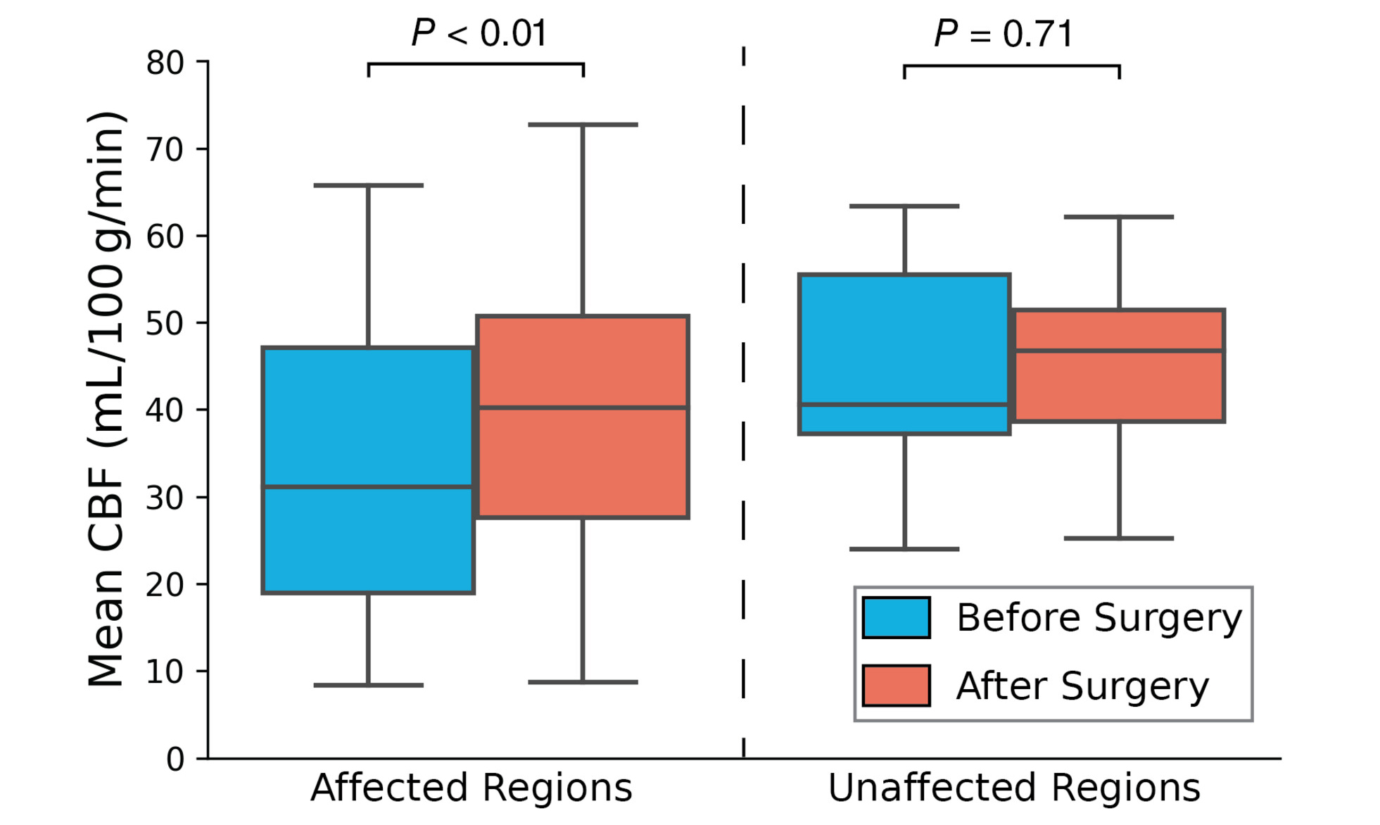
Figure 2 shows the pre- and post-surgical mean CBF of this cohort in affected and unaffected regions by vasculopathy. Overall, the CBF in regions affected by vasculopathy increased significantly after revascularization by 41 ± 31% (p < 0.01). In the unaffected regions, CBF remained unchanged after surgery (p = 0.71).
Discussion
In this work, we investigated hemodynamic changes in 46 pediatric Moyamoya patients within a week after bypass surgery using single-delay PCASL. We demonstrated that the mean CBF in regions affected by vasculopathy increased significantly after bypass surgery. Such improvement in CBF implied restored delivery of essential nutrients and oxygen for brain tissues through revascularization graft, potentially leading to reduced risk for strokes. Our results were consistent with previous observations where CBF in pediatric Moyamoya patients increased significantly 6 months after bypass7. The assessment of short-term CBF changes in our study here provides new information about pediatric hemodynamics after bypass surgery. Evaluating long-term hemodynamic changes in pediatric Moyamoya patients using ASL may provide more comprehensive insights into the prognosis and evaluation of pediatric cerebrovascular disorders, which is an area of our ongoing investigation.Acknowledgements
This work was supported by the American Heart Association (Grant: 23SCEFIA1141920)References
1 Mori S, Sadoshima S, Ibayashi S, Lino K, Fujishima M. Relation of cerebral blood flow to motor and cognitive functions in chronic stroke patients. Stroke 1994; 25: 309–317.
2 Fan Audrey P., Khalighi Mohammad M., Guo Jia, Ishii Yosuke, Rosenberg Jarrett, Wardak Mirwais et al. Identifying Hypoperfusion in Moyamoya Disease With Arterial Spin Labeling and an [15O]-Water Positron Emission Tomography/Magnetic Resonance Imaging Normative Database. Stroke 2019; 50: 373–380.
3 Zhao MY, Armindo RD, Gauden AJ, Yim B, Tong E, Moseley M et al. Revascularization improves vascular hemodynamics − a study assessing cerebrovascular reserve and transit time in Moyamoya patients using MRI. J Cereb Blood Flow Metab 2022; : 0271678X221140343.
4 Rao VL, Prolo LM, Santoro JD, Zhang M, Quon JL, Jin M et al. Acetazolamide-Challenged Arterial Spin Labeling Detects Augmented Cerebrovascular Reserve After Surgery for Moyamoya. Stroke; 0: STROKEAHA.121.036616
5 Steinberg GK, Gooderham PA. Intracranial-Extracranial Bypass surgery for Moyamoya Disease. In: Kalani Y, Nakaji P, Spetzler RF (eds). Neurovascular surgery. Thieme: New York, 2015.
6 Zhao MY, Fan AP, Chen DY-T, Sokolska MJ, Guo J, Ishii Y et al. Cerebrovascular reactivity measurements using simultaneous 15O-water PET and ASL MRI: Impacts of arterial transit time, labeling efficiency, and hematocrit. NeuroImage 2021; 233: 117955.
7 Quon JL, Kim LH, Lober RM, Maleki M, Steinberg GK, Yeom KW. Arterial spin-labeling cerebral perfusion changes after revascularization surgery in pediatric moyamoya disease and syndrome. J Neurosurg Pediatr 2019; 23: 486–492.
Figures