4877
Multi-Tag Time-SLIP Simulation Beyond the General Kinetic Model1Radiology, UC San Diego, La Joll, CA, United States, 2Radiology, UC San Diego, La Jolla, CA, United States
Synopsis
Keywords: Arterial Spin Labelling, Arterial spin labelling, Simulation
Motivation: We identified a gap in understanding multi-tag Time-SLIP MRI dynamics, crucial for advanced perfusion analysis, particularly in varying physiological and pathological states.
Goal(s): Our goal was to develop a comprehensive simulation tool to accurately predict signal changes in multi-tag Time-SLIP ASL MRI across diverse flow conditions.
Approach: We employed MATLAB to simulate numerically non-selective and selective multi-tag Time-SLIP pulse sequences, applying Bloch equations to model tissue-specific magnetization and blood flow perfusion dynamics.
Results: Simulations showed distinct magnetization patterns between odd and even pulse tags, aligning with physiological flow rates.
Impact: This simulation tool enables researchers to customize Time-SLIP MRI protocols for detailed study of fluid dynamics across the body, from blood to CSF flow. It provides a solid foundation for developing more comprehensive ASL-related simulations.
Introduction
Arterial Spin Labeling (ASL) MRI provides a non-invasive tool to probe cerebral blood flow dynamics and blood perfusion, playing a pivotal role in neurovascular research. Time-SLIP (Time-Spatial Labeling Inversion Pulse) [1], a variant of ASL, enables the tracking of labeled fluid through the vasculature and tissues, giving insights into perfusion patterns essential for understanding organ function. The General Kinetic model proposed by Buxton [2] has been instrumental in characterizing blood flow dynamics following a single labeling pulse. However, the more complex physiological scenarios often demand a more elaborate approach. In this work, we present a novel simulation tool designed to elucidate the magnetization evolution in multi-tag Time-SLIP experiments.Methods
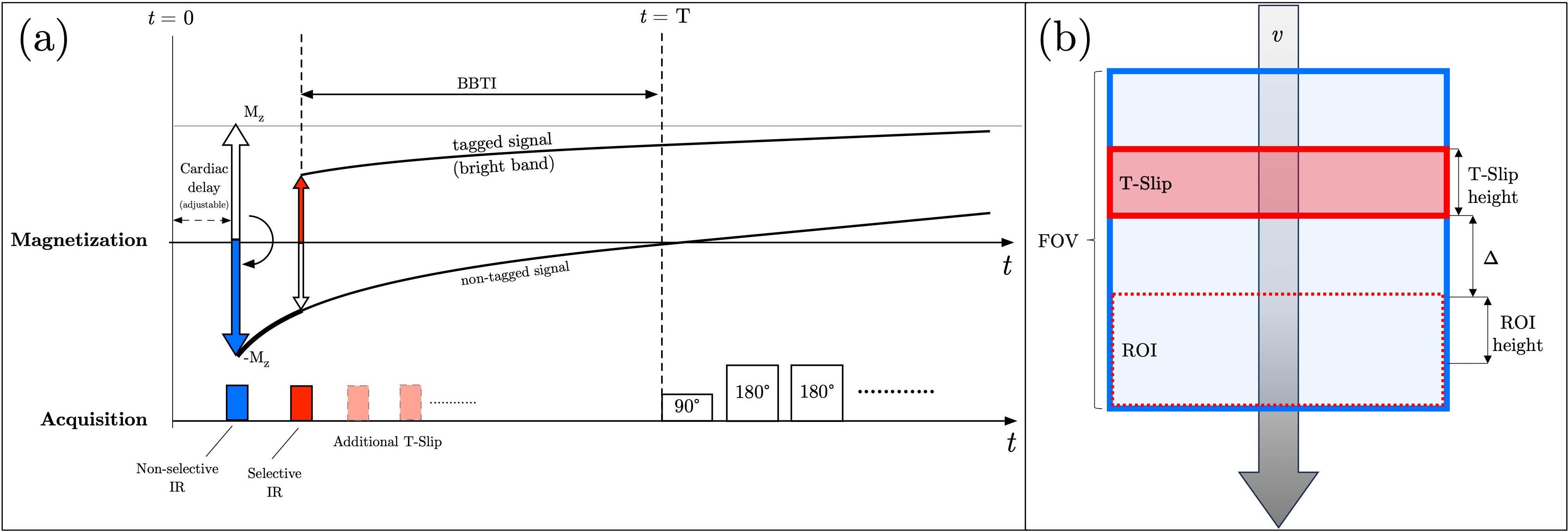
The simulation was implemented in Matlab [3]. Figure 1a illustrates the acquisition and magnetization diagram for a combination of non-selective and selective Time-SLIP pulses. In this representation, only the evolution of magnetization (Mz) for protons exposed to the non-selective (blue) and single selective pulse (saturated red) is demonstrated, a choice made to maintain diagram clarity. Figure 1b shows the simulation's setup: Time-SLIP pulse's geometry, the region of interest (ROI) where the perfusion signal is measured, and the distance between them. Our analysis considers the perfusion signal within this region as a composite of static tissue (experiencing only the non-selective pulse, NS) and fluid subject to both the non-selective inversion pulse and the selective (T-SLIP) pulse. For case with three components: blood (fluid) and bone and muscle (static tissue) signal increase ratio (SIR) can be calculated as follows:$$\mathrm{SIR} (y,t)=\delta (y,t)\cdot\frac{\left| M_{z}^{\mathrm{NS}}(t) - M_{z}^{\mathrm{T-SLIP}} (t) \right|} { \left| M_{z}^{\mathrm{muslce, \, NS}} (\mathrm{TI}_{max}) +M_{z}^{\mathrm{bone, \, NS}} (\mathrm{TI}_{max})+M_{z}^{\mathrm{blood, \, NS}} (\mathrm{TI}_{max})\right|} \quad\quad [Eq.1]$$
Magnetization Mz is derived from the Bloch equations, subject to the following condition: Mz(t+τ)=–Mz(t) when an inversion pulse is applied. Here τ stands for a small time-step denoting point in time right after the inversion RF-pulse (approximated with hard instant inversion) is applied. For the fluid (blood) subject to multi-tag RF pulses the general form for fractions of protons affected by N pulses can be expressed:
$$\delta(y,t)\cdot M_{z}^{\mathrm{blood, \, T-SLIP}} (t) \ = \sum_{i=1}^{N} \delta_{i}(y,t) \cdot M_{z}^{\mathrm{blood}, \,i} (t) \quad\quad [Eq.2]$$
The coordinate dependency y(t) can be described by constant flow:
$$y(t)=y_0+v\cdot t \quad\quad [Eq.3]$$
or any analytical flow function. Using Eq. 1–2, and Bloch equation for the evolution of magnetization Mz the system can be simulated numerically [3].
Results
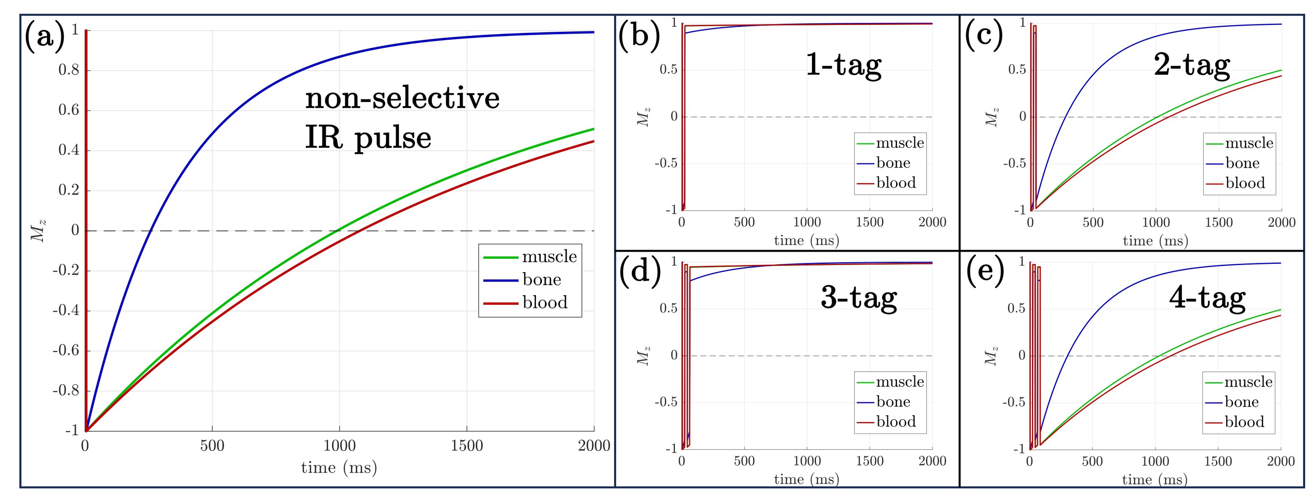
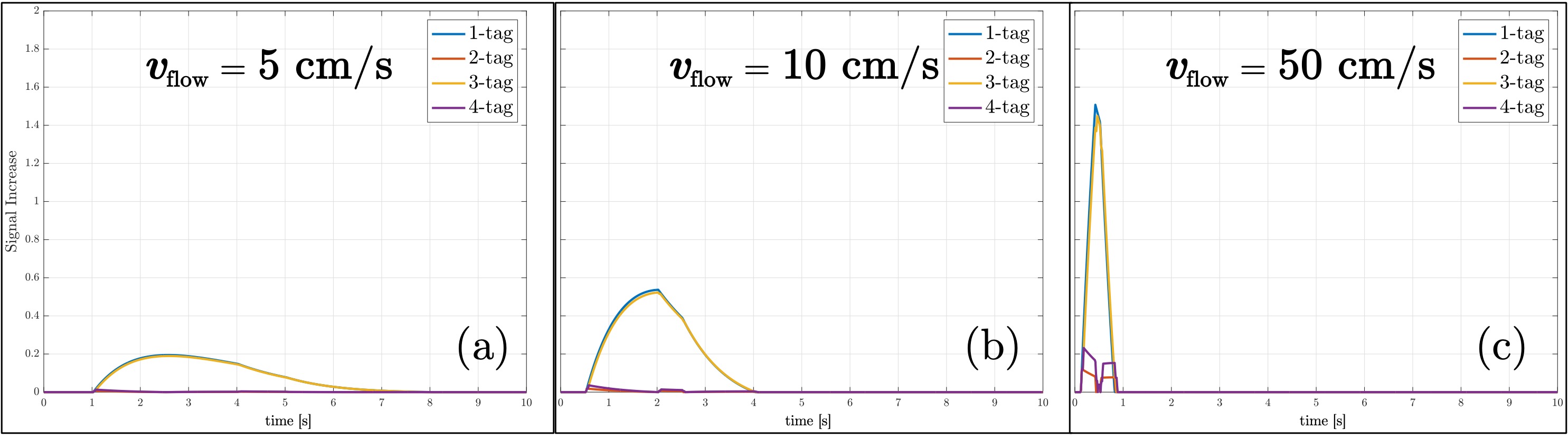
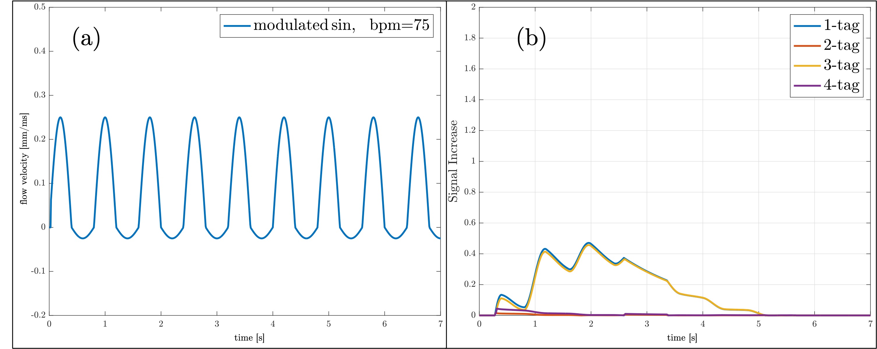
Multiple tag simulations of constant flow were conducted using T1 values of muscle (T1 of 1130ms), bone (mainly fat, T1 of 265ms), and blood (T1 of 1500ms). As shown in Figure 2, the tagged region experiences various T1 recoveries of non-selective IR pulse, 1-tag, 2-tag, 3-tag, and 4-tag pulses. The odd number of tag (1-tag and 3-tag) pulses gives nearly full magnetizations in +Mz, whereas the even number of tag (2-tag and 4-tag) pulses gives an exponential restoration of magnetizations (Mz) as they traverse through the null point. In Figure 3, we consider the impact of constant flow rates on the system. We examined flow rates set at 5cm/s (a) and 10cm/s (b), aligning with the known ranges seen in physiological fluids like cerebrospinal fluid (CSF) and blood. Additionally, we explore a high flow rate of 50cm/s (c) faster flow observed in arterial regions such as the carotid and renal arteries. These assessments are conducted while considering the application of tag pulse(s) positioned 50mm away from the region of interest, a condition often encountered in practical scenarios. Consecutive tag pulses are evenly spaced at intervals of 25ms. The even number of tags (2-tag and 4-tag) resulted in minimal signals, while the odd number of tags (1-tag and 3-tag) yielded higher signal intensities at the tagged region. Results for non-constant flow (sinus function with modulated amplitude) are shown in Figure 4, similar to constant flow higher signals for the odd number of tag pulses at the perfused region are observed.Discussion
The simulation tool introduced in this study offers insights into magnetization evolution during multi-Time-SLIP experiments under both constant and oscillatory flow conditions. A notable difference between odd and even tag pulses was observed. Additionally, the influence of flow rate on magnetization became evident, with results that reflect physiological conditions observed in different regions of the human vasculature. The exploration of non-constant flow, characterized by a modulated sinusoidal function, displayed patterns consistent with those from constant flow scenarios.Conclusions
The multi-tag Time-SLIP simulation offers a deeper understanding of blood flow perfusion and magnetization evolution for ASL MRI. There's indeed potential for future endeavors, such as incorporating more intricate flow models and evaluating the effects of other physiological parameters.Acknowledgements
This work was supported by a grant from Canon Medical Systems, Japan (35938).References
[1] Miyazaki M., et al., Radiology, 248:20-43. (2008).
[2] Buxton RB., et al., Magn Reson Med., 40(3):383-396. (1998)
[3] https://github.com/vmalis/TSLIPsim/tree/main
Figures