4872
The application value of modified time of flight magnetic resonance venography in iliac vein compression syndrome1Radiology, The First Affiliated Hospital of Chongqing Medical University, Chongqing, China, 2Clinical Science, Philips Healthcare, Chengdu, China
Synopsis
Keywords: Contrast Agents, Blood vessels, Time of flight magnetic resonance venography, Iliac vein compression syndrome
Motivation: The use of respiratory-gated TOF-MRV in assessing the degree of iliac vein compression is easily affected by respiratory uniformity, leading to vascular distortion and corresponding false images.
Goal(s): This study evaluated the diagnostic effectiveness of an improved TOF-MRV (mTOF-MRV) in Iliac vein compression syndrome (IVCS) diagnosis by optimizing key parameters, compared with conventional TOF-MRV.
Approach: Objectively evaluated the deviation of vascular stenosis degree between TOF MRV and mTOF MRV using DSA as the standard.
Results: The mTOF-MRV has better image quality and can accurately detect venous stenosis. It can be applied in the diagnosis of IVCS and further assessments after endovascular interventions.
Impact: mTOF-MRV more accurately located iliac vein stenosis without obvious stair-step artifacts and magnetic sensitivity artifacts, thereby meeting the diagnostic and clinical requirements It can be applied in the diagnosis of iliac vein compression syndrome and further assessments after endovascular interventions.
Introduction
Time of flight magnetic resonance venography(TOF-MRV) is a non-invasive diagnostic method for IVCS .The use of respiratory-gated TOF-MRV in assessing the degree of iliac vein compression is easily affected by respiratory uniformity, leading to vascular distortion and corresponding false images[3-5]. This study evaluated the diagnostic effectiveness of an improved time of flight magnetic resonance venography (mTOF-MRV) in IVCS diagnosis by optimizing key parameters, which improved image quality and diagnostic accuracy in comparison with conventional TOF-MRV, using three-dimensional digital subtraction angiography (DSA) as a gold standard.Methods
This retrospective study was approved by our Institutional Review Board and conducted on 69 patients who underwent routine TOF-MRV and 85 patients who received mTOF-MRV,And all patients underwent DSA subsequently.The Philips Ingenia 3.0T digital network architecture MRI scanner and three-dimensional IntelliSpace Portal was used. The MR scan parameters were listed in Table 1. Objectively evaluated the deviation of vascular stenosis degree between TOF MRV and mTOF MRV using DSA as the standard; Subjective evaluation used a 4-point method to assess image quality[6]. Interobserver agreements for objective and subjective evaluations were evaluated by calculating the intraclass correlation coefficient and Cohen's kappa Coefficient, respectively. The paired T-test was used to compare errors in vascular stenosis rates between MRV and DSA. Sensitivity, specificity, positive predictive value and negative predictive value of TOF MRV and m-TOF MRV in diagnosing significant iliac vein compression (stenosis≥50%) were analyzed with DSA as the gold standard. The Mann-Whitney U test was used to compare the subjective scoring of MRV cases. P<0.05 was the threshold for statistical significance.Results
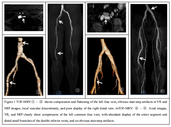
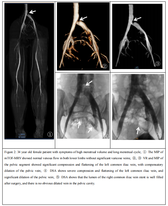
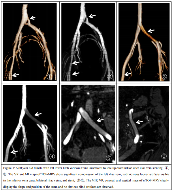
The mean error of stenosis rate for mTOF-MRV was lower than TOF-MRV(0.8% vs1.4%). Using DSA as the gold standard, sensitivity, specificity, positive predictive value and negative predictive value for mTOF-MRV were higher than TOF-MRV((Table 2)). The image score of mTOF-MRV was higher than TOF-MRV (P < 0.01). As shown in Figure 1, mTOF-MRV significantly improved the display of stair-step artifacts and distal veins . In reference to DSA, mTOF-MRV accurately located iliac vein stenosis and clearly displayed the shape and position of the postoperative iliac vein stent (Figure 2). Compared to conventional TOF-MRV, there were no significant stepped artifacts or magnetic sensitive artifacts with mTOF-MRV (Figure 3).Discussion
We systematically optimized the parameters associated with the image quality of lower limb vein TOF-MRV: fast imaging mode, K-space profile order, fat suppression, flip angle, TR, echo time and respiration compensation. Previous studies[4-5] adopted respiratory gating combined with Phase Encoded Artifact Reduction, However, patients with inadequate respiratory coordination are more susceptible to the occurrence of stair-step artifacts. The mTOF-MRV sets the fast acquisition mode as TFE, in which the acquisition time and duration can be controlled according to different imaging purposes to reduce body motion artifacts, thereby eliminating respiratory compensations. In this study, FA was set at 35°, echo time (TE) is set as out of phase, in which the phases of hydrogen protons in fat and water are in the opposite direction, and the signals generated by water and hydrogen protons in the fat in one pixel cancel each other, thereby reducing the signals to suppress the background signals.Conclusion
In conclusion, mTOF-MRV has better image quality and can accurately detect venous stenosis. It can be applied in the diagnosis of iliac vein compression syndrome and further assessments after endovascular interventions.Summary of Main Findings
The mTOF-MRV accurately located iliac vein stenosis, and clearly displayed the shapes and positions of postoperative iliac vein stents without obvious stair-step artifacts and magnetic sensitivity artifacts, thereby meeting the diagnostic and clinical requirements.Tables
Table 1 Comparisons of key parameters before and after improvement of time-leap magnetic resonance angiography| Key parameter | TOF-MRV | mTOF-MRV |
| Field of vision(mm2) | 400 x 240 | 400 x 300 |
| Collectivitin (mm3) | 1.8 x 2.0 x 3.0 | 1.8 x 2.0 x 2.0 |
| Reconstructed voxel (mm3) | 1.25 x 1.25 x 3.0 | 1.0 x 1.0 x 2.0 |
| Reconstruction matrix | 320 | 400 |
| Fast acquisition mode | No | TFE |
| Excitation mode | - | Multishot |
| K-space filling | Cartesian | Low-high |
| Fat suppression | no | SPIR |
| Flip Angle | 90 | 35 |
| Repetition time(ms) | 25 | Shortest |
| Echo time(ms) | 8 | Out phase |
| Water-fat displacement | maximum | User defined |
| Respiratory compensation | PEAR | no |
| Excitation frequency | 1.7 | 3 |
| Scanning time | 2:29 | 3:39 |
Table 2 Diagnostic measures for MRV assessment of IVCS (stenosis≥50%)
| Methods | Sensitivity(%) | Specificity(%) | Positive predictive value(%) | Negative predictive value(%) |
| TOF-MRV | 96.87 | 63.63 | 93.93 | 77.77 |
| mTOF-MRV | 100 | 100 | 100 | 100 |
Acknowledgements
None.References
1. May R, Thurner J. The cause of the predominantly sinistral occurrence of thrombosis of the pelvic veins. Angiology. Oct 1957;8(5):419-27.
2. Lugo-Fagundo C, Nance JW, Johnson PT, et al. May-Thurner syndrome:MDCT findings and clinical correlates. Abdom Radiol (NY).2016;41(10):2026–30.
3. Massenburg BB, Himel HN, Blue RC, Marin ML, Faries PL, Ting W. Magnetic Resonance Imaging in Proximal Venous Outflow Obstruction. Ann Vasc Surg. Nov 2015;29(8):1619-24. Suttmeyer B, Teichgräber U, Thomas A, et al. Non-invasive ECG-triggered 2D TOF MR angiography of the pelvic and leg arteries in an open 1.0-tesla high-field MRI system in comparison to conventional DSA. Biomed Tech (Berl). Feb 2014;59(1):29-37.
4. Harigai M, Okada T, Umeoka S, et al. Non-contrast-enhanced MR venography of the upper limb: a comparative study of acquisitions with fresh blood imaging vs. time-of-flight methods. Clin Imaging. Sep-Oct 2012;36(5):496-501.
5. Lombardi P, Carr JC, Allen BD, Edelman RR. Updates in Magnetic Resonance Venous Imaging. Seminars in interventional radiology. Jun 2021;38(2):202-208. doi:10.1055/s-0041-1729152.
6. Wu G, Yang J, Zhang T, et al. The diagnostic value of non-contrast enhanced quiescent interval single shot (QISS) magnetic resonance angiography at 3T for lower extremity peripheral arterial disease, in comparison to CT angiography. J Cardiovasc Magn Reson. Oct 20 2016;18(1):71. doi:10.1186/s12968-016-0294-6.
Figures