4867
Concurrent optoacoustic tomography and magnetic resonance imaging of resting-state functional connectivity in the mouse brain1ETH Zurich, Zürich, Switzerland, 2University of Zurich, Zurich, Switzerland
Synopsis
Keywords: Hybrid Imaging, Molecular Imaging, Optoacoustic (Photoacoustic) Imaging
Motivation: Interpreting BOLD signals in fMRI studies is a challenging task due to their dependence on multiple factors.
Goal(s): Multiparametric readouts from a recently developed hybrid magnetic resonance optoacoustic tomography system can offer unprecedented capabilities for studying the rodent brain in resting-state conditions.
Approach: Simultaneously acquired multimodal functional data of mice were utilized to link hemoglobin resolved optoacoustic readouts to BOLD.
Results: Multiparametric brain-wide activity were detected under resting-state condition for the first time. Revealed functional connectivity maps and brain networks showed high spatial overlap among the hemodynamic components. BOLD and total hemoglobin functional connectivity were found tightly correlated, compared to oxygenated- and deoxygenated-hemoglobin.
Impact: Hybridization of optoacoustic tomography and MRI is a powerful multi-modal approach due to highly complementary contrasts and capabilities for functional neuroimaging. The more comprehensive functional readings provide unique capabilities for studying neurovascular and neurometabolic coupling mechanisms and related diseases.
Introduction
Functional magnetic resonance imaging (fMRI) has significantly advanced our understanding of brain function, with resting-state fMRI emerging as a vital tool for exploring functional connectivity and brain disorders. However, the interpretation of blood-oxygen-level-dependent (BOLD) signals in fMRI studies is challenging, given their reliance on multiple factors such as blood flow, blood volume, metabolic rate of oxygen, and the baseline physiological state1. Optoacoustic tomography (OAT) provides a non-invasive means to characterize whole-brain cerebral hemodynamics in mice while attaining multiparametric readouts not available with other modalities2, 3. Hybridization of OAT and MRI is a particularly powerful multi-modal approach due to highly complementary contrasts and capabilities for functional neuroimaging4. The comprehensive readouts from a hybrid system can thus offer unprecedented capabilities for studying the rodent brain in resting state conditions.Methods
Bi-modal functional data of mice (N=14, Foxn1nu) were acquired concurrently with the recently developed hybrid magnetic resonance optoacoustic tomography (MROT) scanner3-5 in resting state conditions. Anesthesia was inducted with intraperitoneal injection of a mixture of ketamine (100 mg kg−1 body weight) and xylazine (10 mg kg−1 body weight). The MROT scanner integrates an OAT module, incorporating an MRI-compatible spherical matrix transducer array, an MRI-compatible fiber bundle and customized RF coils, into a 9.4T MRI scanner (Fig. 1a). Resting state fMRI images were obtained by gradient-echo echo-planar imaging acquisitions. The laser wavelength was switched between five distinct wavelengths during the OAT acquisition. OAT images were reconstructed using model-based methods and the oxygenated (HbO), deoxygenated (HbR), and total (HbT) hemoglobin distributions were subsequently estimated using linear spectral unmixing6. Functional OAT and MRI data were processed and analyzed using custom Matlab scripts, SPM12 and CONN (Fig. 1b).Results and Discussion
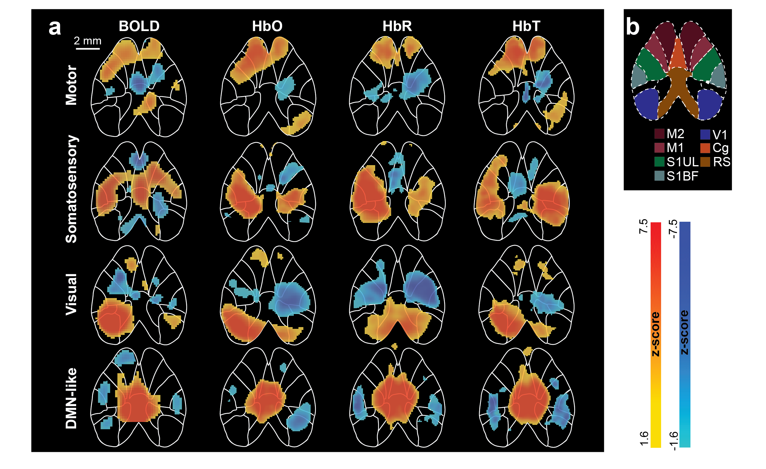
Simultaneously acquired fMRI and OAT data underwent motion correction, smoothing, and spatial normalization. Intermodal coregistration was realized as described elsewhere5. The global signal and motion parameters were regressed out and data were bandpass filtered between 0.01-0.1 Hz for fMRI and 0.04-0.1 Hz for OAT to suppress the unwanted laser fluctuation at ~0.02 Hz. Eight distinct seed regions with a volume of 0.3 x 0.3 x 0.3 mm3 were constructed based on the parcellations according to the Allen mouse brain atlas7. For each seed, Pearson’s correlation coefficient (r) was calculated between the mean time series of the seed volume and all voxels within the brain. Resulting OAT and BOLD functional connectivity maps were thresholded at r= 0.5 and r=0.1, respectively. Strong correlations were observed intra- and inter-hemispherically in motor, somatosensory, visual, parietal and retrosplenial regions of each hemodynamic component (i.e., BOLD, HbO, HbR and HbT) (Fig. 2). The functional connectivity maps exhibited high spatial overlap between the modalities. Interestingly, the negative correlations observable in fMRI functional maps were only reproduced in the HbT channel among the different optoacoustic hemodynamic readings. Additionally, HbT resulted in more localized functional connectivity patterns compared to HbO and HbR, in accordance with previous results4. Group-level (N=14) independent component analysis (ICA) with 20 components was performed separately to BOLD and each OAT hemodynamic components to identify functional brain networks. ICs resembling motor, visual, somatosensory and default-mode networks were commonly identified from HbO, HbR, HbT and BOLD (Fig. 3). Due to the penetration depth limitations of OAT, subcortical regions revealed in the BOLD ICA maps were not observed from OAT hemodynamic components.Conclusion
To date, unraveling the intricate interplay between neurovascular coupling and dynamic physiological states has proven challenging, particularly when relying on single-modality functional neuroimaging data. The enhanced information enabled with simultaneous multi-contrast MROT acquisitions is poised to shed light on the basic mechanisms underlying hemodynamic changes e.g., associated with neuronal activity. Correspondingly, multiparametric brain-wide responses were detected under resting state condition for the first time, facilitating cross-validation and comprehensive readings of complex brain activity patterns. These more comprehensive functional readings provide unique capabilities for studying neurovascular and neurometabolic coupling mechanisms and related diseases.Acknowledgements
The authors acknowledge support from the Swiss National Science Foundation grant 310030_192757.References
1. Blockley NP, Griffeth VE, Simon AB, Buxton RB. A review of calibrated blood oxygenation level-dependent (BOLD) methods for the measurement of task-induced changes in brain oxygen metabolism. NMR Biomed. 2013;26(8):987-1003.
2. Chen ZY, Zhou QY, Dean-Ben XL, Gezginer I, Ni RQ, Reiss M, et al. Multimodal Noninvasive Functional Neurophotonic Imaging of Murine Brain-Wide Sensory Responses. Adv Sci. 2022;9(24).
3. Chen ZY, Gezginer I, Augath MA, Ren WW, Liu YH, Ni RQ, et al. Hybrid magnetic resonance and optoacoustic tomography (MROT) for preclinical neuroimaging. Light-Sci Appl. 2022;11(1).
4. Chen ZY, Gezginer I, Augath MA, Liu YH, Ni RQ, Dean-Ben XL, et al. Simultaneous Functional Magnetic Resonance and Optoacoustic Imaging of Brain-Wide Sensory Responses in Mice. Adv Sci. 2022.
5. Gezginer I, Chen Z, Yoshihara HAI, Dean-Ben XL, Razansky D. Volumetric registration framework for multimodal functional magnetic resonance and optoacoustic tomography of the rodent brain. Photoacoustics. 2023;31:100522.
6. Dean-Ben XL, Buehler A, Ntziachristos V, Razansky D. Accurate Model-Based Reconstruction Algorithm for Three-Dimensional Optoacoustic Tomography. Ieee T Med Imaging. 2012;31(10):1922-8.
7. Wang QX, Ding SL, Li Y, Royall J, Feng D, Lesnar P, et al. The Allen Mouse Brain Common Coordinate Framework: A 3D Reference Atlas. Cell. 2020;181(4):936-+.
Figures
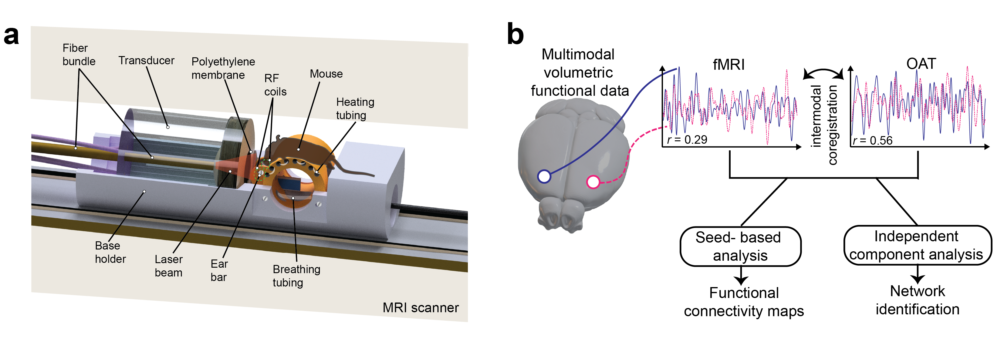
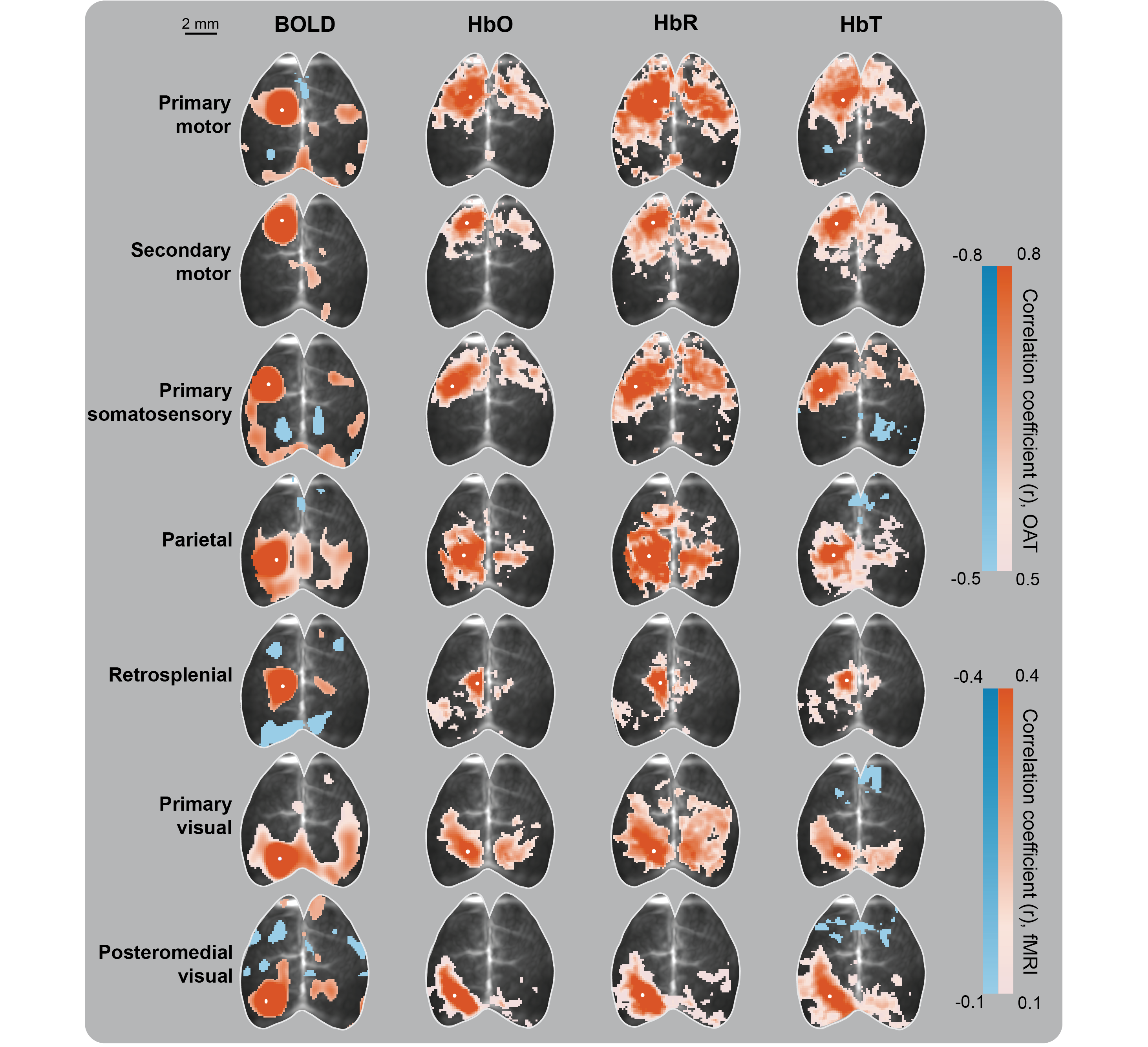
Figure 2. Functional connectivity maps of a representative mouse. Thresholded connectivity maps of eight seed regions were overlaid on the anatomical OAT image of the mouse. Seed centers were shown by white dot.