4863
Hybrid phase-sensitive 4D MRA and perfusion imaging using dynamic arterial spin labeling with stack-of-stars golden-angle radial acquisition1Department of Radiology, Northwestern University, Chicago, IL, United States
Synopsis
Keywords: Arterial Spin Labelling, Pulse Sequence Design
Motivation: Comprehensively characterizing cerebrovascular events including dynamic blood flow patterns and downstream perfusion is important in clinical diagnosis of cerebrovascular disorders.
Goal(s): To develop a time-efficient phase-sensitive ASL technique (PS-ASL) that provides high-quality time-resolved 4D-MRA and perfusion imaging within single scan.
Approach: PS-ASL sequence was designed by combining pCASL and PASL preparations with stack-of-stars golden-angle radial acquisition and a self-constraint low-rank subspace reconstruction. The label and control pulse modules alternated between pCASL and PASL preparations in each TR. Phase-sensitive subtraction between control and label yields 4D-MRA from PASL and perfusion from pCASL.
Results: Both dynamic 4D-MRA and perfusion maps were successfully obtained by PS-ASL.
Impact: The developed PS-ASL technique could be a potential powerful imaging tool in clinical applications, which provides detailed characterization of blood flow from both arterial and capillary beds in a single sequence.
Introduction
Comprehensively characterizing cerebral vasculature and hemodynamics including dynamic blood flow through the cerebral vasculature and downstream tissue perfusion is essential for the diagnosis of cerebrovascular diseases. Multiple sequences are typically required to image different aspects of cerebral hemodynamics, such as MR angiography (MRA) or perfusion, which is time-consuming. Arterial spin labeling (ASL) has shown the capability of capturing both MR angiographic and perfusion contrasts1,2,3,4. In this study, we developed a highly time-efficient phase-sensitive ASL technique dubbed PS-ASL by combining pCASL and PASL preparations with stack-of-stars golden-angle radial acquisition, which provides time-resolved high-resolution 4D MRA and perfusion imaging in a single acquisition.Method
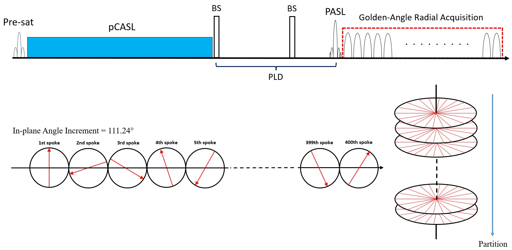
Pulse sequence designThe schematic of the PS-ASL pulse sequence is illustrated in Figure 1. A pCASL labeling pulse train is applied inferior to the imaging slab to label inflowing blood spins at a predefined labeling plane in the neck. Similar to that of conventional 3D pCASL perfusion sequence, a background suppression module consisting of a pre-saturation pulse and two non-selective inversion pulses is employed to enhance the pCASL perfusion signal through suppressing background tissue signal during the acquisition. Right before the imaging readout, a PASL labeling pulse is applied using the STAR labeling scheme5. There is a post-labeling delay (PLD) between pCASL and PASL to eliminate any interference of labeled spins by pCASL and PASL. In each TR, the label and control pulses of pCASL and PASL are alternated, so that the sign of labeled blood signal from each labeling scheme is opposite. In this way, ASL signals from pCASL and PASL can be disentangled using phase-sensitive subtraction from a pair of label/control scans. The SOS golden-angle radial bSSFP readout is used for data acquisition.
MRI acquisition
All MRI datasets were acquired on a Siemens Prisma 3T MR scanner using a 20-channel head coil. The imaging parameters of the PS-ASL sequence included: FOV = 220x220 mm2; Spatial resolution = 1.1x1.1x1.5mm3; TR/TE = 4.34ms/2.17ms; Flip angle = 25o; labeling duration of pCASL = 1.5s, PLD between pCASL and PASL preparations = 700ms, in-plane radial views = 400; 28 slices with total scan time of 4 minutes and 26 seconds.
Image Reconstruction
A previously developed self-calibrated low-rank sub-space reconstruction was used for time-resolved 4D MRA reconstruction6. Specifically, the temporal basis was derived from the k-space center of the control-label k-space subtraction. 4D MRA control and label images were generated using 10 spokes per image frame, corresponding to a temporal resolution of 43ms. Maximum intensity projection (MIP) was performed on the control-label subtraction images to display dynamic MRA. The perfusion image is reconstructed from the central region (96x96 over 208x208) of the k-space, resulting in an effective spatial resolution of 2.29x2.29x6mm3. The GRASP reconstruction with 120 radial spokes per slice was employed to generate perfusion-weighted images7.
Results and Discussion
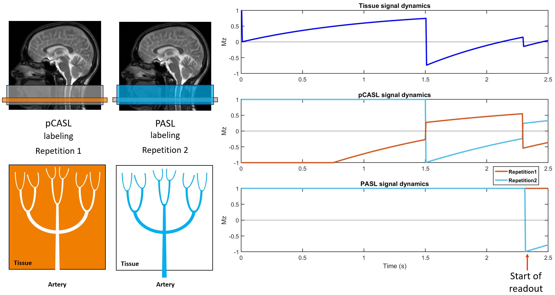
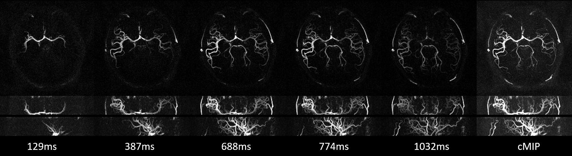
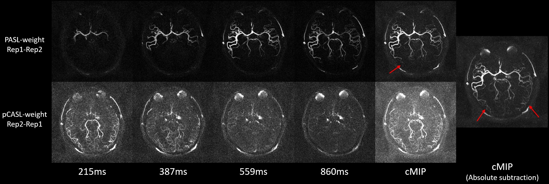
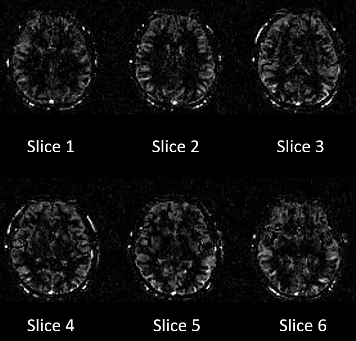
Figure 2 shows the Bloch simulation of the longitudinal magnetization evolution of labeled blood and background tissue in PS-ASL during the 2 repetitions, respectively. The dynamics in the center of the pCASL labeling is simulated in the illustration. Based on the simulation, the sign of ASL signal from pCASL and PASL contrast is reversed from two repetitions (label/control) by alternatively applying the label and control pulse preparations between pCASL and PASL preparations. Figure 3 shows several representative time frames of 4D MRA MIP and collapsed MIP (cMIP) images across all phases. Dynamic blood flow was clearly depicted in all three views. When reversing the label and control image order, no dynamic MRA signal was detected except for some pCASL signal in the distal arteries and posterior cerebral artery at the first phases due to their slow flows as shown in Figure 4. However, there was a clear temporal and spatial separation of ASL MRA from PASL and pCASL (Figure 4), further suggesting there is no interference in ASL signals between PASL and pCASL. A static MRA was generated by absolute subtraction between the label/control measurements, which exhibits both fast flows from PASL and slow distal flows from pCASL, leading to more vascular details compared to cMIP. Perfusion-weighted images were successfully obtained when reversing the subtraction order between label and control acquisitions, as shown in Figure 5. One can appreciate perfusion maps with good image quality.Conclusion
In this study, we have introduced a highly time-efficient phase-sensitive ASL technique for hybrid 4D MRA and perfusion imaging by combining pCASL and PASL preparations with stack-of-stars golden-angle radial acquisition and advanced sub-space low-rank reconstruction. PS-ASL could be a potentially useful ASL technique in clinical applications to comprehensively characterize cerebrovascular events in a single scan.Acknowledgements
This work was partly supported by National Institute of Health (NIH) grants R01NS118019, RF1AG072490, and BrightFocus Foundation A20201411S.References
1. Ferré JC, Bannier E, Raoult H, Mineur G, Carsin-Nicol B, Gauvrit JY. Arterial spin labeling (ASL) perfusion: techniques and clinical use. Diagnostic and interventional imaging. 2013 Dec 1;94(12):1211-23.
2. Haller S, Zaharchuk G, Thomas DL, Lovblad KO, Barkhof F, Golay X. Arterial spin labeling perfusion of the brain: emerging clinical applications. Radiology. 2016 Nov;281(2):337-56.
3. Hernandez‐Garcia L, Aramendía‐Vidaurreta V, Bolar DS, Dai W, Fernández‐Seara MA, Guo J, Madhuranthakam AJ, Mutsaerts H, Petr J, Qin Q, Schollenberger J. Recent technical developments in ASL: a review of the state of the art. Magnetic resonance in medicine. 2022 Nov;88(5):2021-42.
4. Togao O, Hiwatashi A, Obara M, Yamashita K, Momosaka D, Nishimura A, Arimura K, Hata N, Yoshimoto K, Iihara K, Van Cauteren M. 4D ASL-based MR angiography for visualization of distal arteries and leptomeningeal collateral vessels in moyamoya disease: a comparison of techniques. European radiology. 2018 Nov;28:4871-81.
5. Yan L, Wang S, Zhuo Y, Wolf RL, Stiefel MF, An J, Ye Y, Zhang Q, Melhem ER, Wang DJ. Unenhanced dynamic MR angiography: high spatial and temporal resolution by using true FISP–based spin tagging with alternating radiofrequency. Radiology. 2010 Jul;256(1):270-9.
6. Zhao T, Feng L, Yan L. An advanced reconstruction framework in stack-of-stars golden-angle radial non-contrast enhanced 4D MRA with ultra-high temporal resolution. In ISMRM Annual Meeting 2023. ISMRM.
7. Feng L, Grimm R, Block KT, Chandarana H, Kim S, Xu J, Axel L, Sodickson DK, Otazo R. Golden‐angle radial sparse parallel MRI: combination of compressed sensing, parallel imaging, and golden‐angle radial sampling for fast and flexible dynamic volumetric MRI. Magnetic resonance in medicine. 2014 Sep;72(3):707-17.
Figures