4859
Automated Pancreatic Segmentation and Quantitative Calculation Based on nnUnet1East China Normal University, ShangHai, China, 2Department of Radiology, Shanghai East Hospital, Tongji University, Shang Hai, China
Synopsis
Keywords: Diagnosis/Prediction, Segmentation
Motivation: Pancreatic diseases often exhibit spatial non-uniformity. Achieving automated segmentation of different pancreatic regions and conducting quantitative calculations of volume and fat content can effectively assist physicians in diagnosis and treatment.
Goal(s): Developing a segmentation network to achieve the automatic segmentation of the pancreas and to perform quantitative calculations of volume and fat content in diffrent regions.
Approach: Sample acquisition was performed using Dixon sequences, and training was conducted using an improved nnUnet network. Additionally, an automated pancreatic segmentation and quantitative calculation method was developed.
Results: With a training dataset consisting of 800 cases, the network achieved a segmentation Dice coefficient of 0.92.
Impact: To save professional physician annotation time for early detection and diagnosis of pancreatic diseases, as well as for quantifying changes before and after pancreatic treatments, and to assist in clinical drug therapy.
Introduction
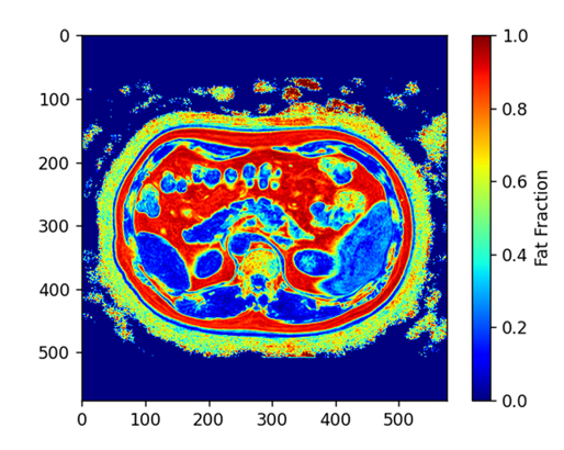
Obesity, biliary tract disorders, and alcohol consumption, among other factors, are contributing to an increasing annual incidence of pancreatic-related diseases[1]. Moreover, phenomena such as fat infiltration, fibroinflammation, and pancreatic cancer often exhibit spatial non-uniformity[2]. Therefore, achieving automated segmentation of different pancreatic regions can effectively assist physicians in diagnosis and treatment.In abdominal organ studies, to quantify related diseases, a common practice among radiologists is to directly use multiple Regions of Interest (ROI) within the pancreas on Fat Fraction (FF) maps and calculate the average fat fraction within these ROIs. However, pancreatic fat infiltration and fibrosis are not uniformly distributed, so delineating ROIs individually may not accurately reflect the condition of pancreatic lesions. Comprehensive delineation is often time-consuming and labor-intensive, leading to computer-assisted annotation becoming the mainstream solution[3].
Methods
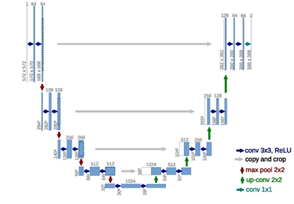
This study employed a cascaded U-Net deep learning network model[4], consisting of a two-layer architecture, for the framework. The first layer network performs coarse segmentation to extract a large-scale pancreatic target area, which is then input into the subsequent U-Net network for precise segmentation. The cascaded segmentation network allows for a more focused learning on the target area, thereby enhancing the segmentation accuracy.In this research, a cascaded nn-Unet network[5] with automated image preprocessing and parameter tuning was used. In the image preprocessing phase, data augmentation techniques such as cropping, resampling, rotation, and intensity normalization were automatically applied based on sample information. During training, learning rate decay was employed to enhance the network's learning capability, and in the post-processing phase, segmentation images were generated by retaining the largest connected region based on prior knowledge.Resluts
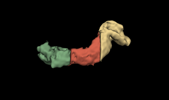
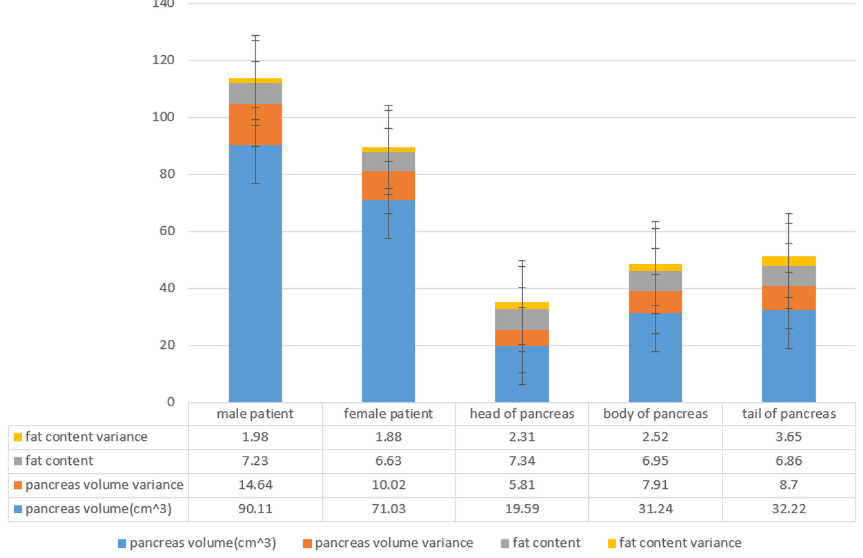
In the quantitative calculation part of this study, Dixon sequences were selected for data acquisition to analyze pancreatic fat content. An enhanced nnUnet network was employed for training a pancreatic segmentation model,with a training dataset consisting of 800 cases, the network achieved a segmentation Dice coefficient of 0.92. An axis-based coordinate localization method was used to achieve automatic segmentation of the pancreatic head, body, and tail.Discussion
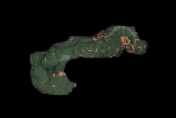
This study demonstrates that the NNUnet network can achieve high-precision segmentation of the pancreatic region on multi-modal m-Dixon sequences. Through extensive sample testing, this work validates the universality and clinical utility of this network model.In recent years, there have been some studies focusing on automatic segmentation and quantitative calculation of the pancreas. For instance, Triay et al. manually segmented the pancreatic head, body, and tail regions using registration methods. However, manual annotation of a large number of samples can be challenging. In this study, we propose an automatic segmentation method based on the body axis that does not require sample annotation and can automatically segment the pancreatic region. We also evaluated the reliability of this method. Furthermore, we calculated pancreatic volume and fat content based on the results of automatic segmentation for a large patient dataset, validating the results against previous studies. Additionally, for certain cases, we observed changes in pancreatic parameters before and after treatment, providing valuable insights for clinical diagnosis.In the field of pancreatic segmentation, it has been observed that previous works often reach a Dice coefficient plateau around 0.92, making it challenging to achieve higher breakthroughs. This issue may arise from the inherent variability in the shape and blurred boundaries of the pancreas, making manual delineation difficult. Moreover, variations in Dice coefficients can be observed when the same physician annotates the same case at different times or when different physicians annotate the same case. Through this method, it is possible to provide a rough assessment of the theoretical limitations of a specific segmentation task.
Conclusion
The nnUnet network can achieve high-precision segmentation of the pancreas. Utilizing the segmentation results, automatic segmentation of the pancreatic head, body, and tail, as well as the calculation of pancreatic volume and fat content, can provide valuable clinical diagnostic and treatment information.Acknowledgements
Thank you to Shanghai East Hospital, Tongji University for providing the relevant MRI data, and gratitude to the radiologists including Kaoming Cao, Renfeng Li, Haiyang Hu, Yiming Jia, Mingmin Shi, Songtao Liang, Wen Xu, and Ziying Yang for their valuable annotations.References
[1]Iannuzzi J P, King J A, Leong J H, et al. Global incidence of acute pancreatitis is increasing over time: a systematic review and meta-analysis[J]. Gastroenterology, 2022, 162(1): 122-134.
[2]Triay Bagur A, Aljabar P, Ridgway G R, et al. Pancreas MRI segmentation into head, body, and tail enables regional quantitative analysis of heterogeneous disease[J]. Journal of Magnetic Resonance Imaging, 2022, 56(4): 997-1008.
[3]Yao X, Song Y, Liu Z. Advances on pancreas segmentation: a review[J]. Multimedia Tools and Applications, 2020, 79: 6799-6821.
[4]Ronneberger O, Fischer P, Brox T. U-net: Convolutional networks for biomedical image segmentation[C]//Medical Image Computing and Computer-Assisted Intervention–MICCAI 2015: 18th International Conference, Munich, Germany, October 5-9, 2015, Proceedings, Part III 18. Springer International Publishing, 2015: 234-241.
[5]Isensee F, Jaeger P F, Kohl S A A, et al. nnU-Net: a self-configuring method for deep learning-based biomedical image segmentation[J]. Nature methods, 2021, 18(2): 203-211.
Figures