4857
High-variability synthetic fat-water MRI dataset for testing the robustness of Deep Learning-based reconstruction models1Centre for Biomedical Engineering, Indian Institute of Technology - Delhi, New Delhi, India, 2Department of Radio Diagnosis, All India Institute of Medical Sciences, New Delhi, India, 3Department of Biomedical Engineering, All India Institute of Medical Sciences, New Delhi, India
Synopsis
Keywords: AI/ML Image Reconstruction, Quantitative Imaging, Fat-water seperation, PDFF, Deep Learning, Fat Quantification, Physics Informed Deep Learning, Synthetic MRI
Motivation: Deep Learning (DL) models have recently been used for fat-water separation in Multi-Echo MRI (ME-MRI). However, DL models may not always be robust and under-perform when not trained with a large and diverse dataset.
Goal(s): This research proposes high-variability synthetic ME-MRI generated using the biophysical model of fat-water separation as a tool for testing the generalizability and robustness of DL-based fat-water separation models.
Approach: High-variability synthetic ME-MRI was used to evaluate the robustness of the recent state-of-the-art DL-based Ad-Hoc Reconstruction (AHR) method for fat-water separation.
Results: The AHR method lacked robustness and synthetic ME-MRIs can be effectively used to test DL models.
Impact: The fat-water maps obtained by processing the Multi Echo-MRI (ME-MRI) are of diagnostic and prognostic value in many diseases. This study investigates the role of synthetic ME-MRIs with high variability in testing the robustness of Deep Learning-based fat-water separation models.
Introduction
Fat-water separation in Multi Echo-Magnetic Resonance Imaging (ME-MRI) signals to produce individual fat-water maps of any region in the body has a diagnostic and prognostic value in many diseases like NAFLD, sarcopenia, and Cancer1,2. Deep Learning (DL) models can perform close to conventional optimization algorithms in fat-water separation3-5, however, DL models may not always be robust and underperform when not trained with a large and sufficiently diverse dataset6,7. This research proposes a high-variability synthetic ME-MRI phantom dataset generated using the biophysical model of fat-water as a potential tool for testing the generalizability and robustness of any fat-water quantification models. High-variability synthetic ME-MRI dataset was created and used to evaluate the robustness of the recent state-of-the-art DL-based Ad-Hoc Reconstruction (AHR) method for fat-water separation8. Unlike conventional DL models, AHR method utilizes the biophysical model to directly reconstruct a batch of ME-MRI-producing fat-water maps and does not require prior training with a large dataset8.Methods
High-Variability Synthetic ME-MRI Dataset Generation:High-variability synthetic ME-MRI dataset was generated using the biophysical model for fat-water separation in equation (1):
$$I_n(x, y)=\left(W(x, y)+F(x, y) \cdot e^{\left(j 2 \pi f t_n\right)}\right) \cdot e^{\left(j 2 \pi \psi t_n\right)} \cdot e^{\left(-R_2^* t_n\right)}\tag{1}$$
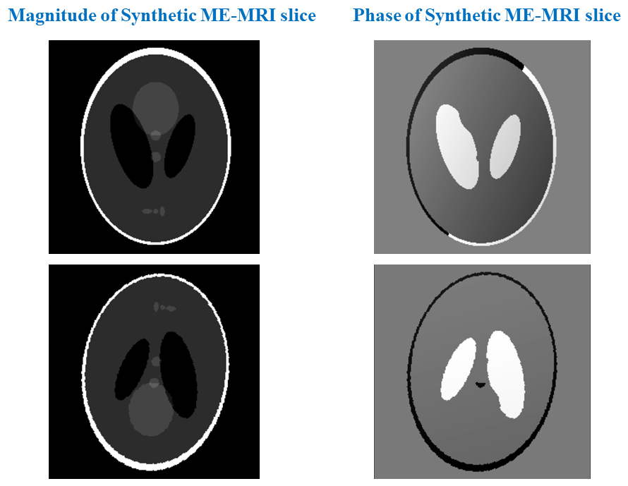
$$$I_n(x, y)$$$ is the signal from $$$n$$$th echo. $$$W$$$ is the water map, $$$F$$$ is the fat map, $$$𝑓$$$ is chemical‐shift frequency between fat and water protons, $$$t_n$$$ is Time for Echo (TE) of $$$n$$$th echo, $$$\psi$$$ is the field inhomogeneity and $$$R_2^*$$$ is inverse of $$$T_2^*$$$. Synthetic ME-MRI (the left side of the equation (1)) was obtained by feeding the values of the variables on the right side of the equation (1), and high variability is emulated by sampling these variables from a very random process. 30 slices of six echo synthetic ME-MRIs were generated. Different intensity values of the digital Shepp-Logan head phantom were chosen as fat and water pixels to generate 30 different $$$F$$$ and $$$W$$$ maps, corresponding $$$R_2^*$$$ maps were generated by linearly scaling the Shepp-Logan phantom slice. For 30 slices of field maps $$$\psi$$$, three random numbers $$$R1$$$, $$$R2$$$ and $$$R3$$$ were sampled from a standard normal distribution and the following equation was used to initialize $$$\psi$$$ at any spatial location $$$(x, y)$$$: $$$\psi(x, y)=R 1+\left(\frac{x}{m}\right) R 2+\left(\frac{y}{n}\right) R 3$$$, where $$$m$$$ and $$$n$$$ are dimensions of the $$$\psi$$$. Values of $$$t_n$$$ were set to 1.7, 4, 6.3, 8.6, 10.9, and 13.2 milliseconds, $$$𝑓$$$ to -223.2 Hz (assuming a 1.5 Tesla B0 magnetic field). Figure 1 presents two representative slices of synthetic ME-MRI generated.
DL-based AHR method for fat-water separation:
The DL network in AHR method takes the ME-MRI as input and produces fat, water, R2* and field inhomogeneity maps as outputs, which are given to the fat-water biophysical model in equation (1) to compute network’s loss8. This loss is minimized on a batch of ME-MRI by back-propagating for 10,000 epochs to produce the individual fat and water maps.
Testing the robustness:
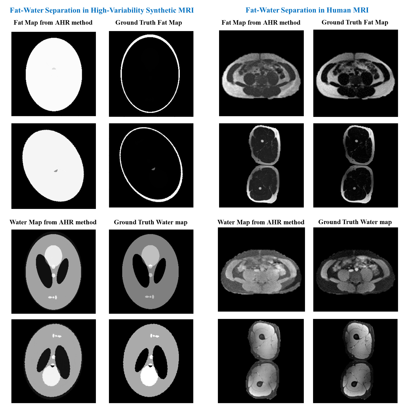
The synthetic high-variability ME-MRI was fat-water separated using the AHR method. To benchmark the performance of AHR in a human subject, six and four echo MRIs of the Thigh and Abdomen, hosted in the 2012 ISMRM fat-water separation workshop9 were also used with the AHR. All the fat-water maps from AHR were compared against the ground truths maps generated by the widely used conventional Graph-Cut method10. Synthetic ME-MRIs were also fat-water separated using golden-section-search method11. Fat-water maps of synthetic ME-MRI from golden-section-search and Graph-cut methods were highly similar with Structural Similarity Index Measure (SSIM) of 0.909±0.002 and 0.892±0.0004. This ensures the robustness of Graph-Cut method in ground truth generation and the accuracy of the synthetic ME-MRI generation process.
Results
The fat-water separation performance of AHR was observed to be poor in synthetic ME-MRI: an average SSIM of only 0.390±0.0006 in water maps and 0.889±0.009 in fat maps. On human subject MRIs, AHR achieved a better average SSIM of 0.845±0.023 and 0.865±0.05 in fat and water maps. Figure 2 compares fat-water maps obtained from synthetic and human subject MRIs using AHR against the ground truth maps.Discussion and Conclusion
The conventional Graph-Cut10 and golden-section-search11 methods performed equally good in both synthetic and in vivo data. Poor performance of AHR method in synthetic ME-MRI shows that the model lacks robustness and has probably not learned sufficiently the underlying mathematical relationship of the parameters. This indicates that the high-variability synthetic data generated using bio-physical models can be used to test the robustness of DL models for any task. These synthetic data could be included in training set of DL models to improve their robustness, which needs further investigation.Acknowledgements
The human subject multi-echo MRI data utilized in this study was hosted in the 2012 ISMRM Fat-Water separation workshop9.References
1. Thomas, E.L., Fitzpatrick, J.A., Malik, S.J., Taylor-Robinson, S.D. and Bell, J.D., 2013. Whole body fat: content and distribution. Progress in nuclear magnetic resonance spectroscopy, 73, pp.56-80.
2. Yip, C., Dinkel, C., Mahajan, A., Siddique, M., Cook, G.J. and Goh, V., 2015. Imaging body composition in cancer patients: visceral obesity, sarcopenia and sarcopenic obesity may impact on clinical outcome. Insights into imaging, 6, pp.489-497.
3. J. W. Goldfarb, J. Craft, and J. J. Cao, “Water-fat separation and parameter mapping in cardiac MRI via deep learning with a convolutional neural network: MR Water-Fat Separation,” J. Magn. eason. Imaging, vol. 50, no. 2, pp. 655–665, Aug. 2019, doi: 10.1002/jmri.26658.
4. J. Cho and H. Park, “Robust water–fat separation for multi‐echo gradient‐recalled echo sequence using convolutional neural network,” Magn. eason. Med., vol. 82, no. 1, pp. 476–484, Jul. 2019, doi: 10.1002/mrm.27697.
5. J. Andersson, H. Ahlström, and J. Kullberg, “Separation of water and fat signal in whole‐body gradient echo scans using convolutional neural networks,” Magn. eason. Med., vol. 82, no. 3, pp. 1177–1186, Sep. 2019, doi: 10.1002/mrm.27786.
6. X. Liang, D. Nguyen, and S. B. Jiang, “Generalizability issues with deep learning models in medicine and their potential solutions: illustrated with cone-beam computed tomography (CBCT) to computed tomography (CT) image conversion,” Mach. Learn. Sci. Technol., vol. 2, no. 1, p. 015007, Mar. 2021, doi: 10.1088/2632-2153/abb214.
7. J. Krois et al., “Generalizability of deep learning models for dental image analysis,” Sci. Rep., vol. 11, no. 1, p. 6102, Mar. 2021, doi: 10.1038/s41598-021-85454-5.
8. R. Jafari et al., “Deep neural network for water/fat separation: Supervised training, unsupervised training, and no training,” Magn. eason. Med., vol. 85, no. 4, pp. 2263–2277, Apr. 2021, doi: 10.1002/mrm.28546.
9. Hu, H.H., Börnert, P., Hernando, D., Kellman, P., Ma, J., Reeder, S. and Sirlin, C., 2012. ISMRM workshop on fat–water separation: insights, applications and progress in MRI. Magnetic resonance in medicine, 68(2), pp.378-388
10. Hernando D, Kellman P, Haldar JP, Liang ZP. Robust water/fat separation in the presence of large field inhomogeneities using a graph cut algorithm. Magn Reson Med. 2010 Jan;63(1):79-90. doi: 10.1002/mrm.22177. PMID: 19859956; PMCID: PMC3414226.
11. Lu W, Hargreaves BA. Multiresolution field
map estimation using golden section search for water-fat separation. Magn Reson
Med. 2008 Jul;60(1):236-44. doi: 10.1002/mrm.21544. PMID: 18581397.
Figures