4855
Classification of Grade II and III Astrocytomas for Multi-modal MRI using Deep Volumetric Attention Networks.1Computing and Electronics, Atlantic Technological University Sligo, Sligo, Ireland, 2Mechanical & Electronic Engineering, Atlantic Technological University Sligo, Sligo, Ireland, 3Computer Science and Applied Physic, Atlantic Technological University Sligo, Galway, Ireland
Synopsis
Keywords: Diagnosis/Prediction, Brain, Volumetric Attention Network, Deep Learning, Astrocytomas, Glioma, Classification
Motivation: Diagnosis and grading of astrocytomas tumour present considerable challenges. Manual grading is time-consuming and error prone. Preoperative MRIs are a useful, yet deep learning presents challenges due to computing limitations and complex architecture.
Goal(s): Study introduces novel multimodal MRI classification for grade II and III astrocytomas, aiming to improve accuracy, reduce complexity, and address interclass homogeneity via attention mechanism.
Approach: Single slice from eight MRI modalities forms a three-dimensional cube. Normalized, iPCA processed, and passed to deep model with volumetric attention network.
Results: The DVA using advanced and traditional MRI information outperforms existing models achieving an overall accuracy of 77% using five-fold cross-validation.
Impact: The proposed multimodal MRI classification approach enhances astrocytoma diagnosis and grading. The deep volumetric attention model improves accuracy, reduces model complexity, and holds potential for trustworthiness impacts in clinical practice.
Introduction
Astrocytomas are the most prevalent malignant primary brain tumors in adults, posing a crucial challenge for clinicians in terms of diagnosis, grading, and localization1. Traditional MRI-based grading of astrocytomas demands a high level of expertise from clinicians and characterization is tedious, time-consuming in nature, as well as it is susceptible to observer errors 2. Recent advancements in computer-aided diagnostic systems (CAD), particularly deep learning models, have eased the burden of manual classification, especially for high-grade (IV) and lower-grade (II and III) astrocytomas using traditional MRI data. However, the classification of grade II and III astrocytomas remains a challenging task when using preoperative MRIs3, primarily due to interclass homogeneity and overlapping information. Deep learning models in this domain often encounter significant computational, complex architecture and memory limitations, demanding extensive training data to achieve higher accuracy levels4. Therefore, this research proposes a multi-modality MRI classification approach using traditional and advanced MRIs for grade II and III astrocytomas, leveraging a 3D deep model following a volumetric attention network (DVA).Method:
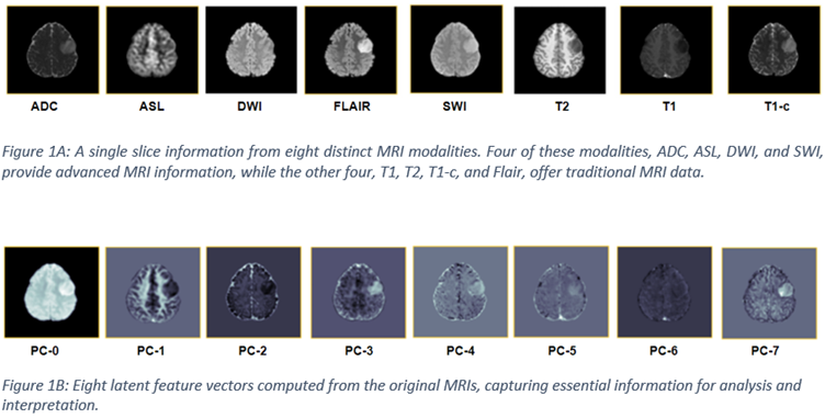
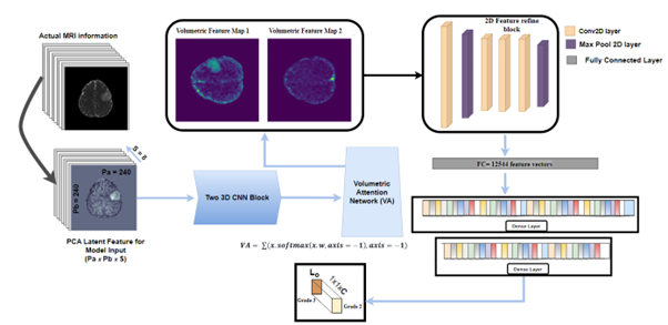
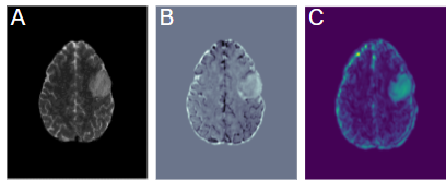
A single MRI slice from each of the eight modalities was collected for 86 patients (46 with grade 2 and 40 with grade 3 astrocytomas) from the University of California San Francisco Preoperative Diffuse Glioma MRI (UCSF-PDGM) dataset5, as illustrated in Figure 1A. The objective is to classify grade 2 and 3 astrocytomas. Initially, the most informative MRI slice from each modality was extracted based on the highest covariance value, using a segmented mask. These informative slices were then stacked together to create a 3D representation with a spatial size of 240 x 240 x 8, incorporating eight unique slices for each patient. To mitigate modality heterogeneity among different slices within the 3D cube, an incremental Principal Component Analysis (iPCA) was performed6, followed by min-max normalization, as depicted in Figure 1B. Subsequently, a novel deep model with a volumetric attention (VA) mechanism was developed to address the classification challenge 7. The model comprises two 3D convolutional layers to extract 3D features, which are then coupled with a volumetric attention mechanism to dynamically emphasize and learn the most informative regions. The attention mechanism reduces the 3D feature maps to two 2D informative feature maps, which are further processed by a series of 2D convolutional layers, as shown in Figure 2. To reduce the overfitting issue, four dropout layers were integrated into the model. The model was then trained and tested using 5-fold cross-validation to achieve optimal accuracy.Result and Discussion:
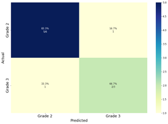
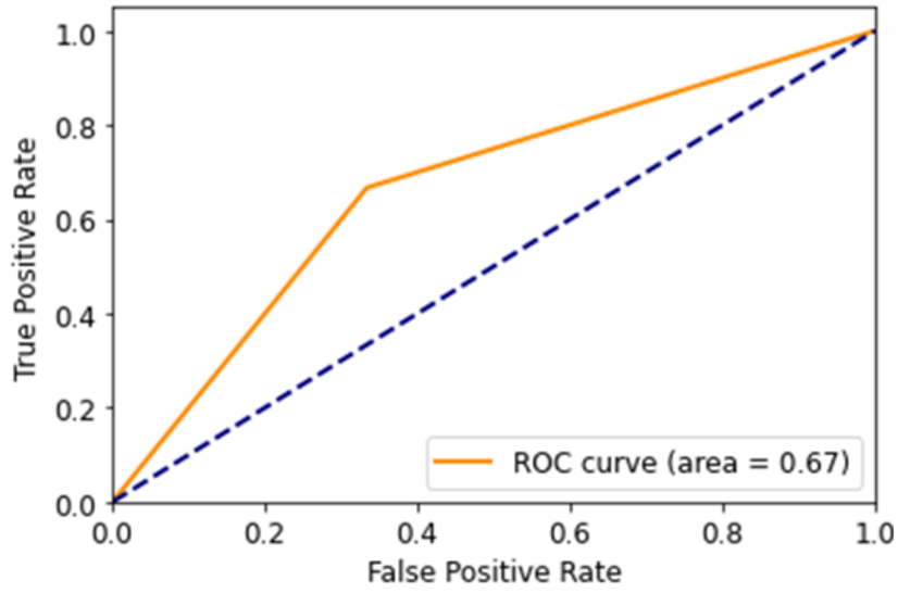
Classifying multimodality grade II and III astrocytomas is a challenging task due to their intensely homogeneous representation. The DVA model has achieved an overall accuracy of 77% for the independent test set in classifying grade II and III astrocytomas. Figure 3 highlights the robustness of the proposed model for nine different patients using a learning rate of 0.005 Figure 3 highlights the robustness of the proposed model for an independent set of nine patients using an SGD optimizer with a learning rate of 0.005. It is worth noting that in Figure 3, one can observe that grade II and III is classified with only one miss-classified sample each. Figure 4 provides insights into the model's performance across all five folds and its fine-tuning for each iteration using mean Receiver Operator Characteristic (ROC) of 0.67. Notably, when compared to contemporary deep models, the proposed attention model requires fewer time and resources to achieve a higher overall accuracy of (0.77) surpassing ResNet8 (0.66), DesNet9 (0.66), Cossim2D10 (0.66), and VggNet11 (0.55). Furthermore, the fusion of advanced and traditional MRI data represents both abstract and functional information. The proposed model effectively identifies these patterns and represents the quality feature map, as shown in Figure 5.Conclusion:
Compared with state-of-the-art models, the proposed deep volumetric attention model achieved promising results for multimodal MRI grade II and III astrocytomas tumour classification. In future, the model entails incorporating transfer learning using volumetric attention model, and patch analysis model. One of the possible future directions also include perturbation based XAI to examine model performance for clinical generalizability and trustworthiness.Acknowledgements
This research was funded by the Connacht-Ulster Alliance (CUA), ATlantic Technological University, Sligo, IrelandBursary reference (PCUAB016). Additionally, this investigation is a component of the COST Action CA18206 Glioma MR Imaging 2.0, endorsed by COST (European Cooperation in Science andTechnology). More details can be found at www.glimr.eu and www.cost.euReferences
1. Giantini-Larsen AM, Pannullo S, Juthani RG. Challenges in the Diagnosis and Management of Low-Grade Gliomas. World Neurosurg. 2022;166. doi:10.1016/j.wneu.2022.06.0742. Tandel GS, Tiwari A, Kakde OG. Performance enhancement of MRI-based brain tumor classification using suitable segmentation method and deep learning-based ensemble algorithm. Biomed Signal Process Control. 2022;78. doi:10.1016/j.bspc.2022.104018
3. Kern M, Auer TA, Picht T, Misch M, Wiener E. T2 mapping of molecular subtypes of WHO grade II/III gliomas. BMC Neurol. 2020;20(1). doi:10.1186/s12883-019-1590-1
4. Thompson NC, Greenewald K, Lee K, Manso GF. The Computational Limits of Deep Learning. 2020;4:2-5.
5. Calabrese E, Villanueva-Meyer JE, Rudie JD, et al. The University of California San Francisco Preoperative Diffuse Glioma MRI Dataset. Radiol Artif Intell. 2022;4(6). doi:10.1148/ryai.220058
6. Rehman A, Khan A, Ali MA, Khan MU, Khan SU, Ali L. Performance Analysis of PCA, Sparse PCA, Kernel PCA and Incremental PCA Algorithms for Heart Failure Prediction. In: 2nd International Conference on Electrical, Communication and Computer Engineering, ICECCE 2020. ; 2020. doi:10.1109/ICECCE49384.2020.9179199
7. Wang X, Han S, Chen Y, Gao D, Vasconcelos N. Volumetric Attention for 3D Medical Image Segmentation and Detection. In: Lecture Notes in Computer Science (Including Subseries Lecture Notes in Artificial Intelligence and Lecture Notes in Bioinformatics). Vol 11769 LNCS. ; 2019. doi:10.1007/978-3-030-32226-7_20
8. Sarwinda D, Paradisa RH, Bustamam A, Anggia P. Deep Learning in Image Classification using Residual Network (ResNet) Variants for Detection of Colorectal Cancer. In: Procedia Computer Science. Vol 179. ; 2021. doi:10.1016/j.procs.2021.01.025
9. Li C, Yu C, Lin H. DesNet: PCB defect detection network based on deformable convolution. In: 2023 IEEE 3rd International Conference on Electronic Technology, Communication and Information, ICETCI 2023. ; 2023. doi:10.1109/ICETCI57876.2023.10176845
10. Luo C, Zhan J, Xue X, Wang L, Ren R, Yang Q. Cosine normalization: Using cosine similarity instead of dot product in neural networks. Lecture Notes in Computer Science (including subseries Lecture Notes in Artificial Intelligence and Lecture Notes in Bioinformatics). 2018;11139 LNCS:382-391. doi:10.1007/978-3-030-01418-6_38/COVER
11. Majib MS, Rahman MM, Shahriar Sazzad TM, Khan NI, Dey SK. VGG-SCNet: A VGG Net based Deep Learning framework for Brain Tumor Detection on MRI Images. IEEE Access. Published online 2021. doi:10.1109/ACCESS.2021.3105874
Figures