4853
MRI-based prediction of cerebral palsy risk in infants aged 6 months to 2 years: a deep learning approach1School of Future Technology, Xi'an Jiaotong University, Xi'an, China, 2Department of Radiology, The First Affiliated Hospital of Xi'an Jiaotong University, Xi'an, China, 3Shaanxi Engineering Research Center of Computational Imaging and Medical Intelligence, Xi'an, China, 4Department of Research and Development, Shanghai United Imaging Intelligence Co., Ltd., Shanghai, China
Synopsis
Keywords: Diagnosis/Prediction, Brain, Cerebral Palsy
Motivation: Early prediction of cerebral palsy (CP) in infants plays a pivotal role in facilitating tailored rehabilitation treatment.
Goal(s): We hope to achieve early prediction of CP in infants aged 6 months to 2 years old based on MRI and deep learning technology.
Approach: We introduce a novel neural network model, known as the "Cerebral Palsy Brain Constraint Residual Network" (CPBC-Resnet), for the automatic prediction of CP risk based on MRI data.
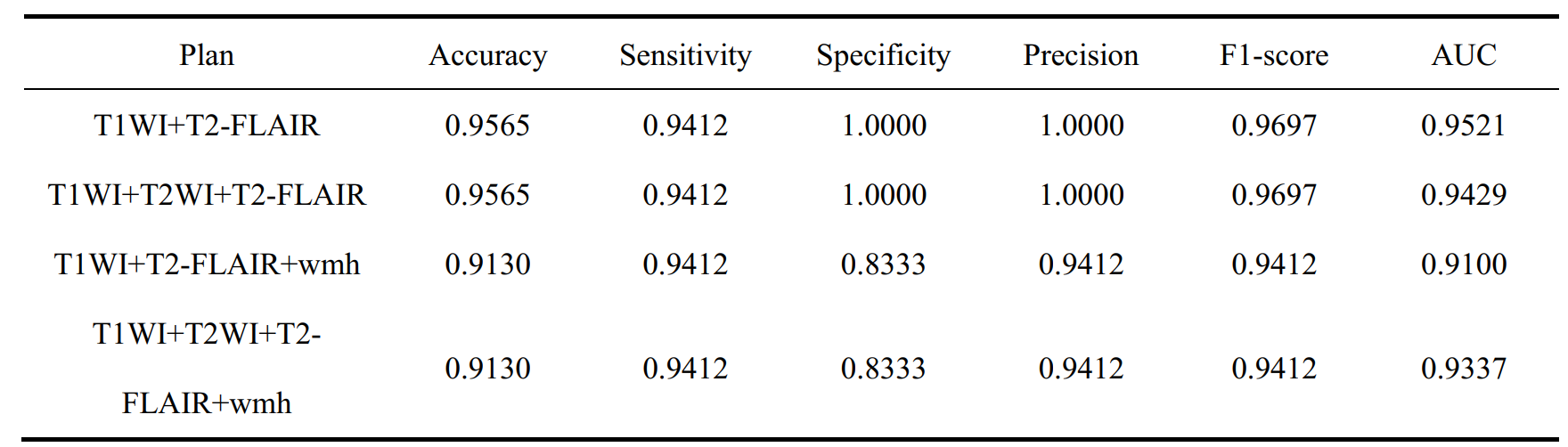
Results: The CPBC-Resnet model exhibits an impressive receiver operating characteristic area under the curve (AUC) of 0.9521, achieving a sensitivity of 94.12% and a specificity of 100%.
Impact: This study streamlines cerebral palsy (CP) imaging diagnostics, reducing physician training costs, and expanding the reach of CP diagnostic technology. It promotes early CP diagnosis and intervention, particularly in areas with underdeveloped medical standards, contributing to overall child health improvement.
Introduction
Cerebral palsy (CP) is a prevalent group of movement disorders, frequently leading to childhood disabilities[1, 2]. The significance of early diagnosis cannot be overstated in the context of CP, yet the current diagnostic paradigm typically identifies cases after the age of 2[3, 4]. Magnetic resonance imaging (MRI) constitutes an indispensable component of the comprehensive assessment, following an initial screening of infants at high risk of CP through behavioral scoring[3]. Conventional diagnosis training based on MRI demands a substantial investment of time and financial resources, with an average sensitivity rate falling below 90%[3]. As of now, deep learning technology has been extensively leveraged in tasks related to image-based disease classification and has consistently exhibited commendable performance[5, 6]. Therefore, the primary objective of this study is to develop a deep learning prediction model utilizing MRI data for the early assessment of CP in infants aged 6 months to 2 years.Methods
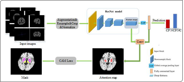
A total of 240 infants (aged 6 months to 2 years) who underwent MRI examinations between April 2013 and April 2019 were included in this study. CP was diagnosed after the age of 2 years based on the follow-up assessments. Therefore, the datasets comprise CP and non-CP cases. We proposed a novel neural network model, CPBC-Resnet, based on the ResNet-50 framework (Fig. 1). Firstly, five crucial brain regions (centrum semiovale, cerebral peduncle, lentiform nucleus, thalamus, and posterior limb of the internal capsule) essential for CP diagnosis were segmented by the uAI Research Portal (United Imaging Intelligence, China, Version: 20230915), in which deep learning segmentation and classification were embedded. Class Activation Mapping (CAM) loss was introduced to guide the network in emphasizing the characteristics of these key brain areas, resulting in the construction of a classification model to distinguish CP and non-CP. Additionally, to leverage diverse sequence characteristics, we employed a combination of different sequences: (1) T1-weighted imaging (T1WI)+T2 fluid-attenuated inversion recovery (T2-FLAIR), (2) T1WI+T2-weighted imaging (T2WI)+ T2-FLAIR, (3) T1WI+ T2-FLAIR+wmh mask, and (4) T1WI+T2WI+wmh mask, with 'wmh' signifying periventricular white matter damage signals segmented by uAI Discover-CSVD (version: R001). The dataset was divided into an 8:2 ratio for training and testing the model, and test metrics from the four schemes were compared to evaluate the performance of each configuration.Results
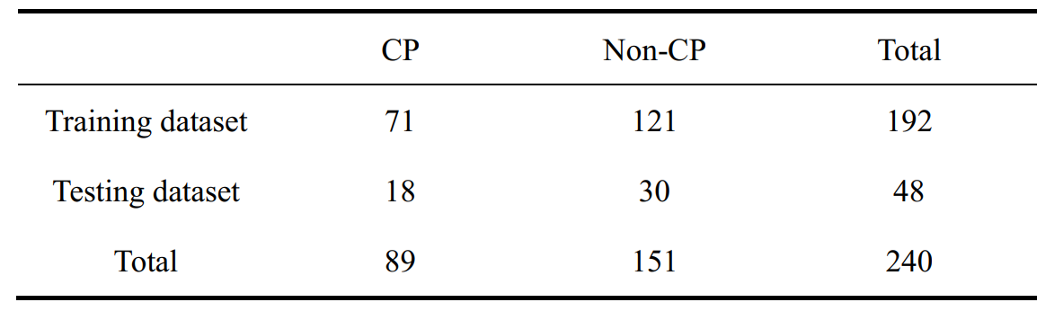
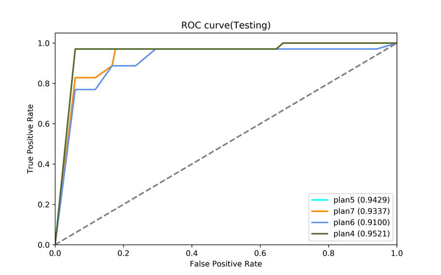
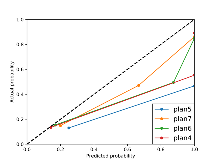
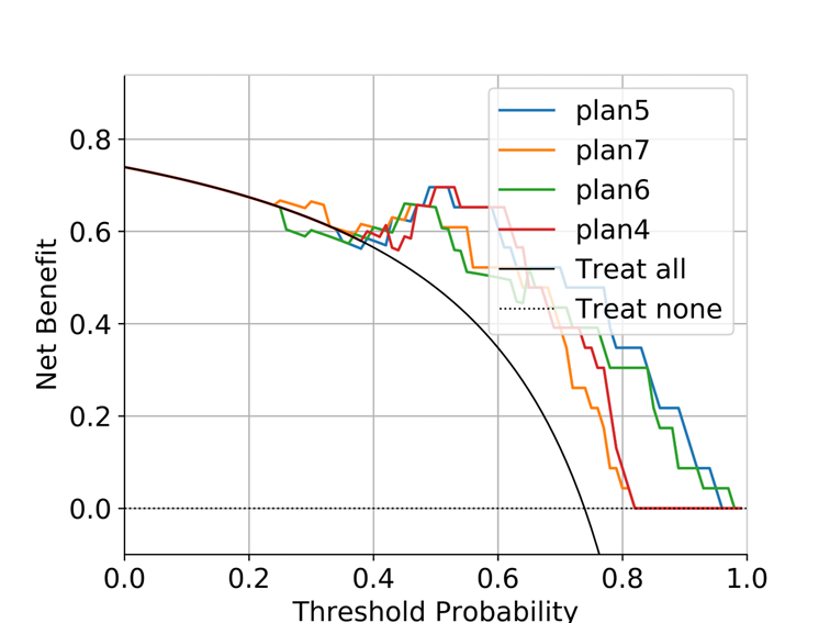
Among the 240 infants, 89 infants were diagnosed with CP, while 151 were classified as non-CP. Of the non-CP cases, 32 cases exhibited white matter injuries, while the remaining 119 did not. Each case included T1WI, T2WI, and T2-FLAIR sequences. The dataset division into training and test sets is detailed in Table 1. The CPBC-ResNet model was evaluated across four different schemes on this dataset, yielding the following results: (1) T1WI+ T2-FLAIR: Accuracy, sensitivity, specificity, and AUC were 95.65%, 94.12%, 100%, and 95.21%, respectively. (2) T1WI +T2WI + T2-FLAIR: Achieving accuracy, sensitivity, specificity, and AUC of 95.65%, 94.12%, 100%, and 94.29%, respectively. (3) T1WI + T2-FLAIR+wmh: This scheme obtained results of 91.30% accuracy, 94.12% sensitivity, 83.33% specificity, and an AUC of 91.00%. (4) T1WI +T2WI + T2-FLAIR+wmh: For this scheme, results included 91.30% accuracy, 94.12% sensitivity, 83.33% specificity, and an AUC of 93.37%. In an overall comparative assessment (refer to Fig. 2 - Fig. 6), the T1WI+FLAIR scheme displayed the most favorable classification performance, although no significant performance disparities were observed among the four schemes.Discussion
In this study, we have devised an intelligent risk prediction model for CP using deep learning technology, aiming to automate the diagnostic process through MRI scans of infants aged 6 months to 2 years. In comparison to conventional CP diagnosis based on MRI, our model exhibits a reduction in training cost and an improvement in diagnostic performance. Our findings demonstrate the model’s robustness when the extracted features are constrained based on five key brain areas, underscoring the critical role of these regions in CP diagnosis[7-11]. Furthermore, the introduction of a periventricular white matter damage mask does not yield enhanced classification performance, affirming the model's proficient feature extraction capabilities without the necessity of incorporating additional artificial features.Conclusion
The CPBC-Resnet model introduced in this study showcases the capacity to leverage MRI scans for early predictions of CP in infants aged 6 months to 2 years. This holds significant practical relevance, as it facilitates early interventions, timely treatment, and comprehensive rehabilitation strategies for infants afflicted by CP.Acknowledgements
This work was supported by the National Natural Science Foundation of China (82272618, 81971581). Please address correspondence to Jian Yang, e-mail: yj1118@mail.xjtu.edu.cn, and Xianjun Li, e-mail: xianj.li@mail.xjtu.edu.cn.References
[1] Sadowska M, Sarecka-Hujar B, Kopyta I. Cerebral Palsy: Current Opinions on Definition, Epidemiology, Risk Factors, Classification and Treatment Options [J]. Neuropsychiatr Dis Treat, 2020, 16: 1505-18.
[2] Martinez-Biarge M, Diez-Sebastian J, Kapellou O, et al. Predicting motor outcome and death in term hypoxic-ischemic encephalopathy [J]. Neurology, 2011, 76(24): 2055-61.
[3] Novak I, Morgan C, Adde L, et al. Early, Accurate Diagnosis and Early Intervention in Cerebral Palsy: Advances in Diagnosis and Treatment [J]. JAMA Pediatr, 2017, 171(9): 897-907.
[4] Morgan C, Fetters L, Adde L, et al. Early Intervention for Children Aged 0 to 2 Years With or at High Risk of Cerebral Palsy: International Clinical Practice Guideline Based on Systematic Reviews [J]. JAMA Pediatr, 2021, 175(8): 846-58.
[5] Chen S, Wu Z, Li M, et al. FIT-Net: Feature Interaction Transformer Network for Pathologic Myopia Diagnosis [J]. IEEE Trans Med Imaging, 2023, 42(9): 2524-38.
[6] Zheng Y, Li J, Shi J, et al. Kernel Attention Transformer for Histopathology Whole Slide Image Analysis and Assistant Cancer Diagnosis [J]. IEEE Trans Med Imaging, 2023, 42(9): 2726-39.
[7] Jiang H, Liu H, Huang T, et al. Structural network performance for early diagnosis of spastic cerebral palsy in periventricular white matter injury [J]. Brain Imaging Behav, 2021, 15(2): 855-64.
[8] Back S A, Riddle A, McClure M M. Maturation-dependent vulnerability of perinatal white matter in premature birth [J]. Stroke, 2007, 38(2 Suppl): 724-30.
[9] Bano S, Chaudhary V, Garga U C. Neonatal Hypoxic-ischemic Encephalopathy: A Radiological Review [J]. J Pediatr Neurosci, 2017, 12(1): 1-6.
[10] Simon-Martinez C, Decraene L, Zielinski I, et al. The impact of brain lesion characteristics and the corticospinal tract wiring on mirror movements in unilateral cerebral palsy [J]. Sci Rep, 2022, 12(1): 16301.
[11] Kuo H C, Ferre C L, Chin K Y, et al. Mirror movements and brain pathology in children with unilateral cerebral palsy [J]. Dev Med Child Neurol, 2023, 65(2): 264-73.
Figures