4843
Early Alzheimer's Detection and Classification using VGG Convolutional Neural Network and Systematic Data Augmentation using MR Images1Carmel High School, Carmel, IN, United States, 2Radiology and Imaging Sciences, Indiana University School of Medicine, Indianapolis, IN, United States
Synopsis
Keywords: Diagnosis/Prediction, Machine Learning/Artificial Intelligence
Motivation: The use of different imaging tools at various hospitals results in varying contrast images. This fragmentation in healthcare prompted me to develop a personalized network that can be trained using hospital imaging database.
Goal(s): The main goal of this project is to predict early stages of Alzheimer's Disease (AD) using Magnetic Resonance (MR) images.
Approach: We applied convolutional neural network (CNN) to the T1 weighted images of AD, publicly available at https://www.kaggle.com/datasets/tourist55/alzheimers-dataset-4-class-of-images. The images were classified into four classes. F1 score and Area Under Curve (AUC) were calculated for the model after training.
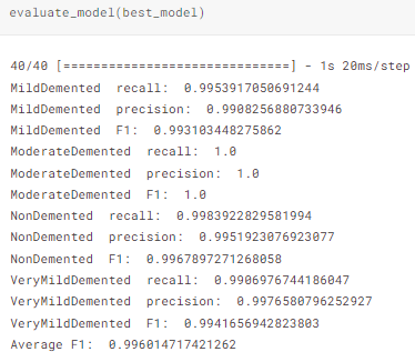
Results: We demonstrated F1 score of 99.60% and AUC 0.994.
Impact: This model could be used to predict AD to other datasets that might help early detection of AD and subsequently improve treatment strategies. With various mice brain scan training, this network can also be used to aid AD researchers.
Introduction
Magnetic Resonance (MR) image analysis can provide differential diagnosis, prognosis, and the progression and stages of Alzheimer’s Disease. Physicians, however, are not always able to detect signs of Alzheimer’s, especially during early stages. This highlights a need for the development of deep-learning network architecture that is able to provide those analysis results and assist humans in tracking Alzheimer’s Disease progression.Alzheimer’s Disease (AD) is a progressive neurological disorder, with more than 50 million people suffering Alzheimer’s globally.1 This puts global cost at over $818 billion dollars, and the number of people affected is projected to reach 151 million by 2050.2 There is no therapeutic measures of curing the disorder, highlighting the need for a way to diagnose AD in its early stages, when the effects are still reversible and manageable with intervention.3 So, we aim to predict AD using convolutional neural network of MR images.
Methods
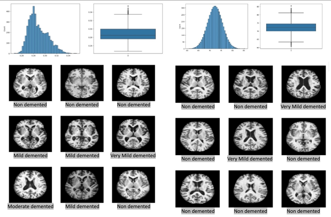
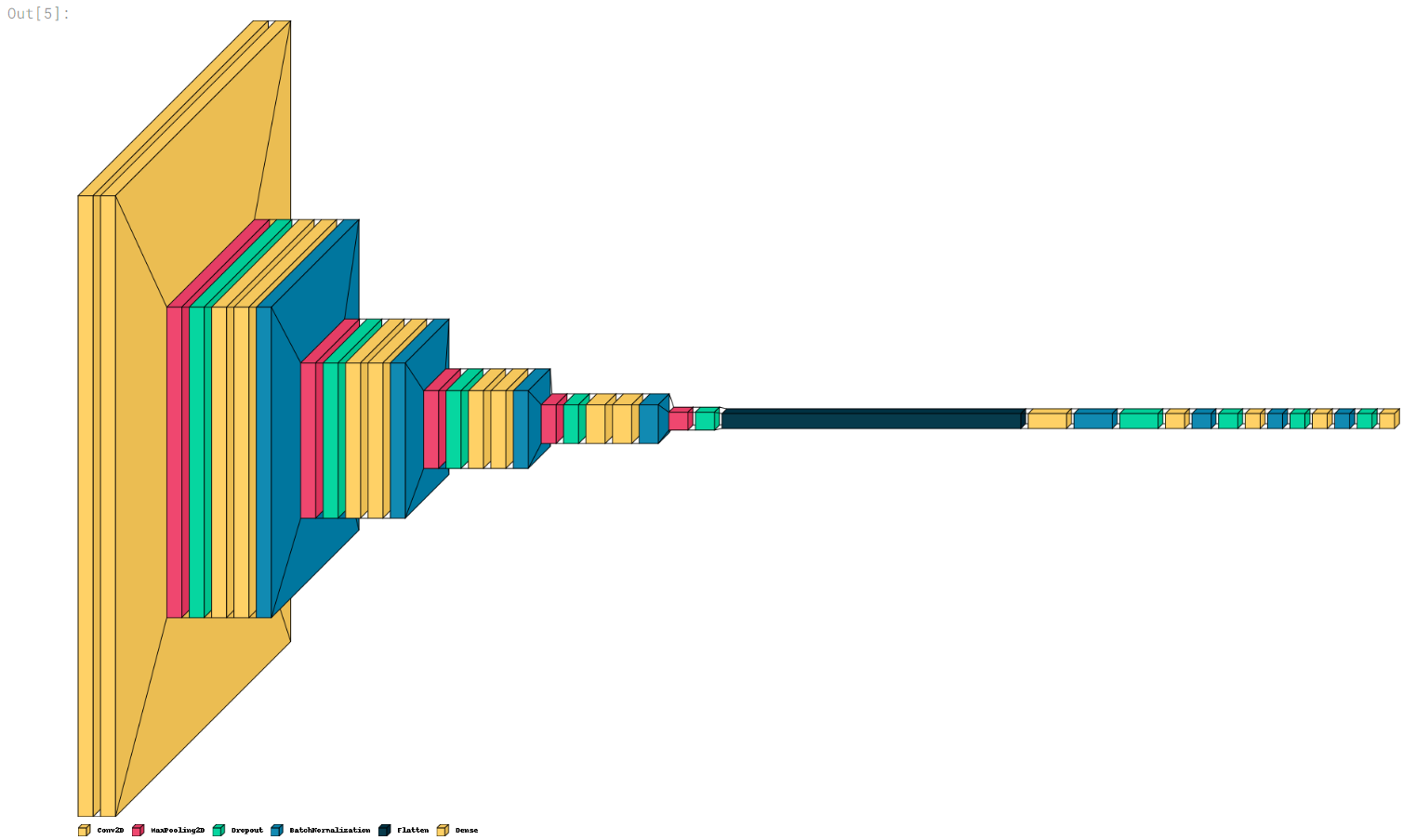
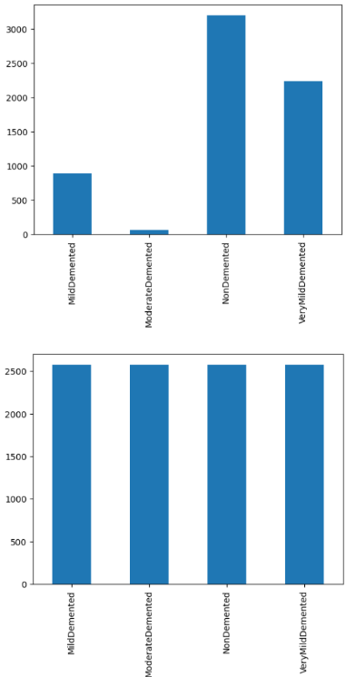
We obtained publicly available AD datasets from Kaggle website https://www.kaggle.com/datasets/tourist55/alzheimers-dataset-4-class-of-images. There were four classes of T1 weighted images of AD, namely mild demented, moderate demented, nondemented and very mild demented. A Visual Geometry Group (VGG) neural network in Keras library was used for training the dataset with Nvidia Tesla P100 GPU. In order to ensure maximum accuracy for our model, a systematic data augmentation network was made. Using imblearn library, outlier images were identified that were then removed under parameters outlined by Synthetic Minority Over-sampling Technique (SMOTE) while the rest were standardized.4 Matplotlib was used to visualize 9 sample images before and after data augmentation which were extracted and inputted into the VGGNet as shown in Figure 1.The VGG architecture is most used in imaging recognition because of its 2D convolution and max pooling layout for oversampling and undersampling.5 We utilized five 2D convolutional layers coupled with leaky ReLU, to prevent “dying ReLU” problems, followed by pooling layer.6 The proposed VGG neural network architecture is shown in Figure 2. Furthermore, four dense layers were added to enhance model accuracy.7 The network was then trained 150 epoch each with the batch size 32. During the training, Adaptive Movement Estimation (Adam) optimizer was used to add weights and biases accordingly in order to improve its accuracy and speed.8 Learning rate was set at 1x10⁻³. The full visual for our VGG architecture can be viewed on Figure 2.
Results
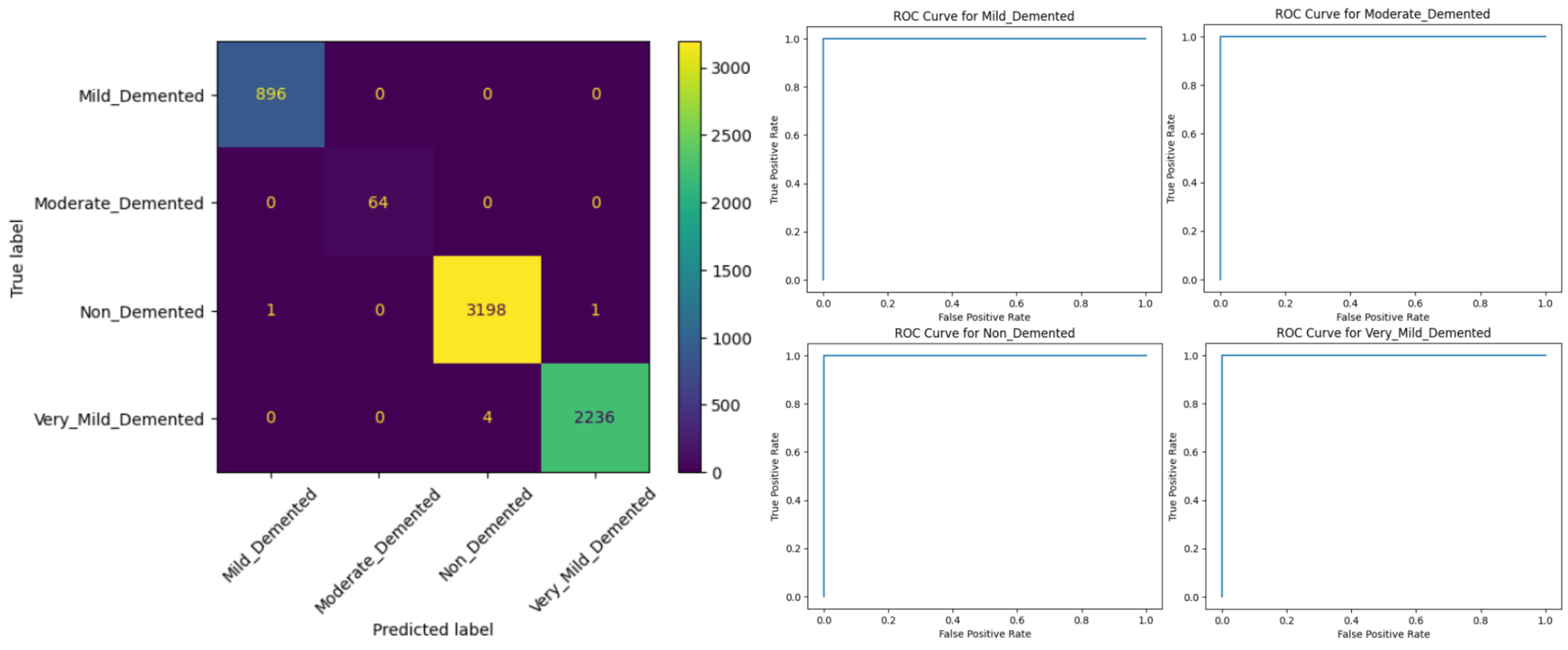
Figure 3 showed the average brightness levels between four categories of images. The discrepancies in the brightness and contrast were adjusted to 2500. Figure 4 demonstrated a 4x4confusion matrix and ROC curves. We found minimal false positive rate (FPR) and false negative rate (FNR), which confirmed the network's accuracy, F1 score, precision, and recall evaluation. The proposed neural network has an AUC value of 0.994 as shown in Figure 4. We obtained a F1 score 99.60% as shown in Figure 5.Discussion
We developed the CNN architecture that used MRI axial images that could accurately classify various staging of AD at 99.60%. Because of phenotypic differences reflected in the images, we were able to predict them accurately. Accurate prediction is essential for proper treatment plan of AD. The AUC value of 0.994 implied excellent prediction. This suggested MR imaging could potentially be used as a non-invasive diagnostic imaging tool for accurate prediction of AD. The outlier removal data augmentation using SMOTE technique reduced the risk of overfitting problem posed by random oversampling. Some potential limitations of this study could be small sample size (n=5212). Future work is recommended using multiple neural network architecture and multiple contrast MR images.Conclusion
This model comes with many uses, as any 2D image dataset can be put into the augmentation network and then VGGNet architecture, any other neurological disorders and mice models of AD scans can be used to train the network. This would allow not only an effective and personalized diagnostic assistance tool for each lab/hospital setting, but it would also assist in research as the network could characterize and track mice models. This would minimize the need for IHC and MSD analysis when comparing the phenotypic manifestation difference between mice models.Future steps will be implemented like training the network with ADNI and OASIS database as well as clinical MRI database from Stark Neuroscience Research Institute to further train the network and develop a personalized diagnostic tool for the physicians at Indiana University. Finally, future testing and fine tuning with different learning rates can be implemented for maximum optimization of our Alzheimer's Disease detection network.
Acknowledgements
We would like to thank Wali Mirza for providing the Nvidia Tesla P100 during the network training and assistance.References
1. Gustavsson A, Norton N, Fast T, Frölich L, Georges J, Holzapfel D. Global estimates on the number of persons across the Alzheimer’s disease continuum. The Journal of the Alzheimer’s Association. July 2, 2022. Accessed October 13, 2023. https://alz-journals.onlinelibrary.wiley.com/doi/10.1002/alz.12694.
2 Bringas S, Salomón S, Duque R, Lage C, Montaña JL. Alzheimer’s disease stage identification using Deep Learning Models. Journal of Biomedical Informatics. July 23, 2020. Accessed October 13, 2023. https://www.sciencedirect.com/science/article/pii/S1532046420301428.
3. Fan D-Y, Wang Y-J. Early intervention in alzheimer’s disease: How early is early enough? Neuroscience bulletin. February 2020. Accessed October 13, 2023. https://www.ncbi.nlm.nih.gov/pmc/articles/PMC6977799/.
4. Lemaitre G, Nogueira F, Aridas CK. Imbalanced-learn: A python toolbox to tackle the curse of imbalanced datasets in Machine Learning. arXiv.org. September 21, 2016. Accessed October 13, 2023. https://arxiv.org/abs/1609.06570.
5. Simonyan K, Zisserman A. Very deep convolutional networks for large-scale image recognition. arXiv.org. April 10, 2015. Accessed October 13, 2023. https://arxiv.org/abs/1409.1556.
6. Gupta R, Birbilis N. cardiGAN: A Generative Adversarial Network Model for Design and Discovery of Multi Principal Element Alloys. ResearchGate. February 2022. Accessed October 13, 2023. https://www.researchgate.net/publication/358306930_cardiGAN_A_Generative_Adversarial_Network_Model_for_Design_and_Discovery_of_Multi_Principal_Element_Alloys.
7. Josephine H, Nirmala AP, Alluri VL. Impact of Hidden Dense Layers in Convolutional Neural Network to enhance Performance of Classification Model. ResearchGate. April 2021. Accessed October 13, 2023. https://www.researchgate.net/publication/350899586_Impact_of_Hidden_Dense_Layers_in_Convolutional_Neural_Network_to_enhance_Performance_of_Classification_Model.
8. Kingma DP, Ba J. Adam: A Method for Stochastic Optimization. arXiv.org. January 30, 2017. Accessed October 13, 2023. https://arxiv.org/abs/1412.6980.
9. Jo T, Nho K, Saykin AJ. Deep Learning in Alzheimer’s Disease: Diagnostic Classification and Prognostic Prediction Using Neuroimaging Data. Frontiers. August 2, 2019. Accessed October 13, 2023. https://www.frontiersin.org/articles/10.3389/fnagi.2019.00220/full.
Figures