4841
Acoustic Noise Optimization of Trapezoid Oscillating Diffusion Encoding Gradient1Department of Biomedical Engineering, College of Biomedical Engineering & Instrument Science, Zhejiang University, Hangzhou, China, 2Center for Intelligent Biomedical Instrumentation, Zhejiang University Binjiang Research Institute, Hangzhou, China, 3MR Collaboration, Siemens Healthcare China, Shanghai, China
Synopsis
Keywords: Gradients, Safety, Gradients Optimization, Acoustic Noise, OGSE
Motivation: Oscillating gradient spin-echo (OGSE) diffusion MRI sequence involves diffusion preparation and EPI readout, both having rapidly switching trapezoid gradient. Consequently, OGSE sequence generates strong acoustic noise that may introduce comfortless patient experience.
Goal(s): Our study aims to develop an optimization framework to suppress the acoustic noise by softening the trapezoid gradient.
Approach: We measured the acoustic noise frequency response function of scanner. Based on linear model of MRI acoustic noise generation, we designed a convex optimization framework to reduce the predicted A-weighting sound pressure level(SPL)
Results: Optimized gradient achieved 14.09dBA SPL reduction according to our FRF based prediction.
Impact: We developed an effective optimization method to reduce the acoustic noise of oscillating trapezoid gradient waveform, and potentially facilitate the clinical applications of OGSE. This optimization framework also has potential to reduce acoustic noise of EPI readout waveform
Introduction
Acoustic noise during MRI scan is mostly generated from gradient coil vibration, which severely affects patients’ comfort or even poses potential safety risks. The impact of noise is particularly pronounced in sequences characterized by rapid switching in the gradient field. The oscillating gradient spin-echo (OGSE) sequence consists of strong and oscillating diffusion gradient and EPI readout gradient to rapidly filling the k-space, and therefore, it suffers from loud acoustic noise compared to other imaging sequences. The Linear Time Invariant (LTI) model and system Frequency Response Function (FRF) clearly describe acoustic noise generation process of the MRI scanner 1. Previous works in acoustic noise optimization have utilized several methods to smooth waveform including low-pass filtering of the gradient waveforms 2, or using spline interpolation and waveform integral constraint to optimize GRE gradient 3. For fast-switching trapezoid gradient like EPI, certain basic frequency sinusoidal waveform was used frequently to replace original waveform to achieve acoustic noise minimization 4,5. This work uses a numerical optimization approach inspired by earlier gradient optimization studies, such as CODE 6 ,GrOpt 7,8 and ODGD 9. In this work, we optimized the gradient waveform to suppress the frequency components that resonate with our measured FRF high response frequency range.Methods
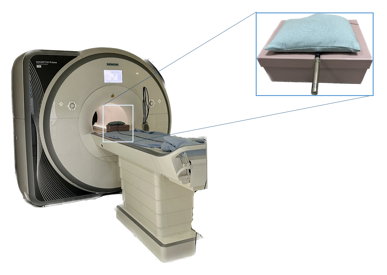
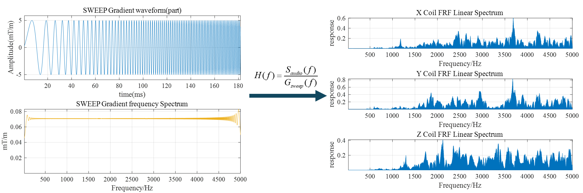
FRF MeasurementThe FRF was measured on a 3T Siemens Prisma scanner, using professional acoustic noise measurement system (microphone, preamplifier, calibrator, multi-channel analyzer), audio sampling frequency is 48000Hz (Fig.1). Based on linear model of noise generation, the expected audio output is $$$s(t)=g(t)*h(t)$$$, where $$$g(t)$$$ is gradient waveform and $$$h(t)$$$ is time domain FRF, $$$*$$$ indicates convolution. In frequency domain, the output audio frequency spectrum is $$$S(f)=G(f) \centerdot H(f)$$$, where $$$H(f)$$$ is the system acoustic FRF (Fourier transform of $$$h(t)$$$). We ran the sweep frequency sequence ranging from 20Hz to 5000Hz to record audio response and calculate the FRF. Fig.2 shows the acquired FRF of the X/Y/Z gradient coils.
Gradient waveform optimization
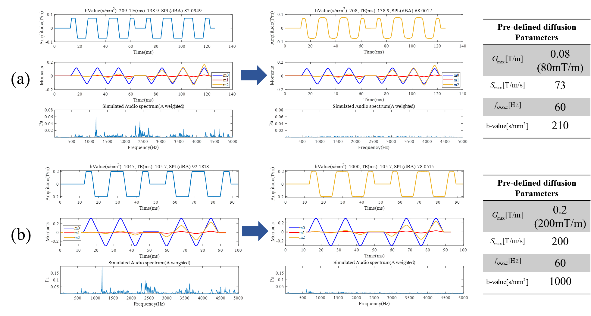
We utilized the optimization framework shown in Fig.3 to minimize the acoustic noise, by reducing the A-weighting Sound Pressure Level (SPL) of gradient waveform. The framework enables us to optimize trapezoidal OGSE waveform with predefined generation parameters (OGSE oscillating frequency $$$f_{O G S E}$$$, b-value $$$b_{t a r g e t}$$$, maximum gradient slew rate $$$S_{m a x}$$$ and maximum gradient amplitude $$$G_{m a x}$$$). The optimization objective function is minimizing the A-weighting SPL, namely $$$RMS(G(f) \centerdot H(f))$$$. The optimization constraints can be categorized into three categories shown in Fig.3. 1) Pulse sequence and hardware constraints were used to guarantee the basic shape of the waveform and feasibility according to the scanner hardware. 2) Extra moments constraint was used to boost motion robustness and guarantee periodicity of the waveform. 3) b-value constraint was used to guarantee the desired diffusion weighting. The optimization problem was solved by interior-point algorithm, using built-in function fmincon in MATLAB Optimization Toolbox.
Results
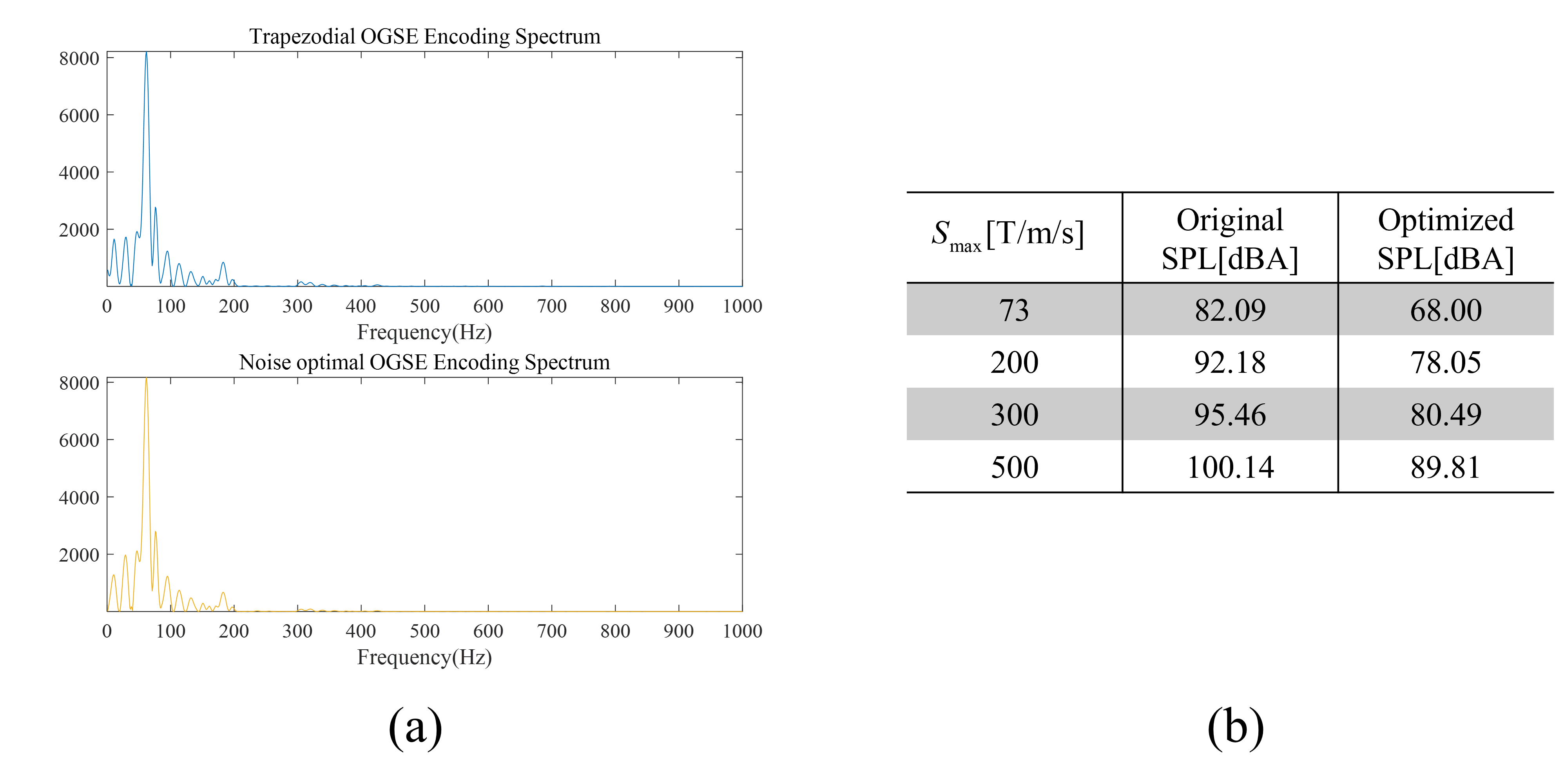
Optimization results in Fig.4 demonstrated that the optimized OGSE waveform was softened and attained reduction in 14.09dBA simulated SPL for result in Fig.4(a), 14.13dBA for result in Fig.4(b). According to Fig.5(a), modulation spectrum (diffusion encoding spectrum) of the OG encoding waveforms was not affected before and after optimization. The solver computational time for Fig.4(a) is 110s, for Fig.4(b) is 143s. We further predicted the A-weighting SPL of OGSE sequence at different slew rates in Fig.5(b), which indicated the potential concerns about acoustic noise in OGSE sequence would become more prominent on high performance gradient MRI system, such as the connectome scanner with SRmax of 300 m/T/s.Discussion and Conclusion
We demonstrated that our optimization framework based on FRF model can effectively soften the gradient waveform to reduce acoustic noise for oscillating diffusion gradient. The OGSE sequences are important to access short diffusion time for microstructural mapping, but it demands for strong and fast-switching gradients, and thus suffered from loud acoustic noise. This problem will become more prominent at high- performance gradient scanners, and needs to be sufficient addressed for its clinical application. Proposed optimization framework also has potential to be utilized in OG-EPI acquisition. Our optimized trapezoidal waveform may address the image quality trade-off issue proposed in work 10. Comparing to sinusoidal EPI readout waveform, the optimized readout waveform closely approximates the original trapezoidal waveform, which means more uniform k-space filling. Further work on both diffusion and EPI readout waveform optimization will be performed with detailed image quality analysis and experimental acoustic noise measurements.Acknowledgements
This work is supported by the National Natural Science Foundation of China (81971606, 82122032), and Science and Technology Department of Zhejiang Province (2022C03057, 202006140)References
1. Hedeen, R. A., & Edelstein, W. A. (1997). Characterization and prediction of gradient acoustic noise in MR imagers. Magnetic Resonance in Medicine, 37(1), 7-10.
2. Hennel, F., Girard, F., & Loenneker, T. (1999). “Silent” MRI with soft gradient pulses. Magnetic Resonance in Medicine, 42(1), 6-10.
3. Heismann, B., Ott, M., & Grodzki, D. (2015). Sequence‐based acoustic noise reduction of clinical MRI scans. Magnetic resonance in medicine, 73(3), 1104-1109.
4. Hutter, J., Price, A. N., Cordero‐Grande, L., Malik, S., Ferrazzi, G., Gaspar, A., ... & Hajnal, J. V. (2018). Quiet echo planar imaging for functional and diffusion MRI. Magnetic resonance in medicine, 79(3), 1447-1459.
5. Zapp, J., Schmitter, S., & Schad, L. R. (2012). Sinusoidal echo‐planar imaging with parallel acquisition technique for reduced acoustic noise in auditory fMRI. Journal of Magnetic Resonance Imaging, 36(3), 581-588.
6. Aliotta, E., Wu, H. H., & Ennis, D. B. (2017). Convex optimized diffusion encoding (CODE) gradient waveforms for minimum echo time and bulk motion–compensated diffusion‐weighted MRI. Magnetic resonance in medicine, 77(2), 717-729.
7. Middione, M. J., Loecher, M., Moulin, K., & Ennis, D. B. (2020). Optimization methods for magnetic resonance imaging gradient waveform design. NMR in biomedicine, 33(12), e4308.
8. Loecher, M., Middione, M. J., & Ennis, D. B. (2020). A gradient optimization toolbox for general purpose time‐optimal MRI gradient waveform design. Magnetic Resonance in Medicine, 84(6), 3234-3245.
9. Peña‐Nogales, Ó., Zhang, Y., Wang, X., de Luis‐Garcia, R., Aja‐Fernández, S., Holmes, J. H., & Hernando, D. (2019). Optimized Diffusion‐Weighting Gradient Waveform Design (ODGD) formulation for motion compensation and concomitant gradient nulling. Magnetic resonance in medicine, 81(2), 989-1003.
10. Liebig, P., Heidemann, R., Tao, Y., Hensel, B., Liu, W., & Porter, D. (2018). fMRI with Low Acoustic Noise using Variable-Blipped EPI with Readout Segmentation. ISMRM 2018
Figures