4840
Measuring CSF net velocity using DENSE at 7T with improved correction for involuntary motion and eddy currents.1Center for Image Sciences, University Medical Center Utrecht, Utrecht, Netherlands
Synopsis
Keywords: Neurofluids, Neurofluids, CSF, Velocity & Flow, Clearance, Brain, Neuro
Motivation: Clearance is important for healthy brain functioning. The ability to measure CSF net velocity would be valuable to gain insight into the underlying mechanisms and pathways of clearance.
Goal(s): To measure CSF net velocities in FH and RL direction whilst accounting for periodic motions, involuntary head motion and eddy currents.
Approach: A multi-slice single shot DENSE acquisition is used to measure CSF displacements over time.
Results: The measured net velocity does not fit the classical view on CSF excretion and absorption locations. Further validation is needed using a moving flow phantom.
Impact: The measured net velocities are about 10 percent of what would be expected. If confirmed in a larger cohort, the results challenge the classical view of main CSF excretion at the choroid plexus and absorption at the sagittal sinus.
Introduction
Understanding brain clearance is important in both healthy and diseased state. Cerebrospinal fluid (CSF) is primarily driven by changes in cerebral blood volume (CBV) resulting from various mechanisms, including heartbeat, respiration and neurovascular coupling1-3. These variations in CBV lead to periodic motion of CSF. However, due to continuous secretion of CSF at the choroid plexus (~ 500 mL/day) and absorption in the sagittal sinus, a net flow would also be expected in the subarachnoid space in the order of 5 µm/s4. Previously, a method to measure these slow velocities in the presence of large periodic motion has been introduced4, using Displacement Encoding of Stimulated Echoes (DENSE)5. The effects of eddy currents (EC) were believed to be minor, given measurements in a relatively small gel phantom. Here, we show similar measurements corrected for EC and sub-voxel involuntary head motion in both Feet-Head (FH) and Right-Left (RL) direction.Methods
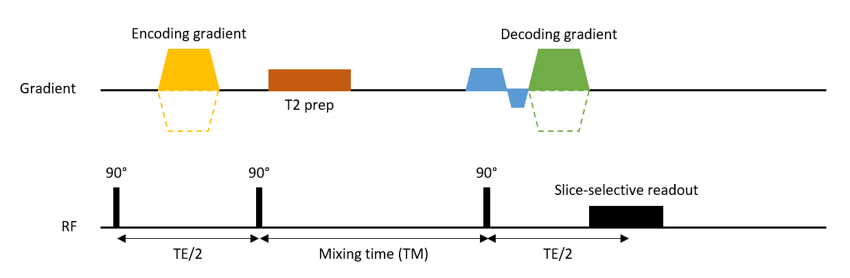
Acquisition CSF motion was assessed in a gel phantom (2% agarose) and in three healthy subjects (2 males, 40±3.4 years), after obtaining written informed consent, on a 7T scanner (Philips Medical) with a 32ch head coil (Nova Medical) using a multi-slice DENSE acquisition (Fig 1)5. A total of 60 slices, divided over two packages, were acquired over 60 repeats using T2 preparation pulses (TE=200 ms) to enhance CSF signal specificity. Displacements were measured over 30 different mixing times (TM=250-1990 ms) with slice permutations to ensure full coverage of the TM-range for all slices. Other MRI parameters were TE/TR = 15 ms/6000 ms (no triggering/gating), coronal FOV of 250x250 mm2, isotropic resolution of 3 mm, SENSE 2.6 (RL), DENC of 125 µm in FH and RL direction. Total scan duration was 12 minutes per encoding direction (6s x 2 packages x 60 repeats). Physiological data was recorded using a pulse oximeter and respiratory belt. Analysis Phase data were corrected for a static gradient, EC in all three orthogonal directions and rotational and translational head motion during the mixing time. The corrected measured phase was modelled as a linear combination of a static phase offset, cardiac and respiratory motion, and a net velocity component as previously described4. Physiological motion was binned into 10 bins where bins were weighted based on the temporal positions with respect to the cardiac and respiratory cycle without any assumptions about the underlying waveform. A voxel-wise least squares fit was performed to estimate the different phase components. Net velocities and physiological motion were determined within a CSF mask after manually removing the ventricles.Results
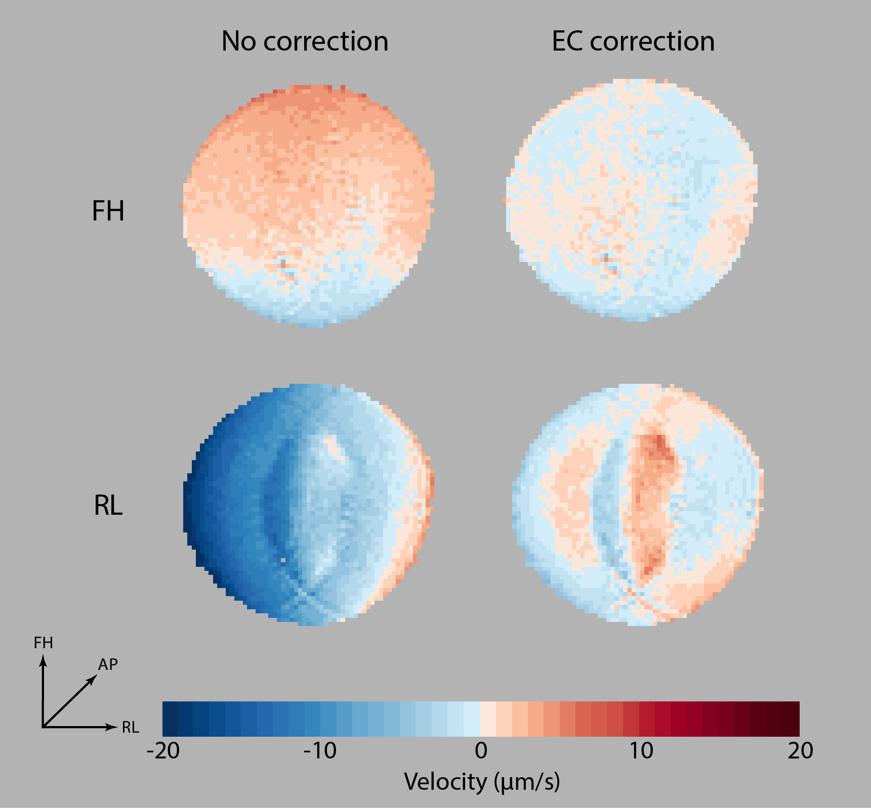
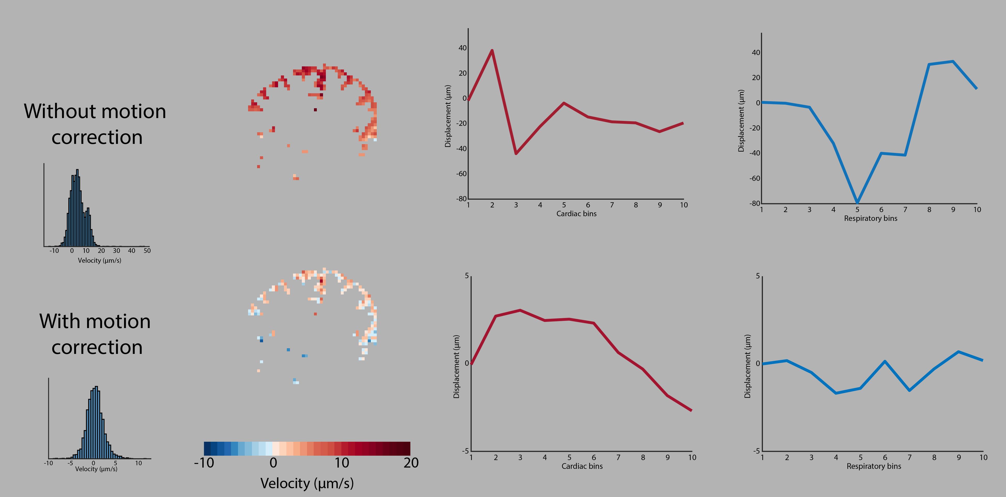
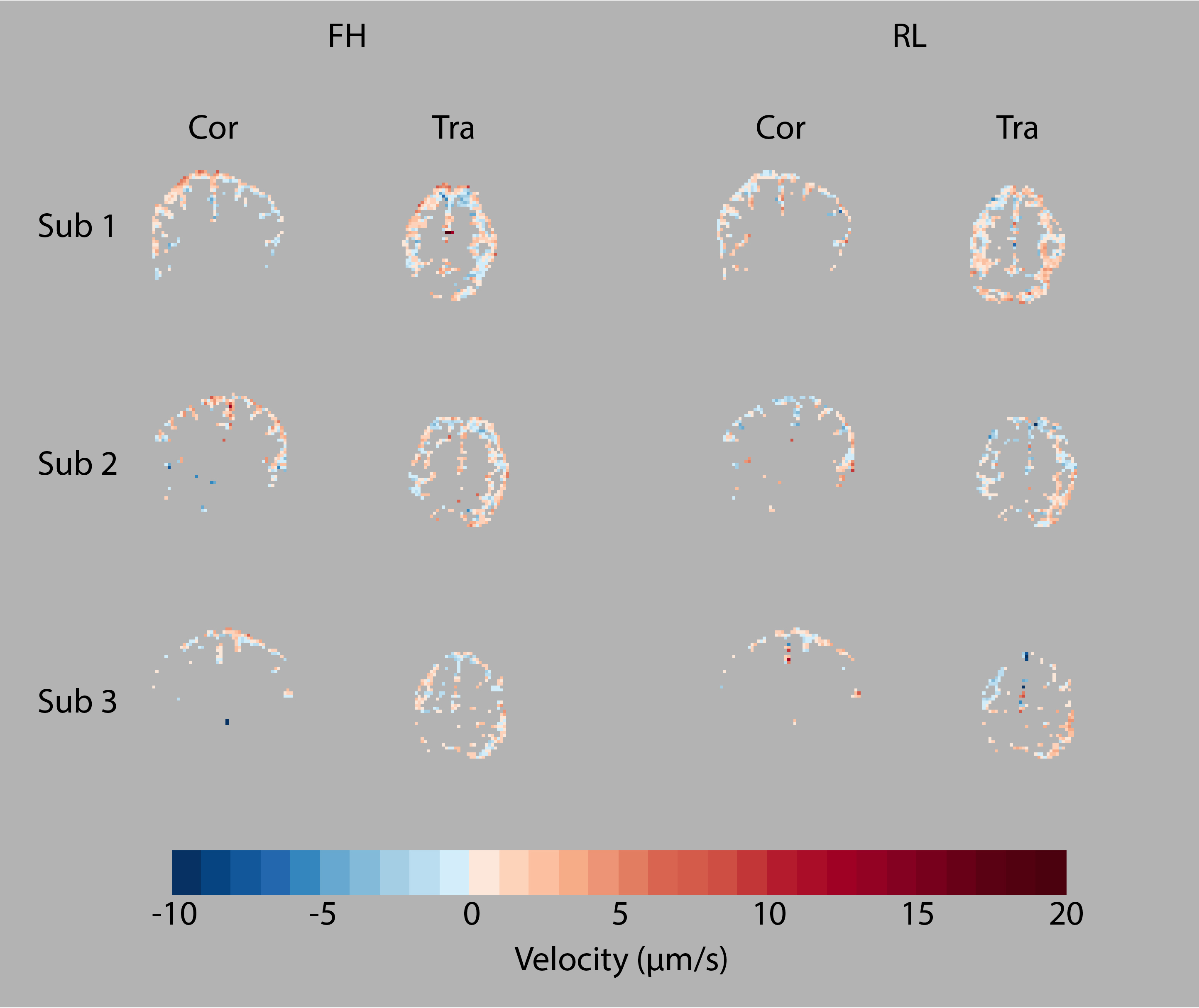
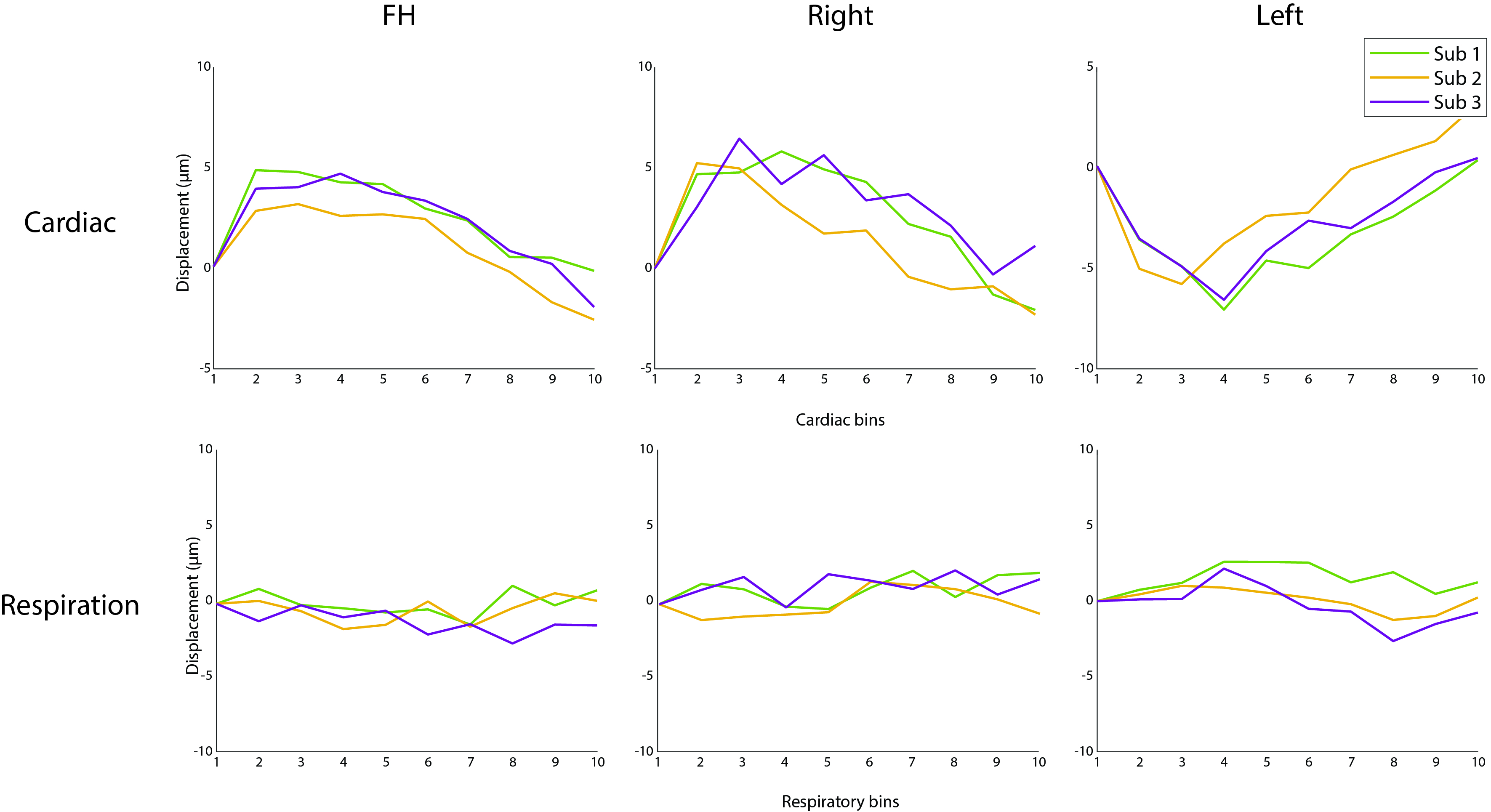
EC correction removed the EC gradients in the net velocity maps of the gel phantom (Fig 2) and the average net velocities were 0.08±0.90 µm/s (FH) and 0.03±1.72 µm/s (RL). Motion correction removed both involuntary motion as well as FH motion as a result of head rolling due to breathing (Fig 3). The average net velocity in FH direction over three subjects was 0.23±0.31 µm/s in the middle transversal part of the subarachnoid space where CSF motion direction is mainly in FH. The average net velocity for the left and right hemisphere were 0.81±0.50 µm/s and 0.46±0.35 µm/s respectively, measured in the upper part of the subarachnoid space (Fig 4). All subjects showed CSF pulsation upward and inward during the systolic phase but no clear CSF motion pattern over the respiratory cycle (Fig 5).Discussion and conclusion
Previously, we did not correct for head motion and EC given the observation that the latter was negligible in a relatively small static phantom. Using a larger phantom, we found that the presence of EC affects the net velocity measurements and, at the periphery of the phantom, is in the order of magnitude of previously measured net velocity values. Head motion corrections affects net velocities and physiological motion. The CSF net velocity found within the subarachnoid space was considerably lower than expected in both FH and RL direction. The intra-subject standard deviations for the velocity distributions were relatively small, indicating good measurement accuracy. The flow measured in the RL direction did not indicate the expected inward motion towards the sagittal sinus. CSF motion over the cardiac cycle shows a typical cardiac curve moving in cranial direction and inwards towards the ventricles. The respiratory cycle seems to have no measureable effect on CSF motion within the subarachnoid space. Validation using a moving slow flow phantom is needed to show that the current method is capable of measuring creep flows while accounting for other confounding factors.Acknowledgements
This research is funded by NWO VICI: Seismology of the brain (#18674)References
1. Mestre, Humberto, et al. "Flow of cerebrospinal fluid is driven by arterial pulsations and is reduced in hypertension." Nature communications 9.1 (2018): 4878.
2. Yamada, Shinya, et al. "Influence of respiration on cerebrospinal fluid movement using magnetic resonance spin labeling." Fluids and Barriers of the CNS 10.1 (2013): 1-7.
3. Williams, Stephanie D., et al. "Neural activity induced by sensory stimulation can drive large-scale cerebrospinal fluid flow during wakefulness in humans." PLoS Biology 21.3 (2023): e3002035.
4. Van der Voort, Elisabeth C., et al. “Assessing CSF secretion by measuring net velocity of CSF in the human subarachnoid space using displacement encoding with stimulated echoes at 7T.” In proceedings of ISMRM 2023 #3191
5. Sloots, Jacob Jan, et al. "Strain tensor imaging: Cardiac-induced brain tissue deformation in humans quantified with high-field MRI." NeuroImage 236 (2021): 118078.
Figures