4836
Multi-parametric Quantitative Magnetic Resonance Imaging Phantom1Biomedical Engineering, Vanderbilt University, Nashville, TN, United States, 2Vanderbilt University Institute of Imaging Science, Nashville, TN, United States, 3Radiology, University of Michigan, Ann Arbor, MI, United States, 4Radiology and Radiological Sciences, Vanderbilt University Medical Center, Nashville, TN, United States
Synopsis
Keywords: Phantoms, Phantoms, multiparametric
Motivation: Currently there is no qMRI phantom that simultaneously reproduces the many contrasts that are present in white matter.
Goal(s): The goal of this work is to characterize a single multiparametric qMRI phantom that exhibits diffusion, magnetization transfer and MET2 properties.
Approach: Aqueous MTF and PVP were combined in varying concentrations and evaluated with both NMR and MRI.
Results: A homogenous, multiparametric qMRI phantom was created with an array of tissue relevant parameters: T2L ranging from 71.9-183 ms, T2s ranging from 13.6-17.7 ms, fs ranging from 0.118-0.177, T1free ranging from 1479-1919 ms and fm ranging from 0.053-0.109.
Impact: A multiparametric phantom allows for improved quality assurance analysis, as well as sequence development by allowing for multiple contrasts to be evaluated simultaneously. Ultimately providing a sample that is controlled, yet more akin to what occurs in biological samples.
Background and Significance
Many tunable quantitative MRI phantoms are available that have characteristics such as monoexponential relaxation1–5, biexponential relaxation6,7, magnetization transfer (MT)8, and diffusion1,2,9,10. Nonetheless, no phantom currently exists that simultaneously reproduces these properties akin to the physiological environment.Aqueous polyvinylpyrrolidone (PVP) is an established diffusion phantom10, and here we demonstrate that it also exhibits multi-exponential transverse relaxation (MET2) similar to myelinated tissue. Another material (69% Cetearyl alcohol, 13.8% SAPDA, 17.2% BTAC), that we’ve named MTF, also impacts water diffusion and exhibits an MT effect. Combined PVP and MTF mixtures are presented here as phantoms that simultaneously possess water diffusion, MT, and MET2 characteristics consistent with white matter and nerve.
Methods
Phantom Preparation: PVP and MTF were combined with deionized water in seven relative mass combinations, including two reference phantoms made from either PVP or MTF alone. Each sample was warmed and mixed to create a pourable solution that was pipetted into NMR or Eppendorf tubes and centrifuged for five minutes at 5,000 rpm. Three independent batches were prepared to assess repeatability.1H NMR: Nonlocalized multiple spin echo (MSE) and selective inversion recovery (SIR) sequences were performed at 4.7 T to assess MET2 and quantitative MT (qMT) characteristics, respectively.
MRI: MSE, SIR and diffusion-weighted spin echo imaging was performed on one batch of phantoms at 7 T for imaging assessments of MET2, qMT, and diffusion.
Results
Analysis of the nonlocalized MSE data produced T2 spectra for each of the five phantoms (Figure 1). Each phantom shows a short-lived signal (T2 < 30 ms) and a spread of longer-lived signals (T2 > 30) ms. The geometric mean T2s of each regime (T2s and T2L respectively) and short-lived signal fraction (fs) are reported in Table 1. Nonlocalized SIR data was analyzed in terms of a coupled two-pool system, resulting in a free proton pool T1 (T1free) and macromolecular proton fraction (fm) (Table 1). Similarly, Figure 2 and Table 2 show the T2 spectra and corresponding MET2 , MT, and diffusion metrics derived from the imaging experiments.Discussion
The nonlocalized data analysis of the two reference phantoms (15%/0% PVP/MTF and 5.4/0% MTF/PVP, Table 1) demonstrates the primary roles of each substance in the combined phantoms. Specifically, the MTF-only phantom exhibited a substantial MT effect (fm = 0.078) but essentially mono-exponential transverse relaxation (fs = 0), while the PVP-only phantom showed a sizeable short-T2 signal (fs = 0.097, T2s = 21 ms) and negligible MT effect (fm = 0.014). When combined, the T2-spectra become somewhat more complicated, but the key T2 and MT metrics follow predictable trends, as shown in Figure 2. In particular, fs varies approximately linearly with the mass ratio of PVP and H20; fm varies almost directly with the mass ratio of MTF and H20, and the dominant R1 = (1/T1) varies linearly with total solute mass fraction.A similar story is told from the imaging experiments (Figure 3 and Table 2) except that fs is more heavily influenced by the MTF. This result is likely a consequence of the limitation of the multiple exponential signal inversion given the lower SNR in the image data (≈300 vs ≈1000 in NMR). Importantly though, the MET2, MT, and diffusion metrics (Table 2) are all within the range of such values found in white matter and nerve, making more or less any combination of PVP and MTF within range of tested concentrations suitable as a multiparametric qMRI phantom.
Finally, we note that these phantoms are derived from relatively inexpensive materials and are easy to make. In particular, no cross-linking step is needed for the MTF to produce a measurable MT effect, and the final products are viscous but pourable. Furthermore, the quantitative properties of these phantoms are stable across a month’s time, and as indicated by the standard deviations across three batches, are repeatable.
Conclusion
In conclusion, MTF and PVP can be combined to create a single, homogenous multi-parametric qMRI phantom with tissue relevant parameters.Acknowledgements
Funding: National Science Foundation Graduate Research Fellowship, Vanderbilt University School of Engineering Faculty Funds.
References
1. Laubach, H. J. et al. A Phantom for diffusion-weighted imaging of acute stroke. Journal of Magnetic Resonance Imaging 8, 1349–1354 (1998).
2. Keenan, K. E. et al. Design of a breast phantom for quantitative MRI. J Magn Reson Imaging 44, 610–619 (2016).
3. Kato, H. et al. Composition of MRI phantom equivalent to human tissues. Med Phys 32, 3199–3208 (2005).
4. Christoffersson, J. O., Olsson, L. E. & Sjöberg, S. Nickel-doped agarose gel phantoms in MR imaging. Acta radiol 32, 426–431 (1991).
5. Blechinger, J. C., Madsen, E. L. & Frank, G. R. Tissue-mimicking gelatin–agar gels for use in magnetic resonance imaging phantoms. Med Phys 15, 629–636 (1988).
6. Horch, R. A. & Does, M. D. Aqueous urea as a model system for bi-exponential relaxation. MAGMA 20, 51–56 (2007).
7. Jones, C., MacKay, A. & Rutt, B. Bi-Exponential T2 Decay in Dairy Cream Phantoms. Magn Reson Imaging 16, 83–85 (1998).
8. Koenig, S. H., Brown, R. D. & Ugolini, R. Magnetization transfer in cross-linked bovine serum albumin solutions at 200 MHz: A model for tissue. Magn Reson Med 29, 311–316 (1993).
9. Wang, X., Reeder, S. B. & Hernando, D. An Acetone-Based Phantom for Quantitative Diffusion MRI. J Magn Reson Imaging 46, 1683–1692 (2017).
10. Pierpaoli, C., Sarlls, J., Nevo, U., Basser, P. J. & Horkay, F. Polyvinylpyrrolidone (PVP) water solutions as isotropic phantoms for diffusion MRI studies. Intl. Soc. Mag. Reson. Med 17, (2009).
Figures
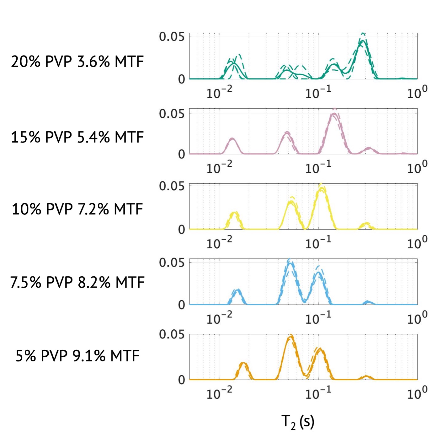
Figure 1: T2 spectra of PVP/MTF samples evaluated via NMR at 4.7 T. Solid lines represent the mean spectra across three separate batches and the dotted lines are the spectra of each individual batch.
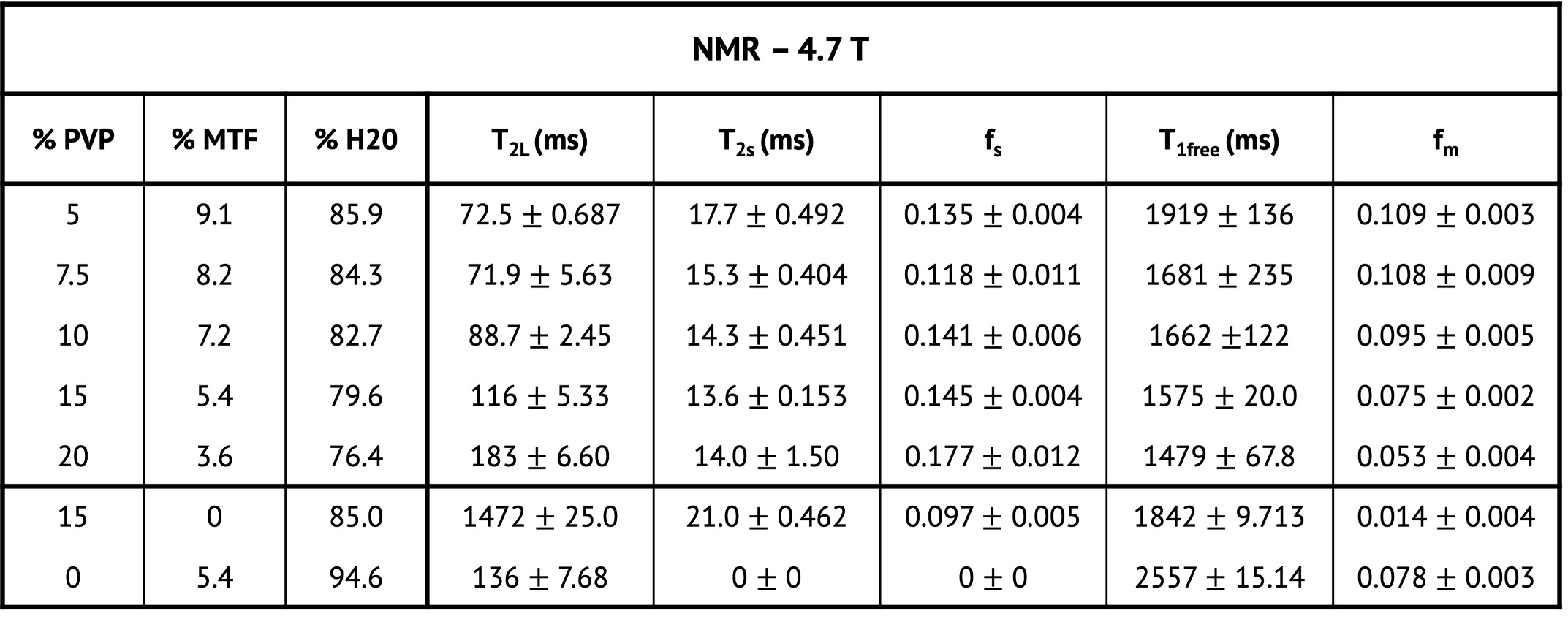
Table 1: Mean parameters ± the standard deviations across three separate batches are reported for the quantitative parameters derived from the data acquired via NMR acquisitions at 4.7 T.
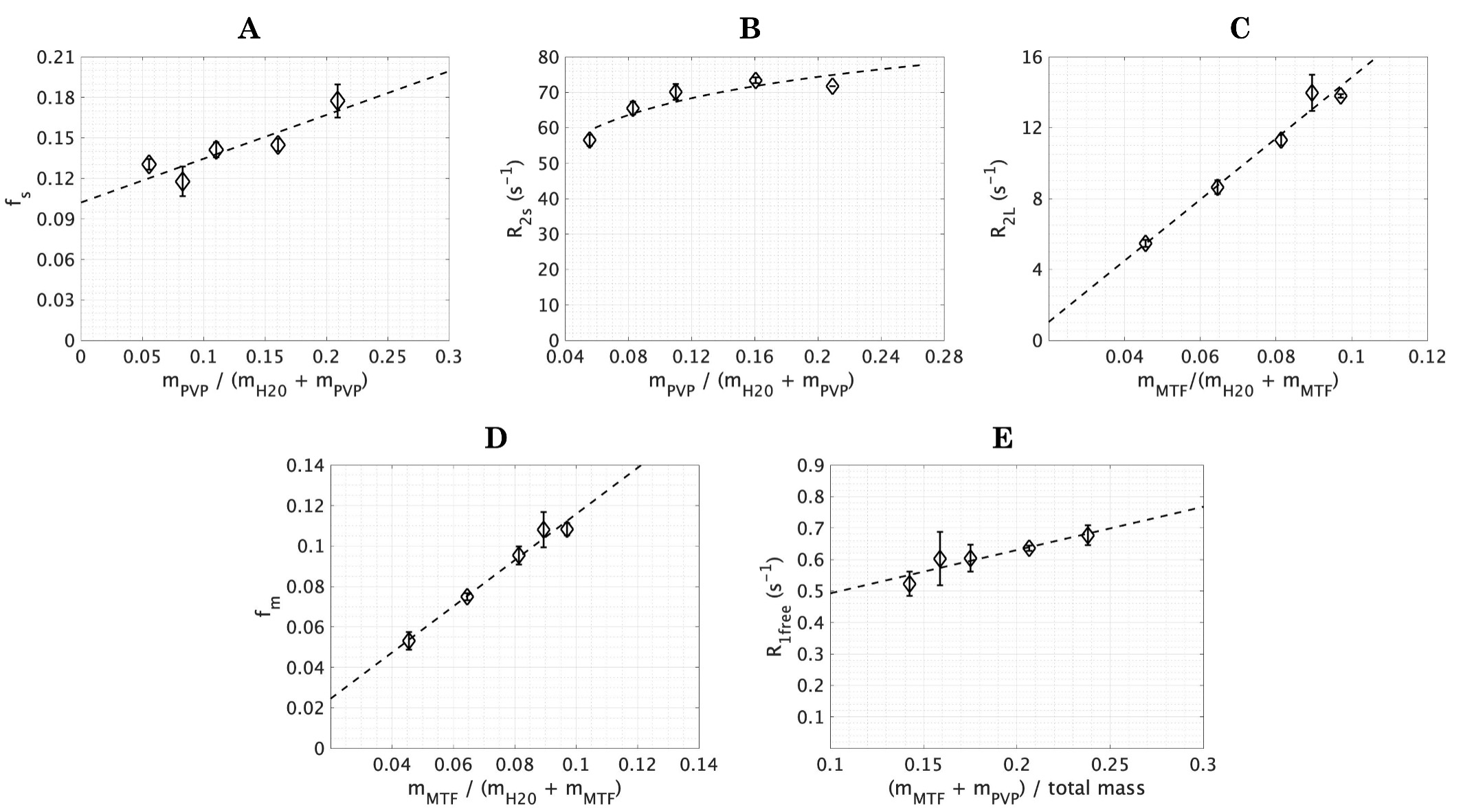
Figure 2: Relationships between the quantitative parameters derived from NMR analyses and phantom composition are demonstrated. A linear fit is shown with the dashed line in A and C-E. A log fit is shown with the dashed line in B. Each data point is represented by the respective shapes and the error bars represent the standard deviations across the three separate batches.
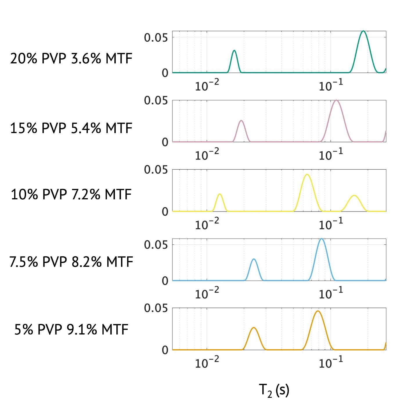
Figure 3: T2 spectra of PVP/MTF samples evaluated via MRI at 7 T.
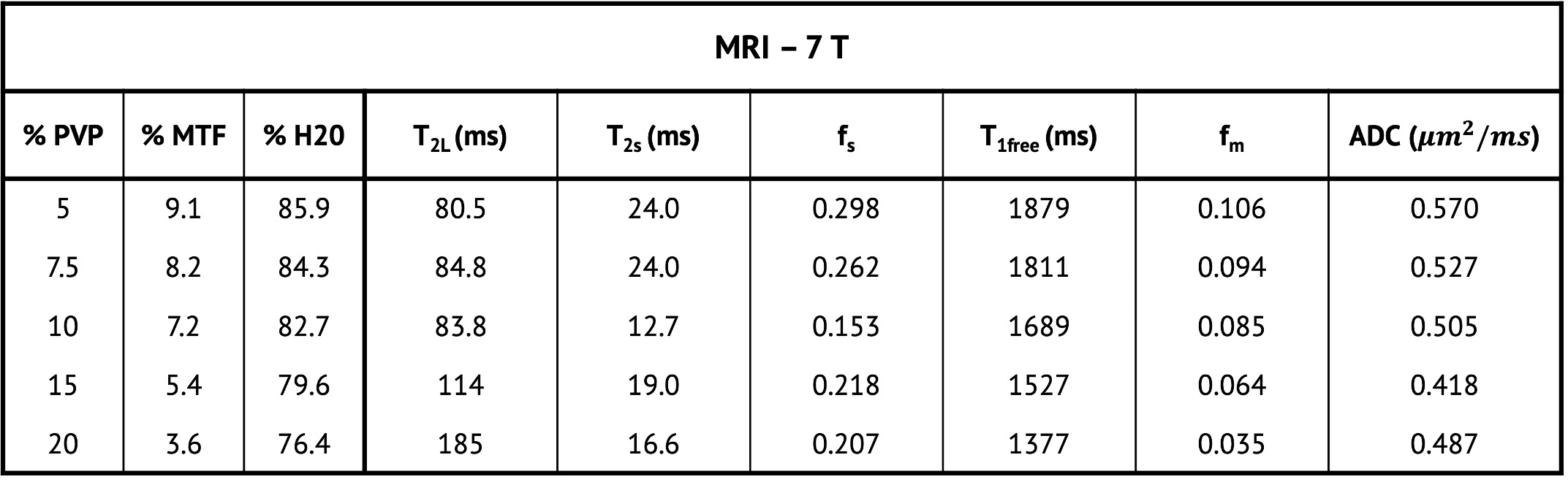
Table 2: Quantitative parameters derived from the data acquired via MRI acquisitions at 7 T.