4835
Elastic modulus of acetabular labrum correlates with UTE apparent spin-spin (T2*) and spin-lattice (T1) magnetic resonance relaxation times1Radiology, University of California San Diego, San Diego, CA, United States, 2Academia-Government-Industry Collaboration, Office of Research and Academia-Government-Community Collaboration Hiroshima University, Hiroshima, Japan, 3Orthopedic, Shiley Center for Orthopedic Research and Education at Scripps Clinic, San Diego, CA, United States, 4Pathology, University of California San Diego, San Diego, CA, United States, 5Radiology, Radiology, University of California San Diego, San Diego, CA, United States, San Diego, CA, United States
Synopsis
Keywords: High-Field MRI, Cartilage, Acetabular Labrum, UTE
Motivation: The acetabulum labrum plays a critical role in the hip function. Medical imaging techniques for detecting labral degeneration may help improve our knowledge of its role in hip osteoarthritis (OA).
Goal(s): This study aimed to explore the correlation between the mechanical properties of the acetabulum labrum and MRI properties.
Approach: The correlations of UTE-T2* and -T1 sequences with the tensile elastic modulus of human acetabular labrum specimens have been investigated.
Results: There was a significant correlation between quantitative UTE-T2*, and UTE-T1 techniques and the mechanical properties of the labrum.
Impact: UTE-T2* and -T1 sequences showed the potential to evaluate acetabulum labrum mechanical assessment, which is needed to improve labrum degeneration detection and monitoring, a primary unmet need in areas where hip osteoarthritis is common and conventional MRI is being implemented.
Background
The acetabular labrum is a C-shaped fibrocartilaginous rim that outlines the bony acetabulum’s ring in the hip joint 1. It is crucial in maintaining normal hip joint biomechanics and function 2,3.Labral damage can lead to abnormal joint mechanics, increasing the risk of osteoarthritis (OA) 7–9. Therefore, a non-invasive assessment of the labrum quality and its mechanical status may help detect OA-related changes in the hip joint.
The labrum appears dark on conventional T1- and T2-weighted MR images due to its short spin-spin (T2) relaxation time 11,12. Therefore, employing ultrashort echo time (UTE) MRI sequences may be suitable for quantitative labrum imaging. Apparent spin-spin (T2*) and Spin-lattice (T1) relaxation times are common quantitative MRI techniques used for musculoskeletal tissue evaluation 13,14, which have been investigated in this study.
Methods
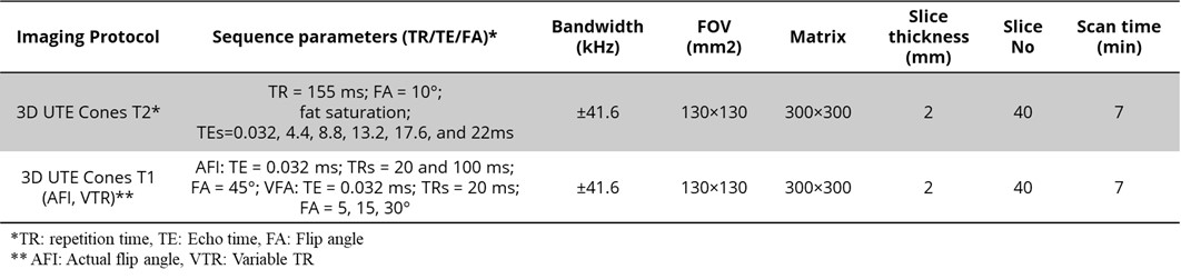
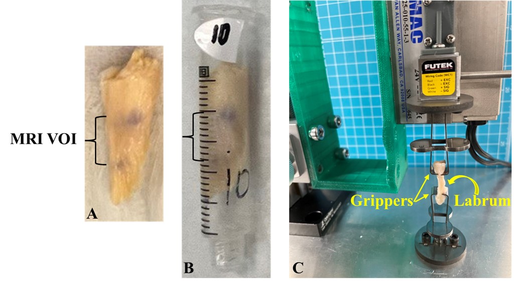
Acetabular labrum specimens were dissected from 12 fresh-frozen human cadaveric hip joints (64.6±11.6 years old at the time of death, 7 female). All specimens were placed separately in 5-ML syringes for MR imaging (Fig 1A-B). The dissected acetabular labrum specimens were soaked in phosphate-buffered saline for 1 hour before scanning. All syringes were filled with perfluoropolyether (Fomblin, Ausimont, USA) to minimize dehydration and susceptibility artifacts during MRI scans.The UTE-MRI scans were performed on a 3T MRI scanner (MR750, GE Healthcare Technologies, USA) using a standard 8 channels transmit/receive knee coil. All MRI images were acquired in the axial plane. Two following sets of 3D UTE Cones MRI sequences were performed, 1) 3D UTE Cones T2* sequence with multiple TEs to measure T2* values and 2) actual flip angle (FA) sequences with variable FA (AFA-VFA)-based 3D UTE Cones to measure T1 values 15. Data acquisition parameters for the two quantitative UTE-MRI protocols are presented in Table 1.
UTE-MRI biomarkers were calculated in three representative slices at the middle of each specimen (volume of interest (VOI) is indicated in Figure 1A) within global regions of interest (ROIs) covering the entire cross-sectional area. For each slice, average signal values within the ROI were used for T2* and T1 measurements using single-component exponential fitting models developed in MATLAB (The Mathworks Inc., Natick, USA).
The stiffness and the elastic modulus (E) of the specimens were measured before MRI scans. The mechanical setup with a representative-mounted acetabular labrum specimen is shown in Figure 1C. Both ends of the specimens were mounted on the stainless-steel grippers of an in-house developed benchtop uniaxial loading device. The initial distance between the fixed and actuated gripper was set to 10 mm. After initial alignment and 0.02 newton (N) preloading to straighten the specimens, a 1-mm tensile displacement (maximum strain = 0.1 N) was applied at a 0.3 mm/sec rate, and the applied force was recorded. The average maximum stiffness and stress were calculated as the maximum force divided by the tissue’s effective length (10 mm) and cross-sectional area, respectively. The average tensile elastic modulus was calculated as the average maximum stress divided by the average maximum strain.
The Spearman’s rank correlations were calculated between average UTE-MRI quantifications and the acetabular labrum mechanical properties. Statistical analyses were performed in SPSS. P values below 0.05 were considered significant.
Results
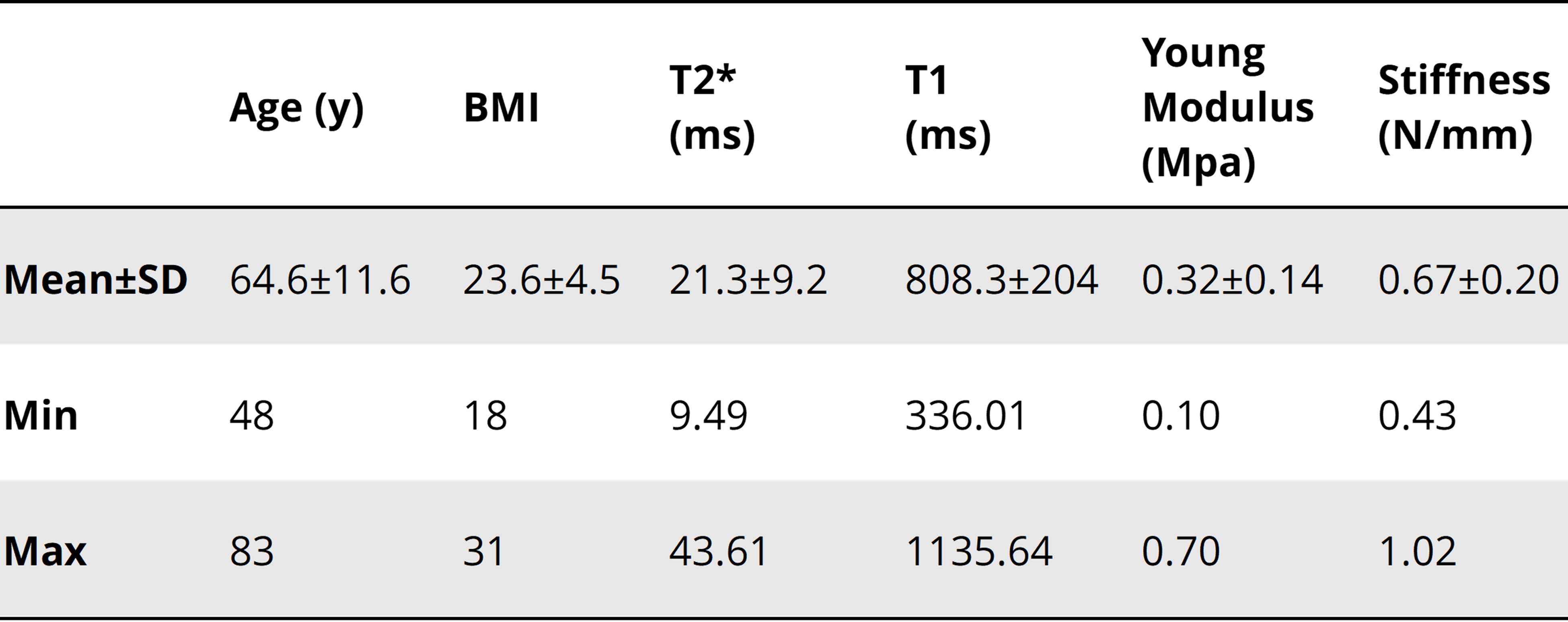
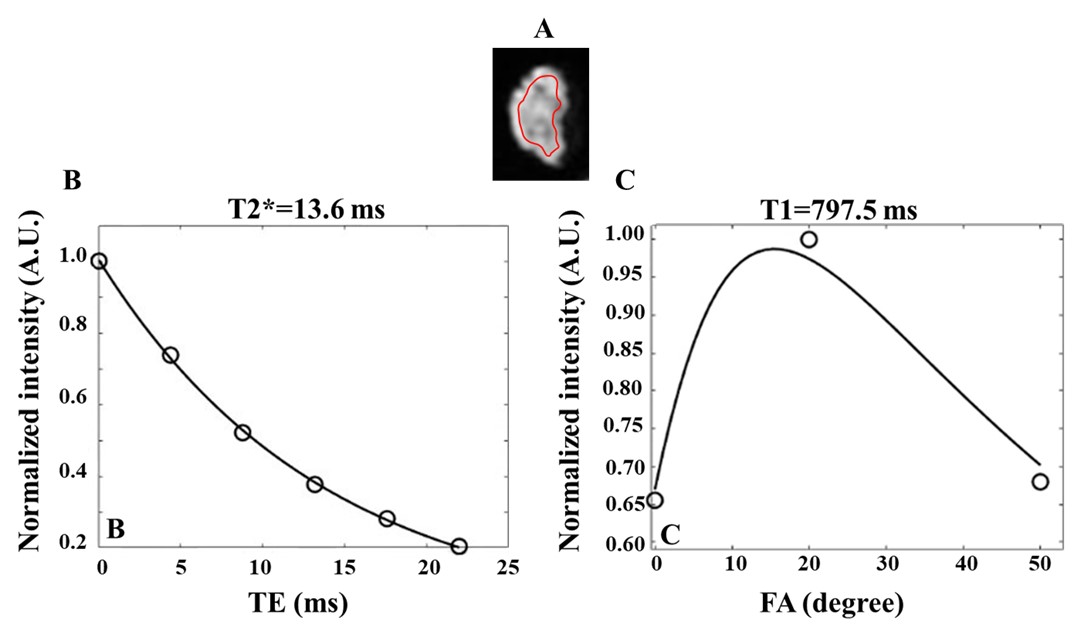
Figure 2 shows single-component exponential T2* and T1 fittings for a representative human acetabular labrum specimen (from a 68-year-old male donor). The high signal obtained by UTE MRI leads to a well-fitting of the actual data points.Table 2 summarizes the mean, standard deviation (SD) of the age, BMI, and UTE-T2* and -T1 values and mechanical properties in the studied acetabular labrum specimens.
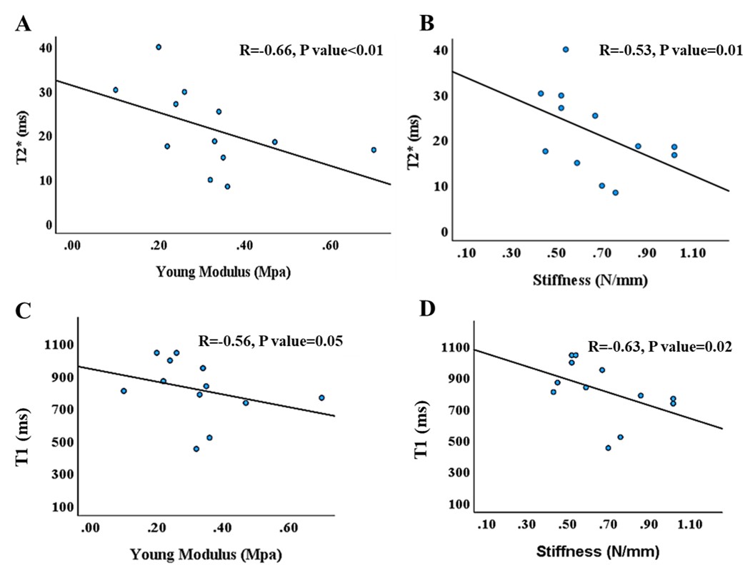
Figure 3 shows the scatter plots, linear regression trendlines, and Spearman’s rank correlation coefficients of the elastic modulus and stiffness on UTE-T2* and UTE-T1 techniques. Elastic modulus showed a significant inverse correlation with UTE-T2* (R=-0.66, P<0.01) and with UTE-T1 (R=-0.56, P=0.05). The stiffness of the specimens showed significant inverse correlations with UTE-T2* (R=-0.53, P=0.01) and UTE-T1 (R=-0.63, P=0.02).
Conclusion
Mechanical properties of the acetabular labrums showed significant inverse correlations with UTE-T2* and UTE-T1. This study highlighted the potential of UTE-MRI techniques for the acetabular labrum mechanical assessment which can help improve labrum degeneration detection and monitoring which requires further investigations.Acknowledgements
The authors acknowledge grant support from NIH (K01AR080257, R01AR079484) and JSPS KAKENHI (JP19K09658, JP18KK0104).References
1. Seldes RM, Tan V, Hunt J, Katz M, Winiarsky R, Fitzgerald RH. Anatomy, histologic features, and vascularity of the adult acetabular labrum. Clin Orthop. 2001;(382):232-240. doi:10.1097/00003086-200101000-000312. Crawford MJ, Dy CJ, Alexander JW, et al. The 2007 Frank Stinchfield Award. The biomechanics of the hip labrum and the stability of the hip. Clin Orthop. 2007;465:16-22. doi:10.1097/BLO.0b013e31815b181f
3. Effects of traction, distension, and joint position on distraction of the hip joint: An experimental study in cadavers — Luxembourg Institute of Health. Accessed October 27, 2023. https://researchportal.lih.lu/en/publications/effects-of-traction-distension-and-joint-position-on-distraction-
4. Seldes RM, Tan V, Hunt J, Katz M, Winiarsky R, Fitzgerald RH. Anatomy, histologic features, and vascularity of the adult acetabular labrum. Clin Orthop. 2001;(382):232-240. doi:10.1097/00003086-200101000-00031
5. Grant AD, Sala DA, Davidovitch RI. The labrum: structure, function, and injury with femoro-acetabular impingement. J Child Orthop. 2012;6(5):357-372. doi:10.1007/s11832-012-0431-1
6. Nogami R, Kaku N, Shimada T, Tabata T, Tagomori H, Tsumura H. Three-dimensional architecture of the acetabular labrum in the human hip joint. Med Mol Morphol. 2020;53(1):21-27. doi:10.1007/s00795-019-00228-3
7. Roemer FW, Hunter DJ, Winterstein A, et al. Hip Osteoarthritis MRI Scoring System (HOAMS): reliability and associations with radiographic and clinical findings. Osteoarthritis Cartilage. 2011;19(8):946-962. doi:10.1016/j.joca.2011.04.003
8. Hip morphology influences the pattern of damage to the acetabular cartilage | Bone & Joint. Accessed October 29, 2023. https://boneandjoint.org.uk/article/10.1302/0301-620x.87b7.15203
9. McCarthy JC, Noble PC, Schuck MR, Wright J, Lee J. The Otto E. Aufranc Award: The role of labral lesions to development of early degenerative hip disease. Clin Orthop. 2001;(393):25-37. doi:10.1097/00003086-200112000-00004
10. Neumann G, Mendicuti AD, Zou KH, et al. Prevalence of labral tears and cartilage loss in patients with mechanical symptoms of the hip: evaluation using MR arthrography. Osteoarthritis Cartilage. 2007;15(8):909-917. doi:10.1016/j.joca.2007.02.002
11. A comprehensive review of hip labral tears | Current Reviews in Musculoskeletal Medicine. Accessed October 29, 2023. https://link.springer.com/article/10.1007/s12178-009-9052-9
12. UTE imaging in the musculoskeletal system - PubMed. Accessed October 29, 2023. https://pubmed.ncbi.nlm.nih.gov/25045018/
13. Clinical use of the partial saturation and saturation recovery sequences in MR imaging - PubMed. Accessed October 29, 2023. https://pubmed.ncbi.nlm.nih.gov/4056131/
14. Du J, Carl M, Bydder M, Takahashi A, Chung CB, Bydder GM. Qualitative and quantitative ultrashort echo time (UTE) imaging of cortical bone. J Magn Reson San Diego Calif 1997. 2010;207(2):304-311. doi:10.1016/j.jmr.2010.09.013
15. Accurate T1 Mapping of Short T2 Tissues Using a Three-Dimensional Ultrashort Echo Time Cones Actual Flip Angle Imaging Variable Repetition Time (3D UTE-Cones AFI-VTR) Method - PMC. Accessed May 12, 2023. https://www.ncbi.nlm.nih.gov/pmc/articles/PMID29314235/
16. Short T2 imaging using a 3D double adiabatic inversion recovery prepared ultrashort echo time cones (3D DIR‐UTE‐Cones) sequence - Ma - 2018 - Magnetic Resonance in Medicine - Wiley Online Library. Accessed October 29, 2023. https://onlinelibrary.wiley.com/doi/10.1002/mrm.26908
Figures