4828
Improved QSM Pipeline to Investigate the Effect of Sickle Cell Anaemia on Brain Magnetic Susceptibility in Tanzanian Children at 1.5 Tesla1Department of Medical Physics and Biomedical Engineering, University College London, London, United Kingdom, 2Department of Radiology & Imaging, Muhimbili University of Health and Allied Sciences, Dar Es Salaam, Tanzania, 3Imaging and Biophysics, Developmental Neurosciences, UCL Great Ormond Street Institute of Child Health, London, United Kingdom
Synopsis
Keywords: Gray Matter, Gray Matter
Motivation: Sickle cell anaemia (SCA) is a major global health burden, but disease mechanisms in the brain are not well understood.
Goal(s): To improve a quantitative susceptibility mapping (QSM) pipeline and apply it to updated data to investigate brain susceptibility differences between SCA patients and controls. To investigate correlations between blood haemoglobin and brain magnetic susceptibility.
Approach: QSM was optimised using denoising/masking approaches. Linear regressions of susceptibility against log(age) were used to compare age-corrected susceptibility in grey matter structures across the age range and correlate with haemoglobin.
Results: Susceptibility increases with age differently for SCA vs controls. Haemoglobin was not significantly correlated with susceptibility.
Impact: This work provides novel insight into the relationship between grey matter magnetic susceptibility and sickle cell anaemia, demonstrating differential trajectories with age between SCA patients and healthy controls. This may support the view of SCA as an accelerated aging syndrome.
Introduction
Sickle cell anaemia (SCA) is a genetic disorder which leads to polymerization, or ‘sickling’, of haemoglobin. It has wide-ranging symptoms including in the brain, causing stroke, pain crises and other neurocognitive complications1. This work aims to improve and build on a prior analysis investigating changes in brain magnetic susceptibility in paediatric SCA patients2. Here, we improved QSM processing and performed a more extensive analysis of the effect of age on brain susceptibility in SCA patients compared to controls. We also investigated the relationship between blood haemoglobin and brain magnetic susceptibility.Methods
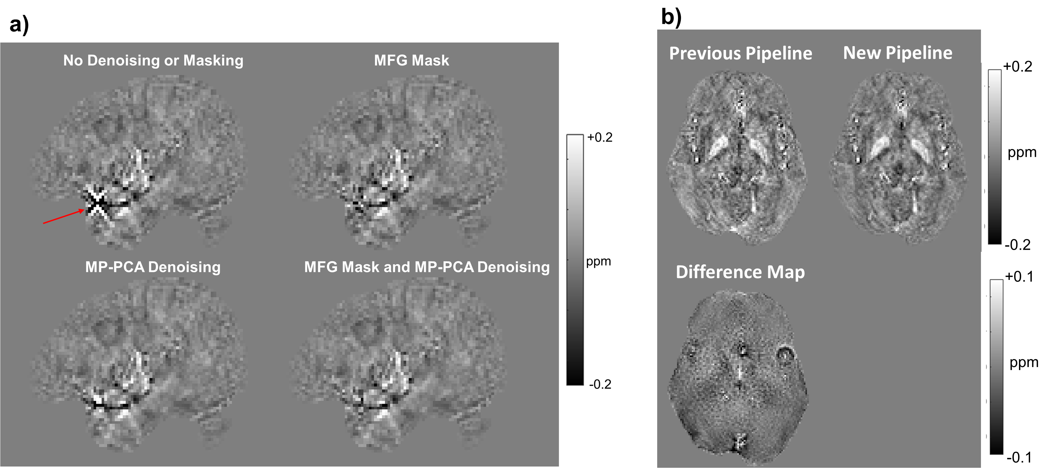
163 children with SCA and 47 healthy controls (aged 13.2 ± 3.8 and 10.9 ± 3.4 years, respectively) were imaged with a T2*-weighted multi-echo 3D GRE sequence and a T1-weighted MPRAGE sequence. All images were acquired at the Muhimbili National Hospital on a 1.5T Phillips Achieva system using either an 8-channel RF coil or a birdcage coil. ME-GRE sequence parameters are shown in Figure 1.A QSM pipeline was optimised based on a previous pipeline2. Key changes to the pipeline are: Tilt correction was introduced to reduce artifacts arising from oblique acquisition3; MP-PCA denoising was applied to reduce structured noise such as motion artifacts4; A magnitude-of-field gradients (MFG) masking approach was applied to reduce streaking artifacts5. This removed voxels with a total field map gradient magnitude more than ten standard deviations greater than the mean gradient magnitude. PDF background field removal was replaced with V-SHARP6, which was observed to better reduce residual background fields.
Region of interest segmentations were obtained jointly from the QSMs and coregistered T1-weighted images using MRICloud6.
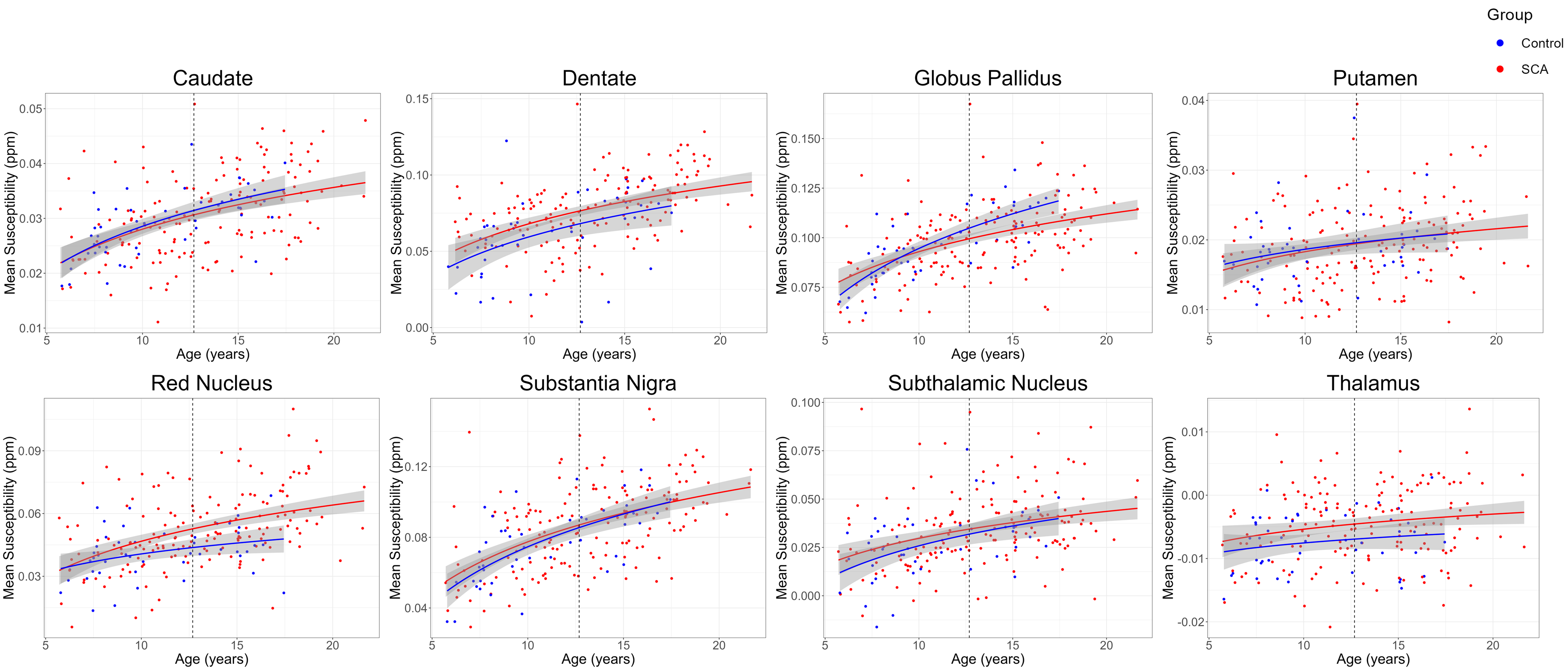
Susceptibility variation with age was investigated by calculating linear regressions of susceptibility as a function of log-transformed age for each region of interest8. This was applied separately to SCA patients and controls, to investigate any effect of SCA status on the age-related progression of susceptibility. The fitted regressions were used to apply age-correction to the susceptibility values. This was done by defining a reference age and transforming each measured susceptibility by adding a correction factor given by the difference of each subject’s log-transformed age to the log-transformed reference age, multiplied by the slope of the linear regression against log(age).
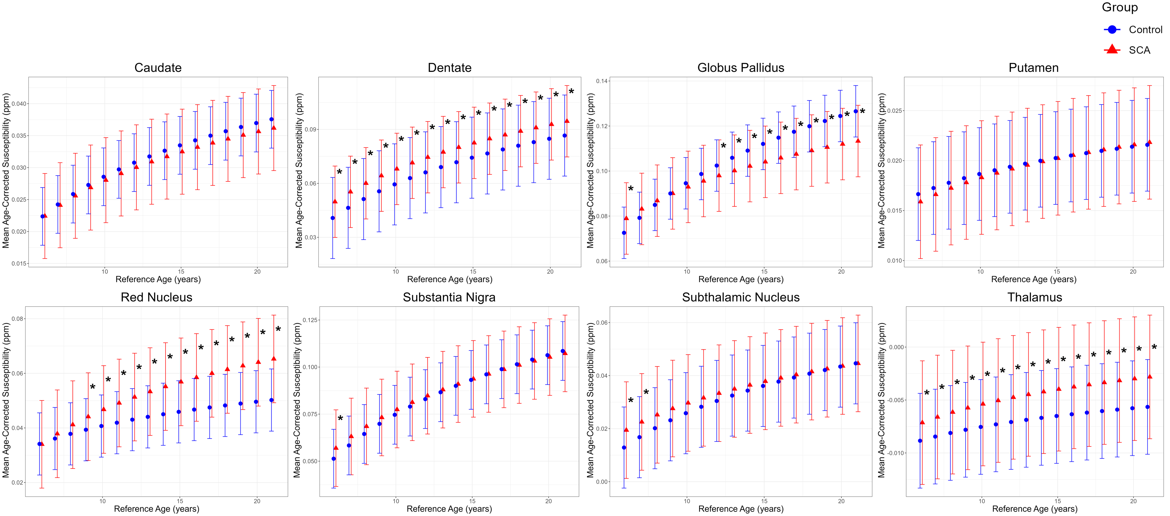
To investigate the effect of the choice of reference age, groupwise comparisons (two-sample t-tests) were carried out between SCA patient and control ROI age-corrected susceptibilities for a range of reference ages that reflected the age range of the cohort (6 to 21 years in steps of 1 year).
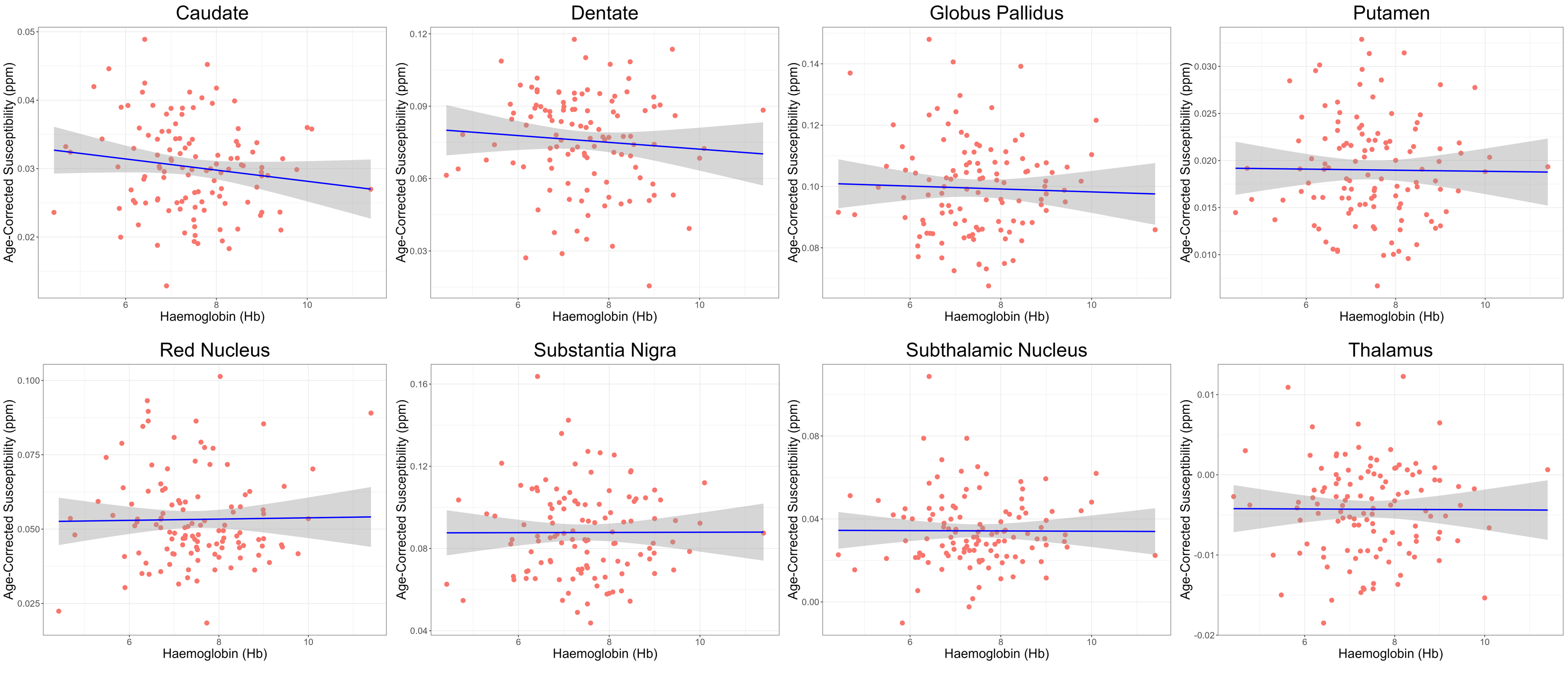
For the 117 SCA patients with blood haemoglobin measurements, linear regressions of age-corrected susceptibility (corrected to 12.7 years - the mean age of the cohort) against blood haemoglobin were calculated.
Results
Figure 2 demonstrates the effect of MP-PCA denoising and MFG-threshold masking on a streaking artifact often seen in QSM. Both techniques reduce the artifact, and combining them appears to reduce streaking most effectively.Figure 3 shows plots of mean ROI susceptibility as a function of age. The ROI mean susceptibility in all regions considered was found to be significantly correlated with log-transformed age in both controls and SCA patients.
Figure 4 shows the groupwise comparisons between age-corrected susceptibility in SCA patients and controls for each ROI as a function of correction reference age. Due to the differing trajectories observed in SCA patients and controls in some regions (e.g globus pallidus), the observed differences vary significantly with the choice of reference age. Other regions (e.g putamen) have very similar trajectories in SCA and controls, so no significant differences are observed across the age range.
Figure 5 shows linear regressions of age-corrected susceptibility against haemoglobin for each ROI. No correlations were significant, but negative trends were observed in the caudate and dentate.
Discussion
Susceptibility increased with age in all regions as expected9. Controls and SCA patients show similar susceptibility trajectories across age in some grey matter ROIs and different trajectories in others. This may relate to changes in iron accumulation in specific regions resulting from disease processes and is consistent with recent understanding of SCA as an accelerated aging syndrome10. A lack of correlation between susceptibility and haemoglobin has been observed in previous work11. Future work will consider relationships between susceptibility and other clinical variables, including cognitive scores and pain episodes.Conclusion
We used an optimised QSM pipeline to investigate changes in grey matter magnetic susceptibility with age in SCA patients and controls. The choice of reference age used to compare age-corrected susceptibility values between groups is important, considering different trends with age between SCA patients and controls. Haemoglobin was not correlated with susceptibility.Acknowledgements
This work is supported by the EPSRC-funded UCL Centre for Doctoral Training in Intelligent, Integrated Imaging in Healthcare (i4health) (EP/S021930/1).References
[1] Piel, F et al. , “Sickle Cell Disease”. New England Journal of Medicine, 376(16) (2017): 1561–1573
[2] Lee et al. , “Investigating Regional Changes in Brain Magnetic Susceptibility in Tanzanian Children with Sickle Cell Anaemia”, Proceedings of the ISMRM Annual Meeting (2023): 7905
[3] Kiersnowski, O et al. “Investigating the effect of oblique image acquisition on the accuracy of QSM and a robust tilt correction method”. Magn Reson Med. 89 (2023): 1791-1808
[4] Doniza, L et al. , “MP-PCA image denoising technique for high resolution quantitative susceptibility mapping (QSM) of the human brain in vivo”, Proceedings of the ISMRM Annual Meeting (2023): 2948
[5] Kiersnowski , O and Shmueli, K, “A Novel Gradient-Based Thresholding Method to Improve Brain Masking for Quantitative Susceptibility Mapping”, BIC-ISMRM Postgraduate Symposium (2023): P2H1
[6] Li, W et al. ,“Quantitative susceptibility mapping of human brain reflects spatial variation in tissue composition”. Neuroimage, (2011): 1645–1656
[7] Li, X et al. “Multi-atlas tool for automated segmentation of brain gray matter nuclei and quantification of their magnetic susceptibility”. Neuroimage, 191 (2019): 337-349
[8] Li W, et al. Differential developmental trajectories of magnetic susceptibility in human brain gray and white matter over the lifespan. Hum Brain Mapp. 35(6) (2014): 2698-2713
[9] Miao, X et al. , “Increased brain iron deposition in patients with sickle cell disease: an MRI quantitative susceptibility mapping study”. Blood, 132 (2018): 1618, 10
[10] Idris, IM et al. , “Sickle cell disease as an accelerated aging syndrome”. Exp Biol Med, 247(4) (2022): 368-374
[11] Murdoch, R, “Developing MRI Quantitative Susceptibility Mapping to Investigate the Effect of Sickle Cell Anaemia on Brain Magnetic Susceptibility”, PhD thesis, University College London, 2022
Figures