4821
The value of IVIM-DWI and DCE-MRI in predicting molecular subtypes of breast cancer1The First Affiliated Hospital of USTC(Anhui Provincial Cancer Hospital), Hefei, China
Synopsis
Keywords: Breast, Cancer, molecular subtypes
Motivation: To analyze the value of imaging examination in accurate diagnosis of breast cancer.
Goal(s): Provide an non-invasive examination for the prediction of molecular subtypes of breast cancer before treatment.
Approach: The quantitative parameters of IVIM-DWI and DCE-MRI in patients with breast cancer without any invasive examination and treatment were compared with pathological molecular subtypes, so as to analyze their diagnostic value in the prediction of molecular subtypes.
Results: DCE-MRI and IVIM-DWI were correlated with immune prognostic factors of breast cancer and had differential diagnostic value for different molecular subtypes.
Impact: DCE-MRI and IVIM-DWI could provide a non-invasive diagnostic method for predicting molecular subtypes of breast cancer and provide a reference for further development of personalized treatment plans.
Introduction
Due to the high heterogeneity of breast cancer at the molecular level, it has been found that the biological behavior, therapeutic response and prognosis of breast cancer could be various even with the same clinical stage and histopathological type1. In the current context of precision diagnosis and treatment, clinicopathologic molecular subtypes were widely used in the accurate diagnosis, protocol formulation and treatment response evaluation of breast cancer. IHC is the gold standard for determination of molecular subtypes. Preoperative puncture biopsy and postoperative pathology are the ways to obtain pathological specimens, so the determination of preoperative molecular subtypes depends on the results of puncture biopsy which is invasive, and has potential risks such as infection, bleeding and needle path transmission and underestimation was common2. Therefore, many scholars have discussed the value of imaging examination in accurate diagnosis of breast cancer before treatment through a large number of studies. The quantitative parameters of DCE and IVIM could reflect the microenvironment of tumor. This study sought to explore the value of IVIM-DWI and DCE-MRI in predicting the molecular subtypes of breast cancer.Methods
A total of 187 patients with breast cancer admitted to our hospital from March, 2019 to December, 2021 were prospectively enrolled in this study. Pathological examination was performed after MRI to observe the expression of ER, PR, HER-2 and Ki-67. The quantitative parameters of IVIM-DWI (ADCstandard, ADCslow, ADCfast, f) and DCE-MRI (Ktrans, Kep, Ve) were measured. SPSS software was used to analyze the relationship between all parameters and the expression of ER, PR, HER-2 and Ki-67 as well as the correlation between all parameters and the prognostic factors of breast cancer. Meanwhile, the differences in parameters of IVIM-DWI and DCE-MRI of different molecular subtypes were compared. Receiver operating characteristic curve (ROC) was plotted for parameters with statistical significance and the area under curve (AUC) was calculated.Results
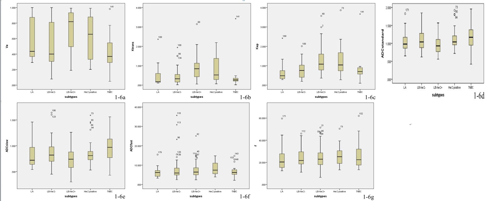
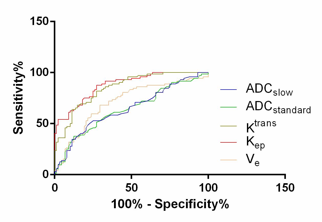
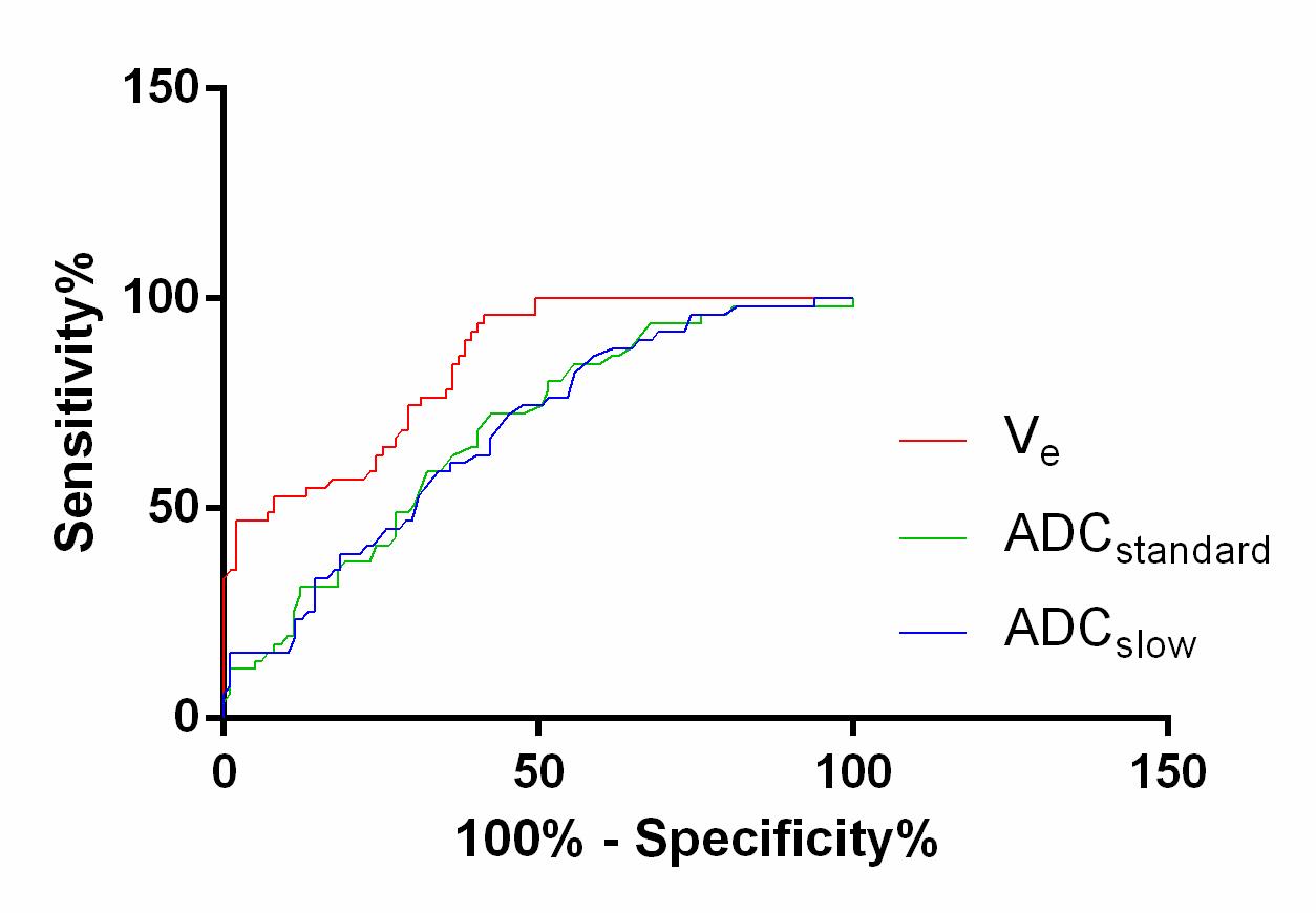
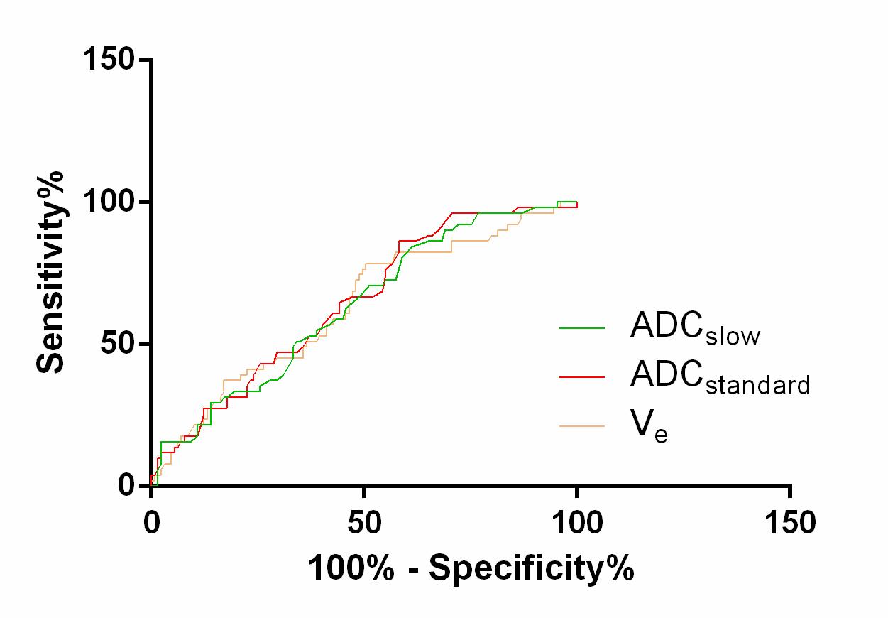
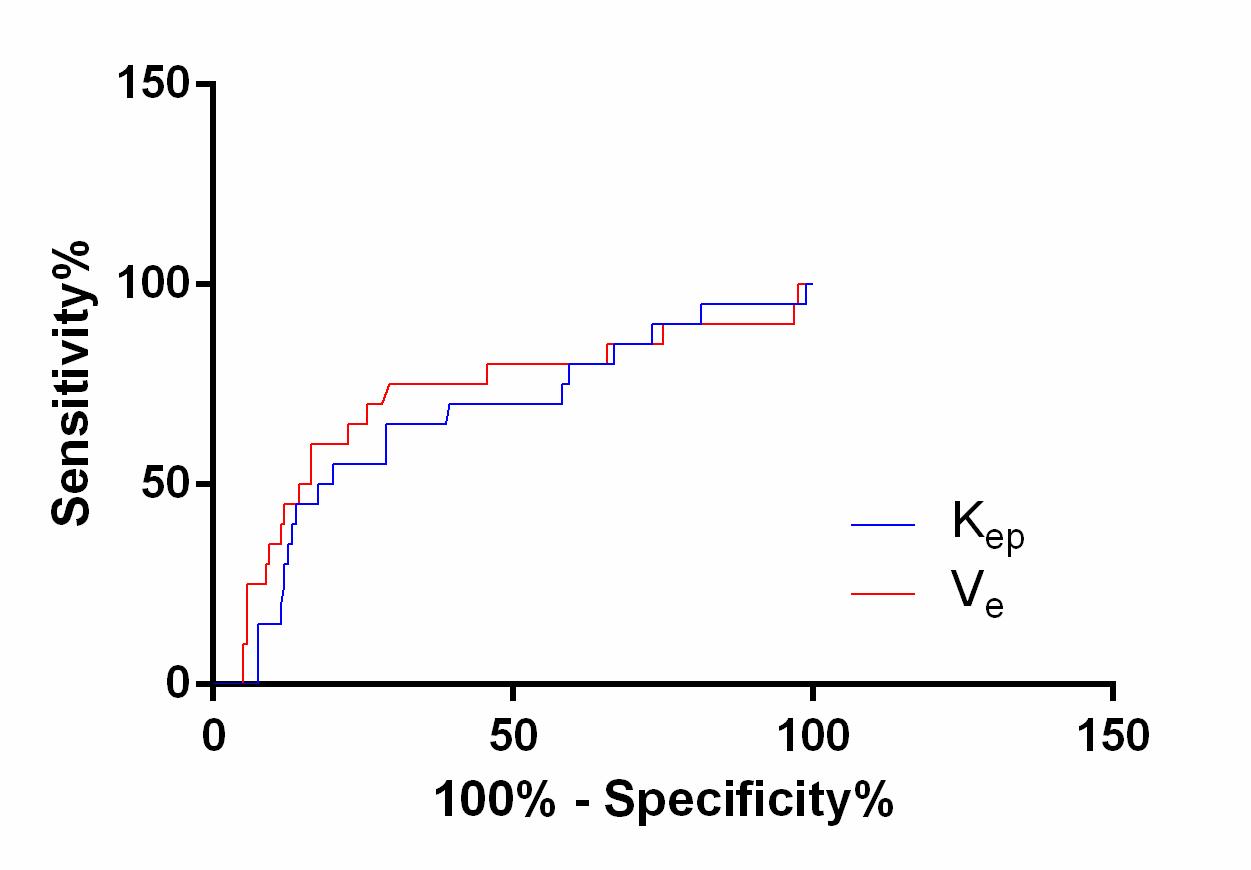
180 cases of breast cancer were included according to the inclusion and exclusion criteria. Containing 15/180 cases of LuminalA (8.33%), 45/180 cases of LuminalB (HER-2-) (25%), 68/180 cases of LuminalB (HER-2+) (37.78%), 30/180 cases of HER-2 over expression (16.67%), and 22/180 cases of triple negative (12.22%). DCE-MRI: Kep and Ktrans showed statistically significant differences between HER-2 positive and negative groups and Ki-67 high and low expression groups, but no statistically significant differences between ER and PR positive and negative groups, while Ve was significantly different between ER, PR, HER-2 positive and negative groups, but was not different between Ki-67 high and low expression groups. Kep still had predictive value for HER-2 status and Ve still had statistical significance for PR status prediction in Logistic multivariate regression analysis. The prediction threshold of Kep (p<0.001, AUC=0.878) and Ve (p<0.001, AUC=0.84) was 0.602 (specificity=87.5%, sensitivity=72.7%) and 0.547 (specificity=96.1%, sensitivity=58.6%), respectively. IVIM: ADCstandard and ADCslow showed statistical significance between positive and negative groups of ER, PR and HER-2, but no statistical significance between high and low expression groups of Ki-67 (p> 0.05). There were no significant differences in ADCfast and f values between positive and negative groups of ER, PR and HER-2, as well as between high and low expression groups of Ki-67. Kep, Ktrans, Ve, ADCstandard and ADCslow showed statistically significant differences among different molecular subtypes of breast cancer.Discussion
Many studies had shown that the occurrence, development and prognosis of breast cancer were closely related to molecular subtypes. In 2021, St Gallen experts agreed that the development of NAC for breast cancer should be determined according to tumor burden and molecular subtypes3-4. Quantitative parameters of DCE and IVIM could reflect the neovascularization and vascular wall permeability in tumors at the molecular level and predict the biological behavior of tumors5-6. In this study, Kep, Ktrans and Ve were significantly different among five different molecular subtypes of breast cancer. Kep has the best diagnostic performance in the prediction of immune prognostic factors of breast cancer, and has the highest value in the differentiation of different molecular subtypes. It was also found that ADCstandard and ADCslow showed statistically significant differences among different molecular subtypes, which was consistent with some reports7-8.Conclusion
IVIM-DWI and DCE-MRI had certain correlation with the expression of ER, PR, HER-2 and Ki-67, as well as had certain predictive value for different molecular subtypes.Acknowledgements
References
1. Horisawa N, Adachi Y, Takatsuka D, et al. The frequency of low HER2 expression in breast cancer and a comparison of prognosis between patients with HER2-low and HER2- negative breast cancer by HR status. Breast cancer. 2022; 29(2):234-241.
2. Jessica AS, Nicole KY, Aimee ES, et al. Concordance of breast cancer biomarker testing in core needle biopsy and surgical specimens: A single institution experience. Cancer Med. 2022; 11(24):4954-4965.
3. Fan R, Chen Y, Nechuta S, et al. Prediction models for breast cancer prognosis among Asian women. Cancer. 2021; 127(11):1758-1769.
4. Burstein HJ, Curigliano G, Th rlimann B, et al. Customizing local and systemic therapies for women with early breast cancer: The St. Gallen International Consensus Guidelines for treatment of early breast cancer 2021. Ann Oncol. 2021;32(10):1216-1235.
5. Lafcı O, Celepli P, Seher Ö P, et al. DCE-MRI radiomics analysis in differenting luminal A and luminal B breast cancer molecular subtypes. Acad Radiol. 2022; 30(1):22-29.
6. Thawani R, Gao L, Mohinani A, et al. Quantitative DCE-MRI prediction of breast cancer recurrence following neoadjuvant chemotherapy: a preliminary study. BMC Med Imaging. 2022; 22(1):182.
7. Fan M, He T, Zhang P, et al. Diffusion-weighted imaging features of breast tumours and the surrounding stroma reflect intrinsic heterogeneous characteristics of molecular subtypes in breast cancer. NMR in Biomedicine. 2018; 31(2):e3869.
8. Suo S, Zhang D, Cheng F, et al. Added value of mean and entropy of apparent diffusion coefficient values for evaluating histologic phenotypes of invasive ductal breast cancer with MR imaging. Euro Radiol. 2019; 29(3):1425-1434.
Figures