4820
DWI Parameters Correlated with Ki-67 Expression, a Prognostic Biomarker, in a Triple-Negative Breast Cancer PDX Mouse Model1Diagnostic Imaging and Nuclear Medicine, Kyoto University Graduate School of Medicine, Kyoto, Japan, 2Institute for Advancement of Clinical and Translational Science (iACT), Kyoto University Hospital, Kyoto, Japan, 3Faculty of Medicine, Kyoto University, Kyoto, Japan, 4Department of Systems science, Graduate School of Informatics, Kyoto University, Kyoto, Japan, 5Cancer Research Institute, Kanazawa University, Kanazawa, Japan, 6Diagnostic Pathology, Nara Medical University, Nara, Japan, 7Diagnostic Pathology, Kyoto University Graduate School of Medicine, Kyoto, Japan, 8Department of Pathology and Tumor Biology, Kyoto University Graduate School of Medicine, Kyoto, Japan, 9Laboratory of Integrative Molecular Medicine, Kyoto University Graduate School of Medicine, Kyoto, Japan, 10Human Brain Research Center, Kyoto University Graduate School of Medicine, Kyoto, Japan, 11CEA-Saclay, Paris-Saclay University, NeuroSpin, Gif/Yvette, France
Synopsis
Keywords: Cancer, Breast
Motivation: The lack of reliable biomarkers for assessing tumor characteristics and the limitations of histological analysis due to tumor heterogeneity led to the exploration of diffusion-weighted parameters.
Goal(s): To investigate the association between DW parameters and Ki-67 expression in triple-negative breast cancer, with a focus on whether they can serve as prognostic biomarkers.
Approach: Seventeen triple-negative breast cancer mice with a PDX model underwent 7T MRI scans, yielding DW images. Advanced analysis evaluated ADC and non-Gaussian diffusion parameters, validated through histological Ki-67 staining.
Results: DWI parameters (S-index, sADC, and ADC0) show strong correlations with Ki67 levels, at short and long diffusion times (9, 27.6ms).
Impact: Promising prognostic biomarkers for triple-negative breast cancer, DWI parameters (S-index, sADC, and ADC0) displayed strong correlations with Ki-67 expression, at short and long diffusion times. This validation through accurate DWI-pathology comparison highlights imaging's pivotal role in advancing breast cancer diagnosis.
Introduction
Breast cancer is a complex disease with several pathological biomarkers that play a crucial role in diagnosis, treatment, and prognosis. Ki-67, widely used to measure cellular proliferation in breast cancer tissue, has been proposed as a prognostic biomarker. The utility of ADC in differentiating Ki-67 positivity/negativity in clinical studies remains inconclusive, raising questions about its effectiveness1. One challenge in clinical studies is that histology can only be obtained from a limited portion of the entire tumor, potentially missing the heterogeneity specific to breast cancer characteristics. In addition, the standardization of Ki-67 is not yet established, and there is a need for establishing imaging biomarkers useful for breast cancer diagnosis, monitoring treatment, and predicting prognosis. Recently, patient-derived xenografts (PDX) have emerged as a valuable model for preserving the natural tumor heterogeneity and microenvironment. They are considered ideal for accurate comparisons between imaging and pathology (see Figure 1). As a result, this study aims to investigate whether DW (diffusion-weighted) parameters are associated with Ki-67 expression in triple-negative breast cancer, where Ki-67 serves as an established prognostic biomarker in breast cancer.Materials & Methods
MRI: 17 PDX (Patient-Derived Xenograft) mice with triple-negative breast cancer in both limbs were established and imaged with 7T MRI scanner (Bruker, Germany).The SE-EPI acquisition parameters were set as follows: resolution 250 x 250μm2, matrix size 100×100, field of view 25×25 mm2, slice thickness 1.5 mm, TE=57ms, TR=2500 ms, 8 averages, 4 segments. IVIM/DW images were acquired using two diffusion times [Td =9/27.6ms with 17 b‑values (0–3000 s/mm2), respectively]. A shifted ADC [sADC200–1500] was calculated using b=200 and 1500s/mm² at both diffusion times. Non‑Gaussian diffusion parameters (ADC0, the virtual ADC at b = 0; K, Kurtosis from non‑Gaussian diffusion) were estimated using the IVIM/Kurtosis model2 and S-index was calculated as previously shown3 using built-in MATLAB software (Mathworks, Natick, MA).Histology: Tumor specimens underwent staining with H&E, Ki-67 (cell proliferation), and CD31(microvessel). For Ki-67 stain, five ROIs were selected on viable parts of cancer, excluding necrosis, and the ratio of Ki-67-positive tumor cells to all cells was assessed using QuPath4. The mean ratio from the five ROIs was recorded as Ki-67.
Pearson correlation coefficient between DWI parameters and Ki-67 positive rate was evaluated using MedCalc Software (Mariakierke, Belgium).
Results and Discussion
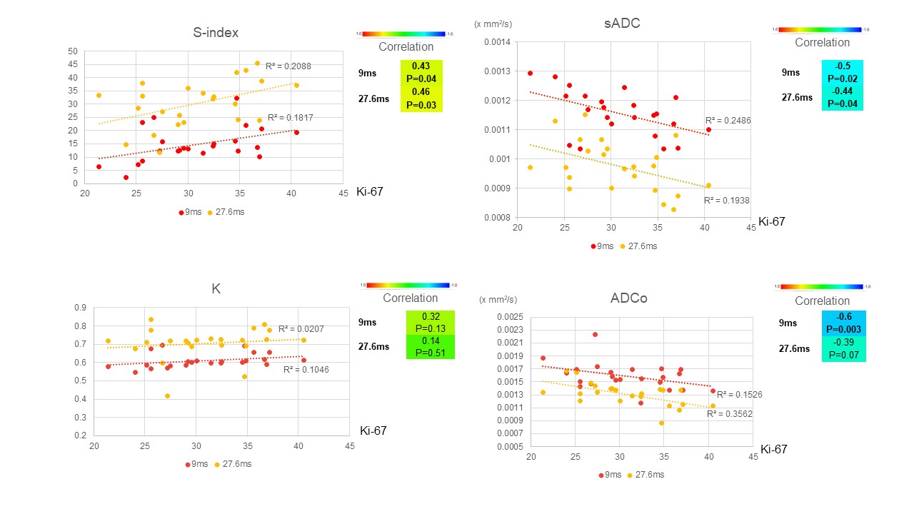
Twenty-three tumors were stained with Ki-67 and analyzed to compare the association between DW parameters and Ki-67. Figures 2 and 3 show the representative triple-negative cancer, indicating central necrosis both in MR images and histology with different Ki-67 levels. Scatter plots in Figure 4 demonstrate the relationship between DWI parameters and Ki-67 and their trends.Overall, at all Ki-67 levels, Sindex increased while sADC and ADCo decreased with the diffusion time, as expected with the increase in diffusion hindrance. The S-index was significantly and positively correlated with the Ki-67 level at both diffusion times. sADC presented an opposite trend, as expected. ADC0 was negatively correlated when Ki-67 level, but correlation was more marked at the shortest diffusion times (9ms). There was no correlation with K (non-Gaussian diffusion index). The results were consistent with earlier findings5. Overall, those results (an increase of S-index and decrease of sADC, sensitive to Gaussian and non-Gaussian diffusion, at short and long diffusion times, decrease of ADC0, Gaussian diffusion marker, at short diffusion times, while kurtosis remain unchanged) suggest that the underlying mechanism for the correlation with Ki67 levels likely comes from the replacement of normal cells by cells with different features such as nuclei where Ki-67 is localized, resulting in different diffusion patterns (e.g. lower intracellular diffusion).
Conclusion
The S-index, sADC and ADC0 might be used as markers of Ki-67 level, a prognostic biomarker in triple-negative breast cancer. Furthermore, those parameters can be shown as parametric maps, which can be used to guide biopsy in heterogeneous tumors. Works remain to investigate further the mechanism of the link between Ki-67 expression and tissue features (e.g., cellularity and intracellular diffusion characteristics such as nuclei where Ki-67 is mainly located), which could be obtained by combining data acquired at short and long diffusion times using models such as IMPULSED 6,7.Acknowledgements
We express our gratitude to Ms. Setsuko Inoue and Dr. Tomomi Nishimura for their invaluable technical support in conducting the animal experiments. This study was supported by AMED grant (23he0422025j0002).References
1. Surov, A., Meyer, H. J. & Wienke, A. Associations between apparent diffusion coefficient (ADC) and KI 67 in different tumors: a meta-analysis. Part 1: ADCmean. Oncotarget 8, 75434–75444 (2017).
2. Iima, M. et al. Quantitative non-Gaussian diffusion and intravoxel incoherent motion magnetic resonance imaging: differentiation of malignant and benign breast lesions. Invest. Radiol. 50, 205–211 (2015).
3. Goto, M., Le Bihan, D., Yoshida, M., Sakai, K. & Yamada, K. Adding a Model-free Diffusion MRI Marker to BI-RADS Assessment Improves Specificity for Diagnosing Breast Lesions. Radiology 292, 84–93 (2019).
4. Bankhead, P. et al. QuPath: Open source software for digital pathology image analysis. Sci. Rep. 7, 16878 (2017).
5. Someya, Y., Iima, M. & Imai, H. Investigation of breast cancer microstructure and microvasculature from time-dependent DWI and CEST in correlation with histological biomarkers. Sci Rep. Nature Publishing Group 12, 1–12 (2022).
6. Xu, J. et al. Magnetic resonance imaging of mean cell size in human breast tumors. Magn. Reson. Med. 83, 2002–2014 (2020).
7. Wu, D. et al. Time-dependent diffusion MRI for quantitative microstructural mapping of prostate cancer. Radiology 303, 578–587 (2022).
Figures
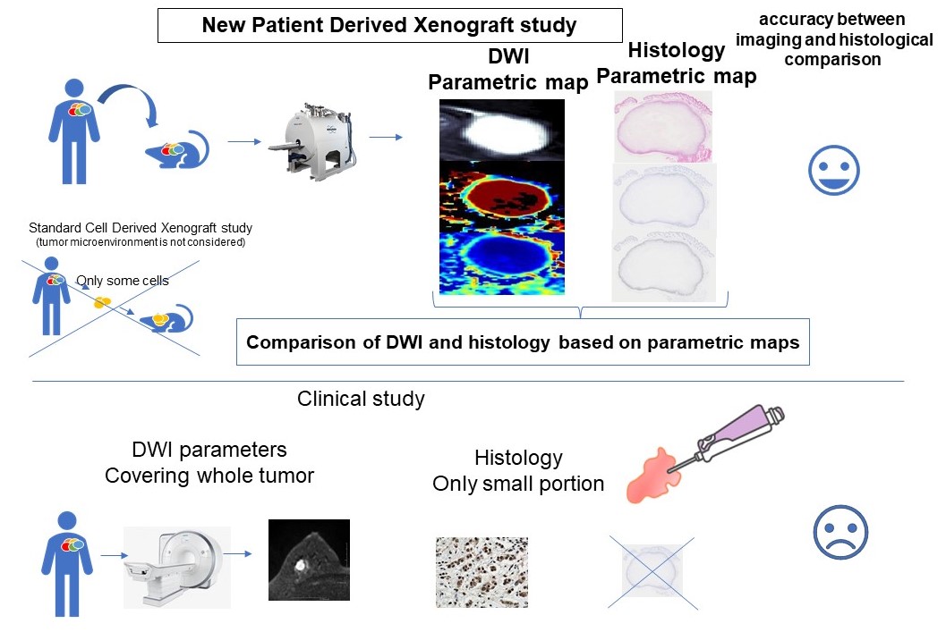
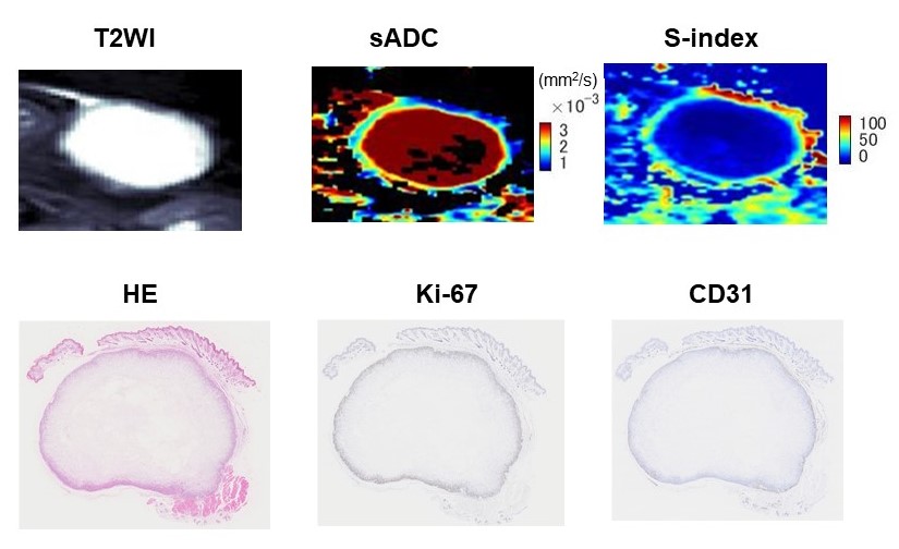
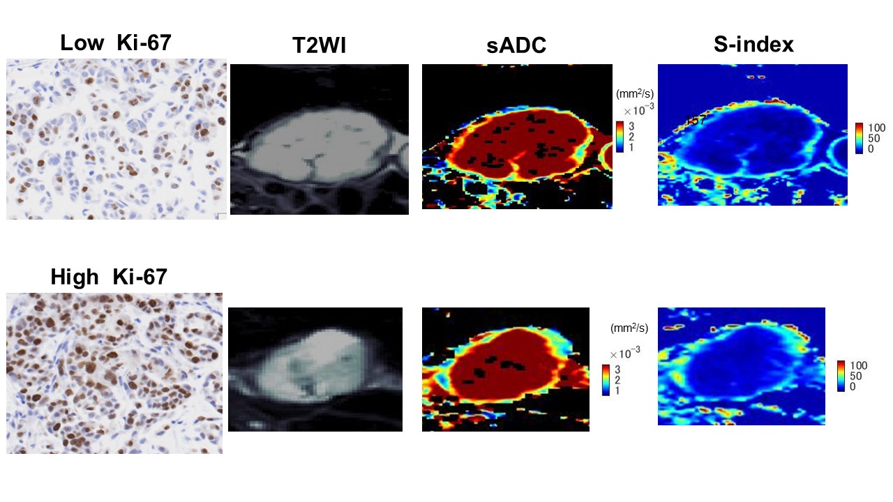
Fig.3: Representative DW images of triple-negative cancer with a different Ki-67 level in a PDX model. The upper tumor shows low Ki-67, with relatively high sADC (1.1 x10⁻³ mm2/s) and low S-index (14.8). The lower tumor exhibits high Ki-67, with relatively low sADC (0.9x10⁻³ mm2/s) and high S-index (37.1). All estimated values are with a diffusion time of 27.6ms.