4815
Test-retest repeatability of renal MRI parameters in healthy volunteers comparing ROI-based and tissue segmentation based image analysis1Department of Diagnostic and Interventional Radiology, University Hospital Tuebingen, Tuebingen, Germany, 2University Tuebingen, Tuebingen, Germany, 3Section on Experimental Radiology, University Hospital Tuebingen, Tuebingen, Germany, 4Siemens Healthineers, Erlangen, Germany
Synopsis
Keywords: Kidney, Kidney
Motivation: Multiparametric MRI of the kidneys is a promising technique for renal diagnostics, but the diversity of imaging protocols and analysis strategies hinders clinical translation.
Goal(s): Our goal was to assess the repeatability of a multiparametric MRI protocol comparing ROI-based and tissue segmentation based analysis.
Approach: Ten volunteers were examined with a multiparametric MRI protocol including ASL, IVIM, BOLD, T1 and T2 mapping twice with one week between visits.
Results: Good repeatability of the multiparametric protocol could be achieved. T1 and T2 values showed less variability compared to perfusion and diffusion related functional parameters. Tissue segmentation showed better repeatability compared to ROI-based analysis.
Impact: Our study demonstrates relatively high repeatability of multiparametric functional MRI of the kidneys. The results of the image analysis methods suggest, that manual segmentation is to be preferred over ROI-based analysis, if automated segmentation is not available.
Introduction
Multiparametric functional MRI of the kidneys is gaining more and more attention with its potential to non-invasively assess functional information and hereby explore renal pathophysiological changes, which have remained difficult to comprehend for many kidney diseases yet. Despite many advances in renal MRI research in the past decades, obstacles remain to clinical translation, one of the biggest being the diversity of MRI protocols and postprocessing and data analysis strategies. In this study, we aim to evaluate a multiparametric renal functional MRI protocol guided by the PARENCHIMA recommendations1 on the test-retest repeatability comparing two different established image analysis techniques: representative ROI-based analysis and manual tissue segmentation.Methods
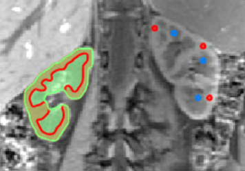
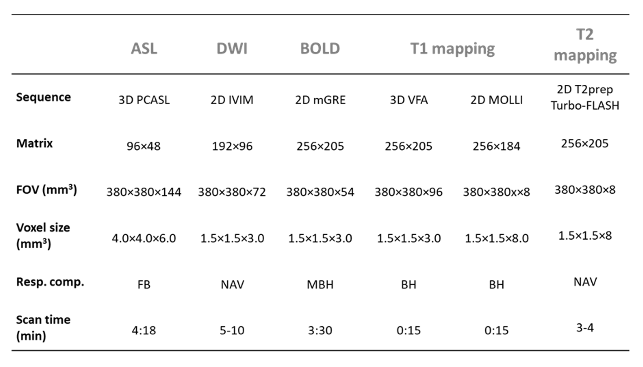
This prospective study was approved by the local ethics committee. Examinations were performed on a 3T MR scanner (MAGNETOM Prismafit, Siemens Healthcare, Erlangen, Germany). For the repeatability study we examined 10 healthy volunteers (31 ± 7 y/o, 5f) twice, with 1 week between examinations. The imaging protocol encompassed arterial spin labeling (ASL), diffusion-weighted imaging (DWI) with intravoxel incoherent motion (IVIM), blood-oxygen-dependent (BOLD) imaging, and T1- and T2 mapping (s. Table 1). Total scan time was approximately 45 minutes. Image analysis was performed using representative ROI-based analysis and manual tissue segmentation (s. Figure 1). For assessment of agreement between scans, the repeatability coefficient (RC), the within-subject coefficient of variation (wCV) and the intraclass correlation coefficient (ICC) were calculated.Results
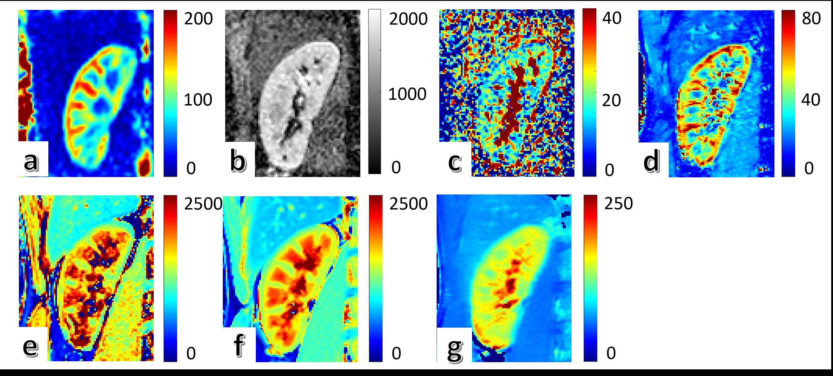
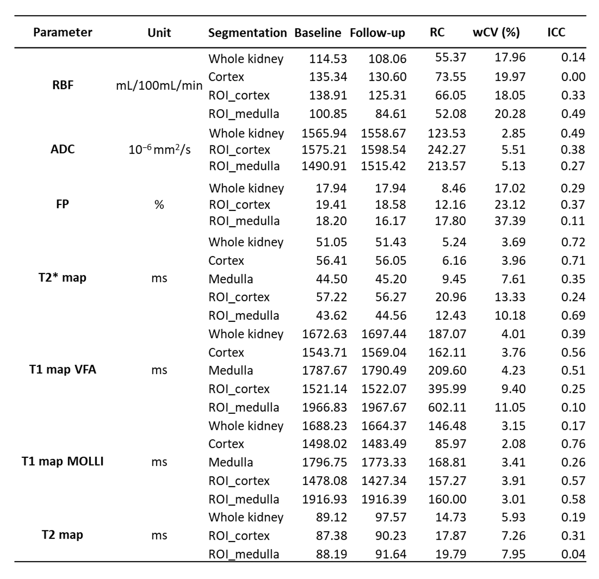
All 10 volunteers could be examined successfully. Examples of the MRI images are shown in Figure 2. Structural tissue parameters (T1, T2) showed wCV (%) from 4 to 11 and ICC from 0.2 and 0.8. Functional parameters (ASL, BOLD and DWI) showed wCV (%) from 3 to 38 and ICC from 0.0 to 0.7. Tissue segmentation could be performed in ASL, BOLD and T1 mapping and provided better repeatability (wCV, 2 to 20%; ICC, 0.0 to 0.7) as compared to ROI-based analysis (wCV, 3-37%; ICC, 0.0 to 0.7). Results are summarized in Table 2.Discussion
The aim of this study was to evaluate the test-retest repeatability of a multiparametric MRI protocol comparing representative ROI-based analysis and tissue segmentation. In comparison to previous studies evaluating the repeatability of functional MRI parameters2–6, our study comprised a broad selection of MRI techniques based on state-of-the-art research. Reasonable test-retest repeatability could be achieved with our protocol. As reported in previous studies5, structural parameters (T1 & T2 map) showed better repeatability compared to functional parameters (ASL, IVIM, BOLD). Overall test-retest repeatability was better for manual tissue segmentation compared to ROI-based analysis. Automated kidney segmentation has been already applied by some research groups5,6 and will be the future, but still needs evaluation7. Despite the amount of work and time required for manual tissue segmentation, our results show that it might still come closest to the “ground truth” and prevent rater bias and variability.Conclusion
The applied multiparametric functional MRI protocol including ASL, IVIM, BOLD, T1 and T2 mapping could provide relatively high test-retest repeatability potentially allowing applications in clinical cases. Manual tissue segmentation achieved better repeatability compared to ROI-based analysis and should be therefore recommended, if automated segmentation is not available. Further studies are needed to evaluate typical deviations and uncertainties of measured values compared to disease related effects.Acknowledgements
This study was funded by the Wilhelm Sander Foundation (Wilhelm Sander-Stiftung, Munich, Germany).References
1. Mendichovszky I, Pullens P, Dekkers I, et al. Technical recommendations for clinical translation of renal MRI: a consensus project of the Cooperation in Science and Technology Action PARENCHIMA. Magn Reson Mater Phy. 2020;33(1):131-140.
2. Li L-P, Thacker J, Li W, et al. Consistency of Multiple Renal Functional MRI Measurements Over 18 Months. Journal of magnetic resonance imaging : JMRI. 2018;48(2):514-521.
3. Rankin AJ, Allwood-Spiers S, Lee MMY, et al. Comparing the interobserver reproducibility of different regions of interest on multi-parametric renal magnetic resonance imaging in healthy volunteers, patients with heart failure and renal transplant recipients. Magnetic Resonance Materials in Physics, Biology and Medicine. 2020;33(1):103-112.
4. Kline TL, Edwards ME, Garg I, et al. Quantitative MRI of kidneys in renal disease. Abdom Radiol. 2018;43(3):629-638.
5. Boer A de, Harteveld AA, Stemkens B, et al. Multiparametric Renal MRI: An Intrasubject Test-Retest Repeatability Study. Journal of magnetic resonance imaging : JMRI. 2021;53(3):859-873.
6. Cox EF, Buchanan CE, Bradley CR, et al. Multiparametric Renal Magnetic Resonance Imaging: Validation, Interventions, and Alterations in Chronic Kidney Disease. Frontiers in physiology. 2017;8:696.
7. Zollner FG, Kocinski M, Hansen L, et al. Kidney Segmentation in Renal Magnetic Resonance Imaging - Current Status and Prospects. IEEE Access. 2021;9:71577-71605.
Figures