4814
Quantitative Characterization of Breast Lesion Using Compartmentalized Model: Intravoxel Incoherent Motion and Restriction Spectrum Imaging1Department of Radiology, Tongji Hospital, Tongji Medical College, Huazhong University of Science and Technology, Wuhan, China, 2MR Collaboration, Central Research Institute, United Imaging Healthcare, Shanghai, China
Synopsis
Keywords: Diffusion Analysis & Visualization, Multimodal
Motivation: Conventional diffusion-weighted MRI with ADC in clinical breast imaging protocol may not accurately reflect the authentic diffusion.
Goal(s): To quantitatively comparison the diagnostic utility of the compartmentalized diffusion-weighted model-intravoxel incoherent motion (IVIM) and restriction spectrum imaging (RSI) in the differential diagnosis of breast lesions.
Approach: The Mann-Whitney U-test and ROC analysis were used to evaluate the diagnostic efficacy of each parameter and model in differentiating breast lesions.
Results: Quantitative parameters derived from three-compartment RSI model have great promise as imaging indicators in differentiating breast lesions compared to bi-exponential IVIM model. Additionally, the hybrid model integrating IVIM and RSI achieves the superior diagnostic performance.
Impact: The integration of IVIM and RSI would likely lead to a new perspective for the characterization of breast lesions, thus have potential clinical utility in the further application of non-contrast-enhanced breast MR screening.
Introduction
Breast cancer accounts for the highest proportion among women diagnosed with cancer worldwide (1). Accurate characterization is crucial for generating the personalized management strategies of patients with breast cancer. Conventional diffusion-weighted MRI (DW-MRI) with apparent diffusion coefficient (ADC) calculating from mono-exponential model for assessment the cellularity, based on the Gaussian distribution of water molecules diffusion, has been extensively used in clinical setting (2). However, given the effects of blood microcirculation and the complexity of breast tissue microstructure, ADC value may not accurately reflect the authentic diffusion. Compartmentalized models, which separates each voxel into multiple compartments on the basis of tissue microstructural or diffusion properties, can partially address these limitations of conventional DWI (3). One of the compartmentalized models-Intravoxel Incoherent Motion (IVIM), which divided the diffusion signal into two distinct components: microcirculation perfusion and tissue diffusivity, can characterize both cellularity and vascularity (4,5). Another compartmentalized model is Restriction Spectrum Imaging (RSI), in which the diffusion signal is modeled as a mixture of three compartments corresponding to intracellular restricted, extracellular hindered, and free water pools. Through the application of generalized linear estimation technique and extended b-value, the underlying tissue microstructure can be reflected (6,7). Therefore, this study was implemented to quantitatively comparison the diagnostic utility and added value of the two compartmentalized models for the differentiation of breast lesions.Methods
This study prospectively enrolled 152 patients with 157 histopathologically-verified breast lesions (41 benign lesions and 116 malignant lesions). Each subject underwent a bilateral breast MRI examination using a 3.0T MR imager. The regions of interest (ROIs) of the whole lesion volume and healthy tissue were delineated by two experienced radiologists and the parameter values of Mono-exponential (apparent diffusion coefficient-ADC), IVIM (true diffusion coefficient-Dt, pseudo-diffusion coefficient-Dp, and perfusion fraction-f), and RSI (signal contributions: restricted diffusion-C1, hindered diffusion-C2, free diffusion-C3, product-C1C2, and corresponding signal fractions: F1, F2, F3, F1F2) were calculated subsequently. The Mann-Whitney-U-test was used for pairwise comparisons in three different tissue types (malignant tumors, benign lesions, and healthy tissues). The ROC analysis was used to evaluate the diagnostic efficacy of each parameter and model in differential diagnosis.Results
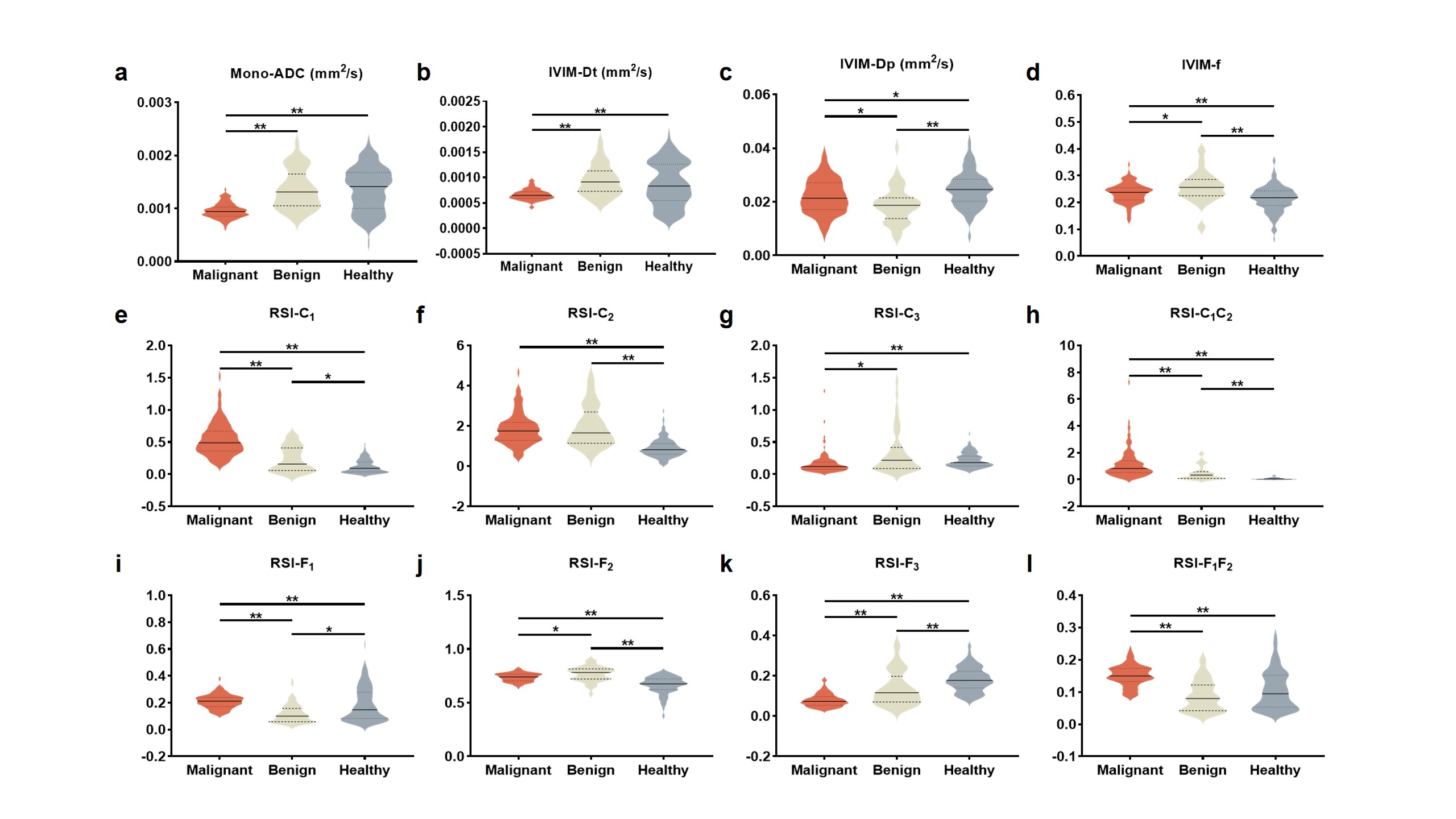
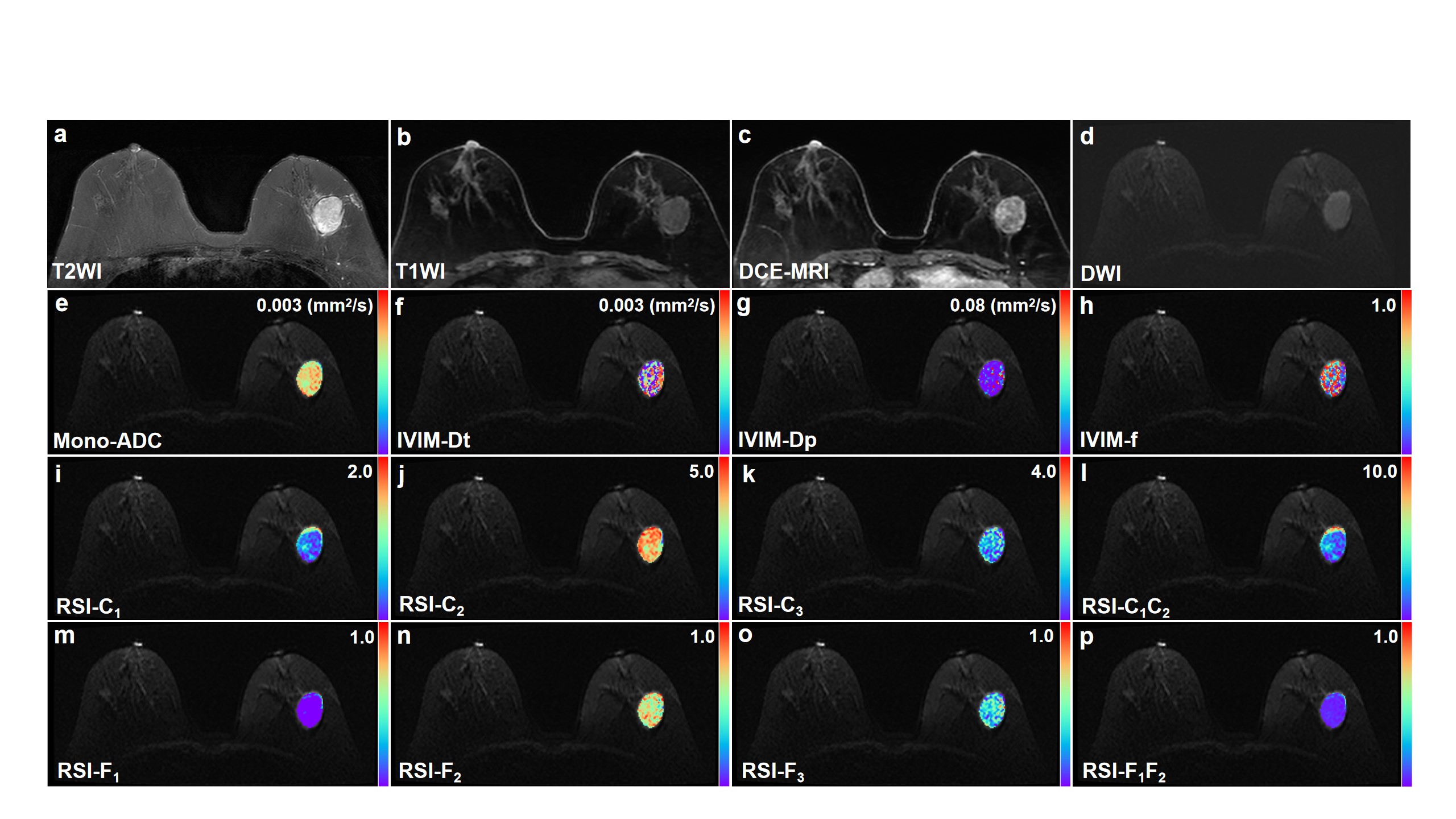
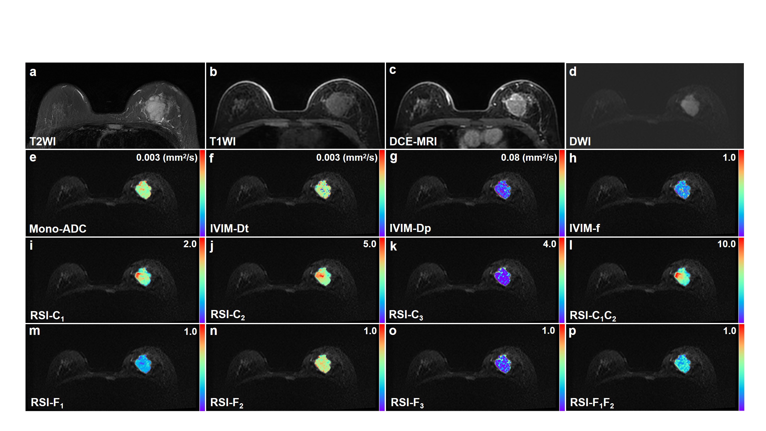
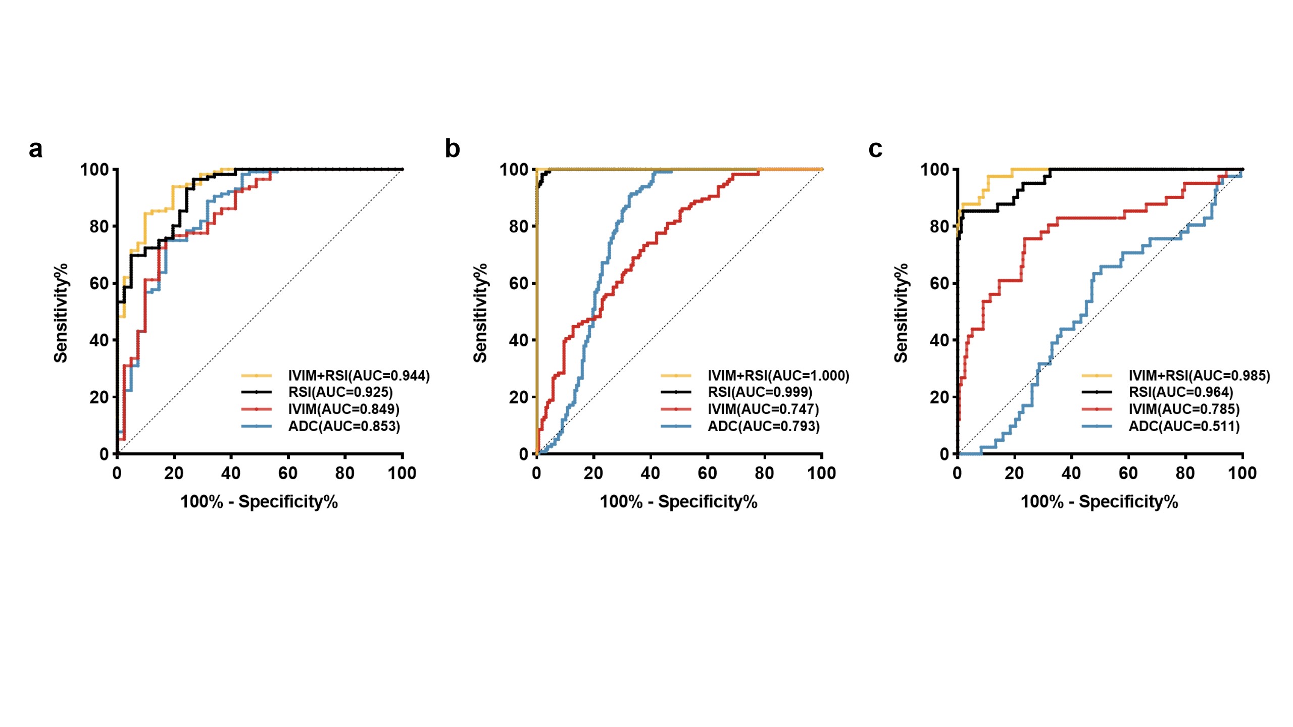
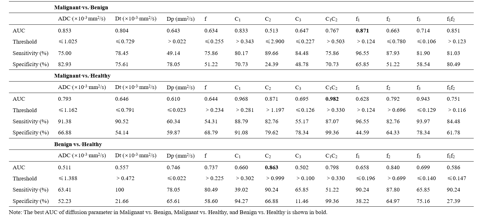
1. Almost all quantitative diffusion parameters showed significant differences for distinguishing malignant tumors from both benign lesions (other than C2) and normal tissues (all parameters) (all P < 0.0167). The parameters derived from IVIM (Dp, f) and RSI (C1, C2, C1C2, F1, F2, F3) showed significant differences in the comparison of benign lesions and healthy tissues (all P < 0.005) (Figure 1). Representative images of malignant tumor and benign lesion were depicted in Figure 2 and Figure 3.2. As shown in Table 1 and Figure 4, RSI derived parameters-F1, C1C2, and C2 values yielded the highest AUCs for malignant vs. benign, malignant vs. healthy and benign vs. healthy (AUCs = 0.871, 0.982, and 0.863, respectively). Furthermore, the hybrid diagnostic model (IVIM + RSI), using binary logistic regression, exhibited the highest diagnostic efficacy for the pairwise comparison (AUCs = 0.944, 1.000, and 0.985, respectively).
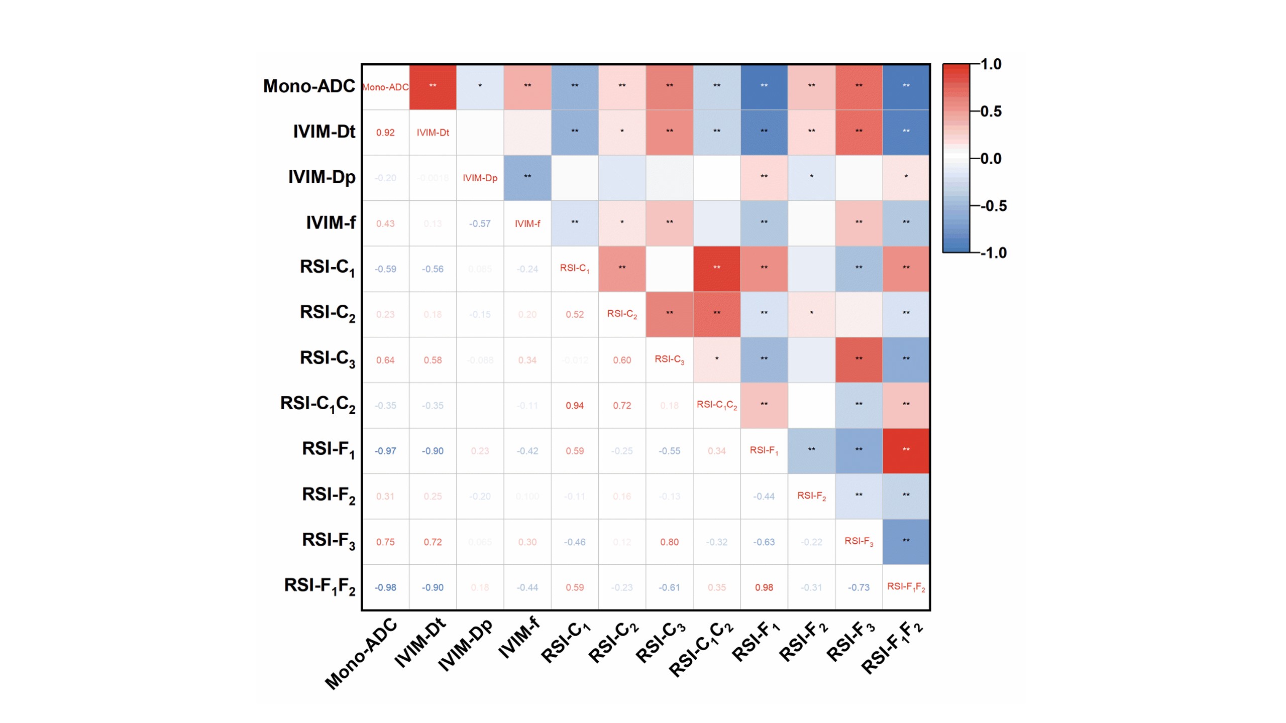
3. There were strong negative correlations between ADC and F1, F1F2 (r = -0,974 and -0.984, respectively, all P < 0.01) and strong positive correlation between ADC and Dt (r = 0.922, P < 0.01) (Figure 5).
Discussion
Significantly higher C1, C1C2, F1, F1F2 and lower ADC, Dt, C3 and F3 were found in the malignant tumors compared to benign lesions and normal tissues. This possibly occurs due to the hypercellular tissue density, substantial synthesis of macromolecular substances and increased necrotic substances in malignant lesions, leading to reduced extracellular space for the diffusion of free water molecule. Discordantly to the preliminary publications (6,8), there are significant differences in some of the parameters of IVIM and RSI between benign lesions and normal tissue in our research. This may be partly related to the differences in delineation of health control ROIs, pathological types of benign lesions, and diffusion coefficients of each compartment. Notably, when we combined the IVIM and RSI, the best diagnostic efficacy for the differential diagnosis could be achieved. This may be result from the reason that hybrid model has capability to simultaneously explore the cellularity, vascularity, and microstructure of breast tissue by making the best of the full b-value spectrum information.Conclusion
Quantitative parameters derived from the three-compartment RSI model have great promise as imaging indicators for the differential diagnosis of breast lesions compared with bi-exponential IVIM model. Additionally, the hybrid model integrating IVIM and RSI achieves the superior diagnostic performance in characterizing breast lesions, thus have potential clinical utility in the further application of non-contrast-enhanced breast MR screening and reducing unnecessary biopsies.Acknowledgements
No.References
1. Siegel RL, Miller KD, Wagle NS, Jemal A. Cancer statistics, 2023. CA Cancer J Clin 2023;73(1):17-48.
2. Mendez AM, Fang LK, Meriwether CH, et al. Diffusion Breast MRI: Current Standard and Emerging Techniques. Front Oncol 2022;12:844790.
3. Tang L, Zhou XJ. Diffusion MRI of cancer: From low to high b-values. J Magn Reson Imaging 2019;49(1):23-40.
4. Baxter GC, Graves MJ, Gilbert FJ, Patterson AJ. A Meta-analysis of the Diagnostic Performance of Diffusion MRI for Breast Lesion Characterization. Radiology 2019;291(3):632-641.
5. Arian A, Seyed-Kolbadi FZ, Yaghoobpoor S, Ghorani H, Saghazadeh A, Ghadimi DJ. Diagnostic accuracy of intravoxel incoherent motion (IVIM) and dynamic contrast-enhanced (DCE) MRI to differentiate benign from malignant breast lesions: A systematic review and meta-analysis. Eur J Radiol 2023;167:111051.
6. Besser AH, Fang LK, Tong MW, et al. Tri-Compartmental Restriction Spectrum Imaging Breast Model Distinguishes Malignant Lesions from Benign Lesions and Healthy Tissue on Diffusion-Weighted Imaging. Cancers (Basel) 2022;14(13).
7. Qin Y, Tang C, Hu Q, et al. Quantitative Assessment of Restriction Spectrum MR Imaging for the Diagnosis of Breast Cancer and Association With Prognostic Factors. J Magn Reson Imaging 2022.
8. Jin YN, Zhang Y, Cheng JL, Zheng DD, Hu Y. Monoexponential, Biexponential, and stretched-exponential models using diffusion-weighted imaging: A quantitative differentiation of breast lesions at 3.0T. J Magn Reson Imaging 2019;50(5):1461-1467.
Figures