4813
Gadolinium-Loaded Melanin-Induced Magnetic Resonance Imaging and Thermoradiotherapy in Glioblastoma Multiforme1Departments of Magnetic Resonance, The First Affiliated Hospital of Harbin Medical University, Harbin, China, 2Philips Healthcare, Beijing, China
Synopsis
Keywords: Tumors (Pre-Treatment), Tumor, Melanin Nanoparticle; Glioblastoma Multiforme; Magnetic Resonance Imaging; Thermoradiotherapy
Motivation: Glioblastoma multiforme(GBM) is characterized by highly invasive growth, resulting in poor treatment efficacy, high recurrence rate and a short life expectancy.
Goal(s): Maximizing removal of tumor tissue, while minimizing damage to brain function areas and enhancing treatment effectiveness holds significant clinical significance. This necessitates precise delineation of GBM edge and ntegration of multiple treatment strategies.
Approach: T1WI mDIXON and T1 mapping was performed on a 3.0T MR scanner. Multi-level tumor models will be established to assess the potential ability of Gd-MNP-PEGs and underlying molecular mechanisms of thermoradiotherapy.
Results: Gadolinium-loaded and PEGylated Melanin nanoparticles performed notable photothermal effect, radiosensitization and the ability of MRI.
Impact: Gd-MNP-PEGs was formed and utilized for integration of diagnosis and treatment to precisely define the boundaries of GBM and enhance the therapeutic outcome.
Introduction
Glioblastoma multiforme (GBM) is the most common and lethal type of central nervous system tumors in adults, characterized by outspread invasion, limited therapeutic options and a poor overall survival [1-3]. The peritumoral region of GBM is composed of infiltrating tumor cells and vasogenic edema, making it challenging to delineate the boundary of tumor on contrast-enhanced MRI [4]. Moreover, tumor heterogeneity and genetic diversity constrain the effectiveness of current standard of care including maximum safe craniotomy, adjuvant radiation and chemotherapy with temozolomide [4]. Thus, it is vital to precisely define the boundaries of GBM and enhance the therapeutic effect. Melanin is an endogenous photothermal agent with high biosecurity. The most comment for clinical application is Gadolinium-based contrast agent, which also possesses radiosensitization capabilities. Herein, we loaded Gadolinium on melanin to develop multifunctional nanotheranostic Gadolinium-loaded and PEGylated Melanin nanoparticles (Gd-MnP-PEGs) which could be utilized for imaging diagnosis, radiosensitization, and photothermal therapy.Methods
Gd-MNP-PEGs were obtained according to the previous method [5]. Transmission electron microscopy (Hitachi, Japan), dynamic light scattering (BI-90Plus, Brookhaven, USA), Ultraviolet-visible-near-infrared spectra (LAMBDA 1050+, PerkinElmer, USA), FT-IR spectra (Nicolet iS20, Thermo Scientific, USA), inductively coupled plasma-mass spectrometry (7800, Aglient, USA) were used to characterize Gd-MNP-PEGs. The subcutaneous tumor model of GBM was established and Gd-MNP-PEGs aqueous solution (1mg/ml) was injected into the caudal vein. MRI was performed on a 3.0T MR scanner (Ingenia Elition; Philips Healthcare, Best, The Netherlands) with an 8-channel wrist coil. Imaging sequences include T1WI mDIXON and T1 mapping. The scanning parameters were shown in Table 1. The axial and coronal images of different time points for subcutaneous tumor model were collected to observe the tumor signal and its metabolism. Cell counting kit-8 assay, colony formation assay, and Western blotting analysis were utilized to explore the outcomes and mechanisms of thermoradiotherapy. All obtained data were analyzed using unpaired t-test, one way anova, and LSD test in SPSS v26.0 (SPSS, Inc.) to identify the statistical significance among the control and experimental groups. Mean–standard deviation presented the data after three independent experiments at least. P-value of <0.05 indicated statistical significance.Result
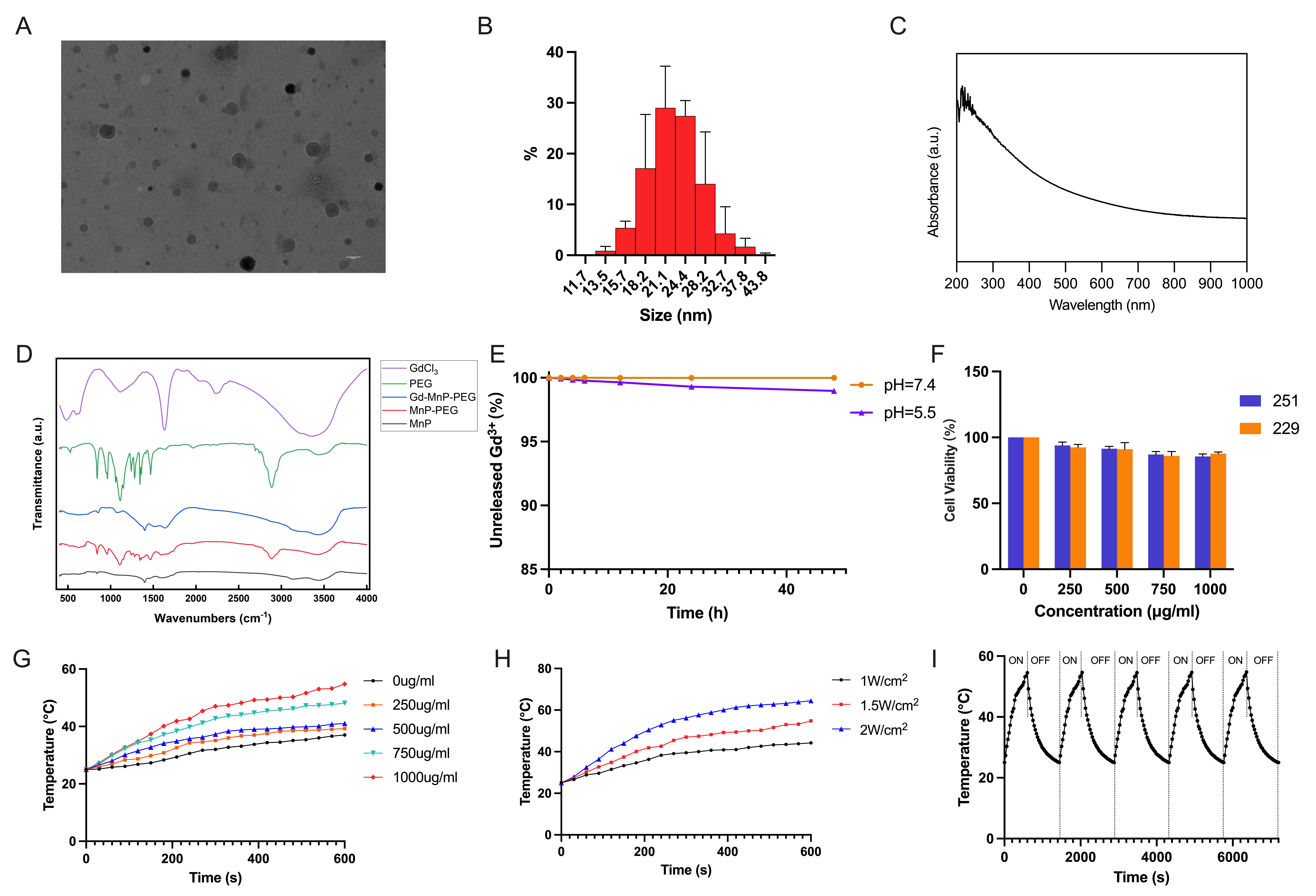

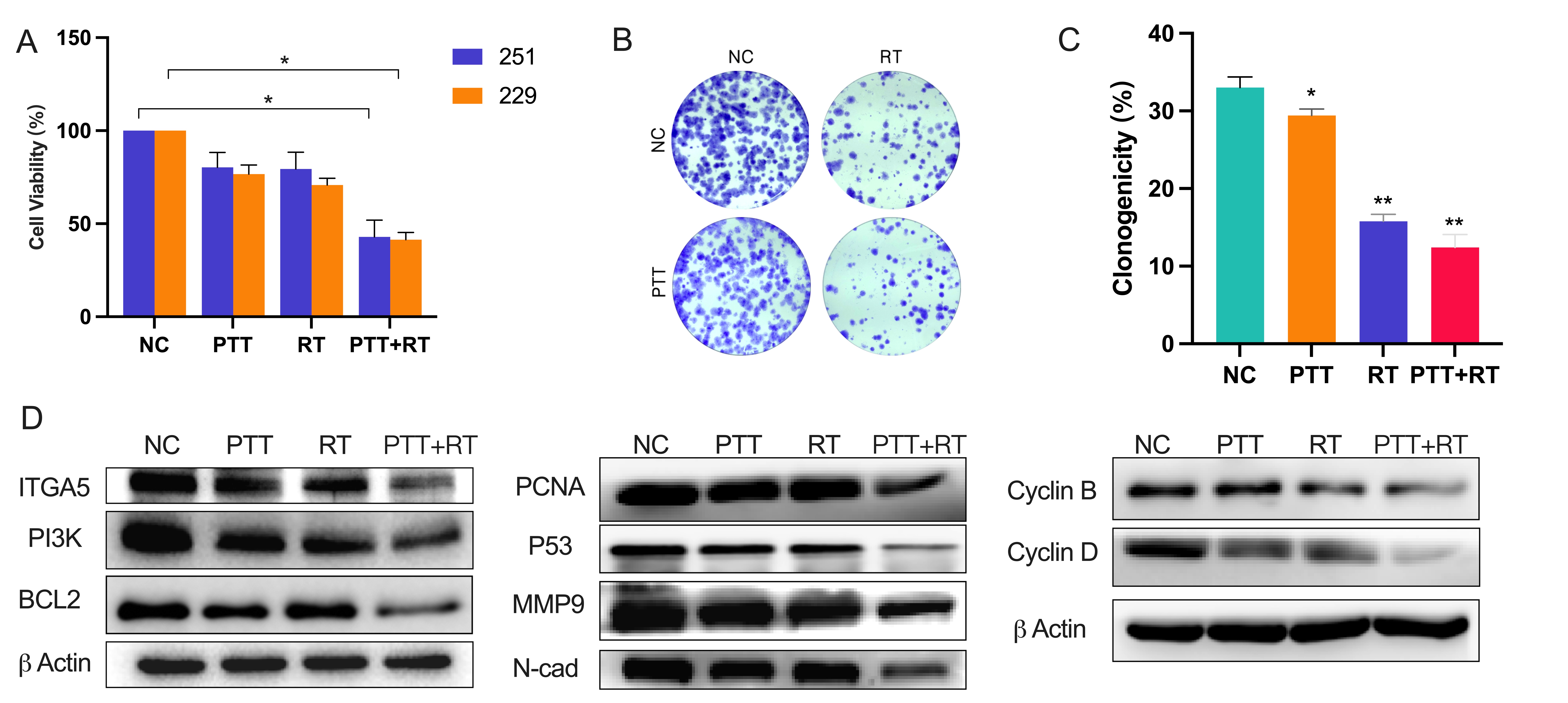
Gd-MNP-PEGs presented small size (21.1nm, Figure 1A, B), good absorbance performance (Figure1C), high chelation stability(Figure 1E) and negligible cytotoxicity estimated by CCK-8 assay (Figure 1F). FTIR analysis indicated successful PEG modification of the surface of the MNPs and chelation of Gd3+(Figure 1D). The nanoparticle exhibited higher r1 relaxivity (7.7013 mM−1 s−1) than clinically approved Gadobutrol (6.6367 mM−1 s−1) at 3.0 T MR scanning (Figure 2A, B). After the nanoparticles were injected into vein in mice with subcutaneous GBM, a dramatic increase in signal of the tumor was observed at 0.5 hr by T1WI mDIXON, while the tumor exerted remarkable signal enhancement at 6 hrs, showing excellent detection sensitivity (Figure 2D), then nanoparticles were excreted through renal and hepatobiliary pathways (Figure 2C, E). Moreover, the nanoparticles demonstrated outstanding photothermal conversion ability, photostability and radiosensitization ability (Figure 1 H-I). The thermoradiotherapy inhibited the proliferation of GBM (Figure 3 A-C). Western blotting analysis indicated that thermoradiotherapy inhibit proliferation, migration, invasion, and cell cyclocyte-related proteins, as well as relieve the resistance to anoikis of GBM (Figure 3D).Discussion
Gd-MNP-PEGs demonstrated excellent MRI contrast enhancement, with a higher r1 relaxivity compared to clinically approved Gadobutrol. When the nanoparticles administered intravenously to mice, the signal enhancement of subcutaneous GBM reached the peak at 6h after injection and was subsequently excreted through the renal and hepatobiliary pathways. Melanin is an endogenous photothermal agent with the pyrrole and benzene rings that grant it excellent photothermal conversion capabilities [6]. It exhibits a high photothermal conversion efficiency of 40%, which is superior to many other reported photothermal agents [7]. In addition, the abundant groups on the surface of melanin-like NPs contribute to the further functional modifications for enhanced imaging or treatment [8]. Thus, melanin nanoparticles exhibit the potential to be the priority for PTT. The metal centers containing high-Z elements (e.g. Gd) can serve as radiosensitizers because of their strong X-ray attenuation ability [9]. Gd is also utilized in contrast-enhanced MRI, which has been applied in clinical practice. Therefore, current study developed Gd-MNP-PEGs to improve the MRI capabilities of melanin and reduce the cytotoxicity associated with Gd3+. Meanwhile, the nanoparticle possess strong NIR absorption and X-ray conversion efficiency, demonstrating excellent photothermal effect and radiosensitization ability. This enables effective treatment of deep-seated tumor with minimal side effects when subjected to low-dose laser and X-rays, which achieved the purpose of integrating diagnosis and treatment.Conclusions
The small-size Gd-MNP-PEG is a promising candidate as a multifunctional nanoparticles for the detection and treatment of GBM.Acknowledgements
NoneReferences
1. Sun X, Klingbeil O, Lu B et al. BRD8 maintains glioblastoma by epigenetic reprogramming of the p53 network. Nature, 2023, 613: 195-202.
2. Huang W, Zhong Z, Luo C et al. The miR-26a/AP-2α/Nanog signaling axis mediates stem cell self-renewal and temozolomide resistance in glioma. Theranostics, 2019, 9: 5497-5516.
3. Tong L, Li J, Li Q et al. ACT001 reduces the expression of PD-L1 by inhibiting the phosphorylation of STAT3 in glioblastoma. Theranostics, 2020, 10: 5943-5956.
4. Marenco-Hillembrand L, Wijesekera O, Suarez-Meade P et al. Trends in glioblastoma: outcomes over time and type of intervention: a systematic evidence based analysis. J Neurooncol, 2020, 147: 297-307.
5. Sun J, Li X, Chen A et al. A Dual-Modality MR/PA Imaging Contrast Agent Based on Ultrasmall Biopolymer Nanoparticles for Orthotopic Hepatocellular Carcinoma Imaging. Int J Nanomedicine, 2019, 14: 9893-9904.
6. Yue Y, Zhao X. Melanin-Like Nanomedicine in Photothermal Therapy Applications. Int J Mol Sci. 2021 Jan 1;22(1):399.
7. Wang H, Mu X, He H et al. Cancer Radiosensitizers. Trends Pharmacol Sci. 2018,39(1):24-48.
8. Yue Y, Zhao X.Melanin-Like Nanomedicine in Photothermal Therapy Applications. Int J Mol Sci, 2021, 22: undefined.
9. Huang N, Qian A, Zou Y et al. viaImmunogenic radiation therapy for enhanced anti-tumor immunity core-shell nanocomposite-mediated multiple strategies. Theranostics, 2023, 13: 4121-4137.
Figures