4811
Influencing factors of high-grade pivot shift in ACL injuries based on 3T MRI1School of Sports Medicine and Health, Chengdu SportUniversity, Chengdu, China, 2GE HealthCare MR Research, Beijing, China, 3Department of Radiology, Sichuan Province Orthopedic Hospital, Chengdu, China
Synopsis
Keywords: Tendon/Ligament, Tendon/Ligament, Anterior cruciate ligament injury . Joint instability·Anterolateral ligament · Predictive factors
Motivation: Anterior cruciate ligament (ACL) reconstruction surgery often fails to fully restore the natural knee joint function.
Goal(s): Investigate if factors like anterolateral ligament abnormalities (ALL), lateral and medial posterior tibial slope (LPTS and MPTS), lateral and medial collateral ligament injury (LCL and MCL), and intraoperative lateral and medial meniscus injuries (LM and MM) are linked to high-grade pivot shift in ACL injuries.
Approach: Case-controlled subjects were imaged using 3T MRI to analyze the effects of these factors.
Results: LPTS, ALL, and LM injuries in ACL cases may be high-risk factors associated with high-grade pivot shift.
Impact: In future treatment of ACL injuries, it may be necessary to consider the impact of these risk factors and improve surgical strategies, to improve outcomes, facilitate recovery, and minimize the occurrence of long-term complications.
Introduction
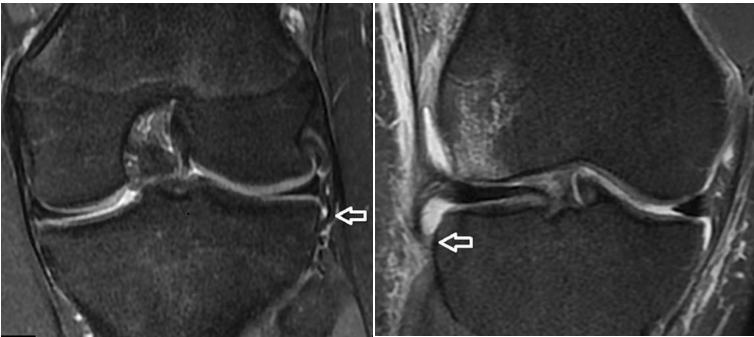
ACL tear is one of the most common knee injuries. Current reports on ACL reconstructive surgery mentioned the inability to reproduce the dynamic function of the native knee after surgery1. The pivot shift test is a physiologic test widely used by clinicians, and its high positivity is often associated with postoperative rotational instability of the knee, graft failure, and the subsequent need for revision surgery. There are many factors that contribute to axial knee instability, such as associated bony risk factors, peripheral ligament injuries, and meniscal injuries2. In current studies, 1.5T MRI was used as the tool of choice for identifying and assessing the injury rate of each factor3. However, there is still disagreement on the overall rate of visualization of fine structures. For example, in a study of assessing ALL visibility using 1.5T MRI, the ALL ligaments were visualized in 51%4. In this study, we utilized 3T MRI for enhanced detection and precise data and investigated high-risk factors associated with high-grade pivot shift in ACL injuries to aid orthopedic surgeons in refining surgical strategies for patients with high axial displacement, potentially improving surgical outcomes and reducing postoperative knee instability.Methods
Patients: A retrospective analysis was conducted on patients who underwent MRI diagnosis of ACL rupture and surgical treatment in our hospital from January 2016 to October 2022. After IRB-approved written informed consent was obtained, all subjects were scanned on 3T MRI (SIGNA Architect, GE Healthcare). Patients were grouped based on the results of tibial translation tests under general anesthesia performed by the same experienced physician. The tibial translation test was performed under general anesthesia before surgery and was classified as grade 0 to grade III according to the International Knee Documentation Committee (IKDC) consensus5. Grade 0 and grade I patients were divided into the normal group A (48 cases), grade II patients were assigned into the group B (60 cases), and grade III patients were divided into the group C (49 cases).Imaging parameters: The MR scan included coronal and sagittal T1 FSE (Auto TR=822ms, resolution=0.5×0.7×3.0mm3, TE=Min Full, FOV=160×160mm2, slices=18, BW=390.6Hz/Px), and three-plane PD-weighted imaging (Auto TR=800ms, resolution=0.6×0.6×0.6mm3, TE=Minimum, ETL=48, FOV=160×160mm2, slices=188, BW=466.4Hz/Px).
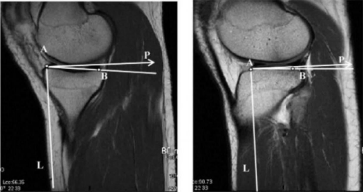
Data processing: Basic data (gender and age), MRI-based effects (including lateral and medial posterior tibial slope (LPTS and MPTS), lateral and medial collateral ligament injury (LCL and MCL), anterolateral ligament abnormalities (ALL)), and intraoperative lateral and medial meniscus injuries (LM and MM) were collected from each group. In all cases, the knee images were analyzed blind by two radiologists with at least 5 years of experience in diagnostic imaging of the musculoskeletal system. In cases of discordant conclusions between the two radiologists, a third radiologist of equal or greater experience was consulted for evaluation. The consensus of at least two out of the three radiologists was considered the final result.
Statistical analysis: The collected data were processed with the SPSS26.0 software and subjected to univariate and multivariate logistic regression analyses.
Results
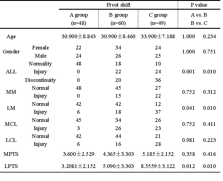
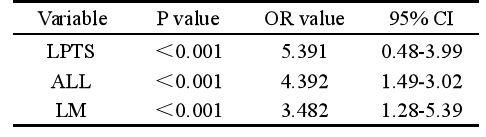
As shown in Table 1, the results showed that there was no significant correlation between the incidence of high pivot-shift ACL injury and gender, age, MPTS, LCL, MCL and MM injury in the three groups (P>0.05); and that there was a significant correlation between the incidence of high pivot-shift ACL injury and LPTS, ALL, and LM injury (P<0.05). As shown in Table 2, the logistic regression analysis showed that the incidence of ACL injury in patients with high tibial translation was significantly correlated with LPTS, ALL injury, and LM injury (P<0.05).Discussion
Previous studies have shown that patients with a high degree of axial displacement after acute ACL injury have a poorer postoperative prognosis and are more likely to have residual laxity of the knee6. However, the potential risk factors for high axial displacement are controversial. It has been shown that the potential risk factors for ACL injury are not solely attributable to the ACL itself, but may also be related to the "multiple mechanisms" of ACL injury7. Our study showed that there was a significant correlation between the incidence of high-grade pivot shift in ACL injury and LPTS, ALL, and LM injury.Conclusions
For ACL injuries, LPTS, ALL, and LM injuries may be high-risk factors leading to high-grade pivot shift.Acknowledgements
No acknowledgement found.References
1. Marshall T, Oak S R, Subhas N, et al. Can the Anterolateral Ligament Be Reliably Identified in Anterior Cruciate Ligament-Intact and Anterior Cruciate Ligament-Injured Knees on 3-T Magnetic Resonance Imaging? Orthop J Sports Med, 2018, 6(9): 2325967118796452.
2.Nielsen E T, Stentz-Olesen K, De Raedt S, et al. Influence of the Anterolateral Ligament on Knee Laxity: A Biomechanical Cadaveric Study Measuring Knee Kinematics in 6 Degrees of Freedom Using Dynamic Radiostereometric Analysis .Orthop J Sports Med, 2018, 6(8): 2325967118789699.
3.Tavlo M, Eljaja S, Jensen J T, et al. The role of the anterolateral ligament in ACL insufficient and reconstructed knees on rotatory stability: A biomechanical study on human cadavers .Scand J Med Sci Sports, 2016, 26(8): 960-6.
4. Puzzitiello R N, Agarwalla A, Zuke W A, et al. Imaging Diagnosis of Injury to the Anterolateral Ligament in Patients With Anterior Cruciate Ligaments: Association of Anterolateral Ligament Injury With Other Types of Knee Pathology and Grade of Pivot-Shift Examination: A Systematic Review. Arthroscopy, 2018, 34(9): 2728-38.
5. Helito P V P, Helito C P, Rodrigues M B. Anterolateral ligament MRI of the knee in ACL injuries: MRI abnormalities association with instability . European Radiology, 2022. 36(6): 1679-86.
6.Claes S, Bartholomeeusen S, Bellemans J. High prevalence of anterolateral ligament abnormalities in magnetic resonance images of anterior cruciate ligament-injured knees . Acta Orthopaedica Belgica, 2014, 80(1): 45-9
7. Helito P V P, Bartholomeeusen S, Claes S, et al. Magnetic Resonance Imaging Evaluation of the Anterolateral Ligament and the Iliotibial Band in Acute Anterior Cruciate Ligament Injuries Associated With Segond Fractures .Arthroscopy, 2020, 36(6): 1679-86.
Figures