4809
Improving PI-RADS rating with Zoomed Diffusion-Weighted Imaging in Deep Learning CAD Systems1Institute of Diagnostic and Interventional Radiology, Shanghai Sixth People’s Hospital Affiliated to Shanghai Jiao Tong University School of Medicine, Shanghai, China, 2Department of Radiology, Guangdong Provincial People’s Hospital (Guangdong Academy of Medical Sciences), Southern Medical University,, Guangzhou, China, 3Guangdong Provincial Key Laboratory of Artificial Intelligence in Medical Image Analysis and Application, Guangzhou, China, 4MR Application Development, Siemens Shenzhen Magnetic Resonance Ltd., Shenzhen, China, 5Digital Health, Siemens Healthineers Digital Technology (Shanghai) Co., Ltd., Shanghai, China, 6Digital Development, Siemens Digital Medical Technology (Shanghai) Co., Ltd., Shanghai, China, 7Digital & Automation, Siemens Shanghai Medical Equipment Ltd., Shanghai, China, 8DI CT Collaboration, Siemens Healthineers, Shanghai, China, 9MR Application Predevelopment, Siemens Healthineers AG, Erlangen, Germany, 10Digital & Automation Innovation, Siemens Healthcare GmbH, Erlangen, Germany, 11Department of Radiology, 989th Hospital of The Joint Logistic Support Force of the Chinese People's Liberation Army, Henan Province, China
Synopsis
Keywords: DWI/DTI/DKI, Prostate, Cancer; PI-RADS rating; DL-CAD; Zoom-DWI
Motivation: The clinical accuracy of the Prostate Imaging Reporting and Data System (PI-RADS) rating by deep-learning-based computer-aided diagnosis (DL-CAD) models need further enhancement for improved prostate cancer (PCa) detection and fewer unnecessary biopsies.
Goal(s): This study aimed to achieve more precise PI-RADS rating for PCa lesions by using zoomed diffusion-weighted imaging (z-DWI) in DL-CAD models.
Approach: We compared the diagnostic performance and PI-RADS rating of DL-CAD using advanced z-DWI vs. conventional DWI and extended this analysis to radiological practice.
Results: z-DWI improved the PI-RADS rating of PCa lesions by DL-CAD based on superior diagnostic performance compared with conventional DWI.
Impact: Deep-learning-based computer-aided diagnosis using zoomed diffusion-weighted imaging provides more accurate PI-RADS rating than conventional DWI, correlating MRI-detected lesions with prostate cancer (PCa) from biopsy. This can help minimize unnecessary biopsies for benign lesions while facilitating timely PCa treatment.
Introduction
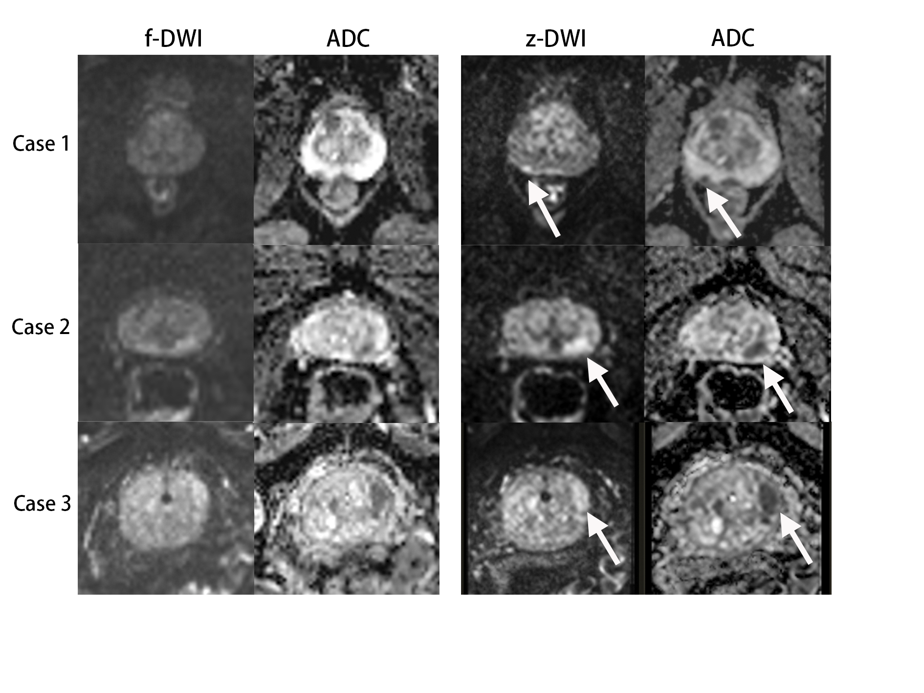
Deep-learning-based computer-aided diagnosis (DL-CAD) systems using magnetic resonance imaging (MRI) sequences have been gradually applied to prostate cancer (PCa) lesions detection [1-3].Diffusion-weighted imaging (DWI), as a dominant component of prostate MRI, provides functional information to detect tumor lesions [4, 5]. However, traditional full-field-of-view DWI (f-DWI), limited by distortions, susceptibility artifacts, and spatial resolution, results in low true positive rates and ambiguous rating in DL-CAD detection of PCa[6]. Recently, zoomed DWI (z-DWI), a new technique for DWI acquisition that only covers a specific region of interest has been proposed in order to reduce geometric distortions and susceptibility artifacts and to achieve higher spatial resolution. Previous studies have demonstrated the potential of z-DWI in enhancing the diagnostic performance of radiologists and radiomic models in detecting PCa lesions [7, 8]. This study aimed to (1) compare the diagnostic performance of DL-CAD based on f-DWI or z-DWI, (2) analyze the difference in PI-RADS rating provided by DL-CAD based on the 2 DWI techniques, and (3) extend this comparison to radiological practice to prove the clinical value of z-DWI.
Method
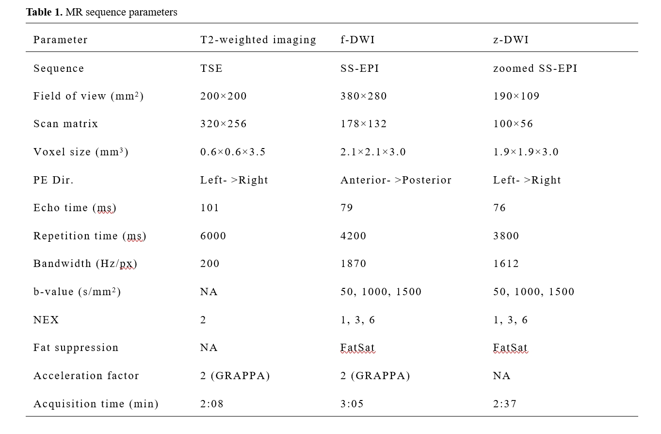
This study included 424 lesions from 306 patients undergoing MRI on a 3T system (MAGNETOM Skyra, Siemens Healthineers, Erlangen, Germany) for clinically suspected PCa, followed by ultrasound-guided targeted biopsies of suspicious lesions (PI-RADS category ≥3) and systematic biopsies. Inclusion criteria were complete MRI sequences, biopsy records, and clinical information. Exclusion criteria included (i) incomplete MRI sequences or severe artifacts; (ii) treatment history for PCa; (iii) biopsy performed within six months prior to MRI examination; (iv) ≥2 week interval between MRI and biopsy; (v) unavailability of the pathological diagnosis.f-DWI scans were acquired using conventional full FOV single-shot echo-planar imaging and z-DWI scans using a zoomed single-shot echo-planar imaging research application which enables acquisition of a reduced FOV while avoiding infolding artifacts due to a tilted 2D excitation scheme [9]. Detailed parameters of MRI sequences are presented in Table 1. MRI images were categorized into 2 groups: T2WI + f-DWI and T2WI + z-DWI, and uploaded to a research DL-CAD (MR Prostate AI v1.2.5; October 2020; Siemens Healthineers) for PCa detection and PI-RADS rating (3, 4, or 5). Two experienced radiologists, blinded to previous MRI reports and other clinical information, reinterpreted the MRI images together and gave PI-RADS rating for each lesion by using PI-RADS v2.1, based on the DL-CAD (f-DWI) and DL-CAD (z-DWI) results, respectively. To decrease recall bias, the images in the 2 groups were interpreted at 4-week intervals. The diagnostic performances of DL-CAD alone and radiologists using DL-CAD were evaluated by free-response receiver-operating characteristics (FROC) and alternative free-response receiver-operating characteristics (AFROC) analysis. The differences in PI-RADS categories between DL-CAD and radiologists using DL-CAD based on various DWI technologies were recorded. The 95% confidence interval (CI) and comparisons of areas under the curve (AUCs) were determined using the method proposed by DeLong et al [10].Results
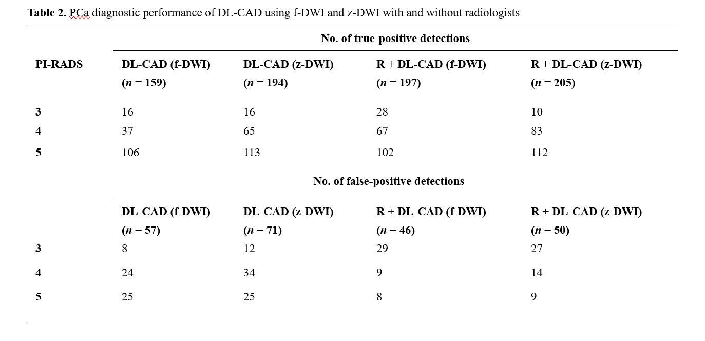
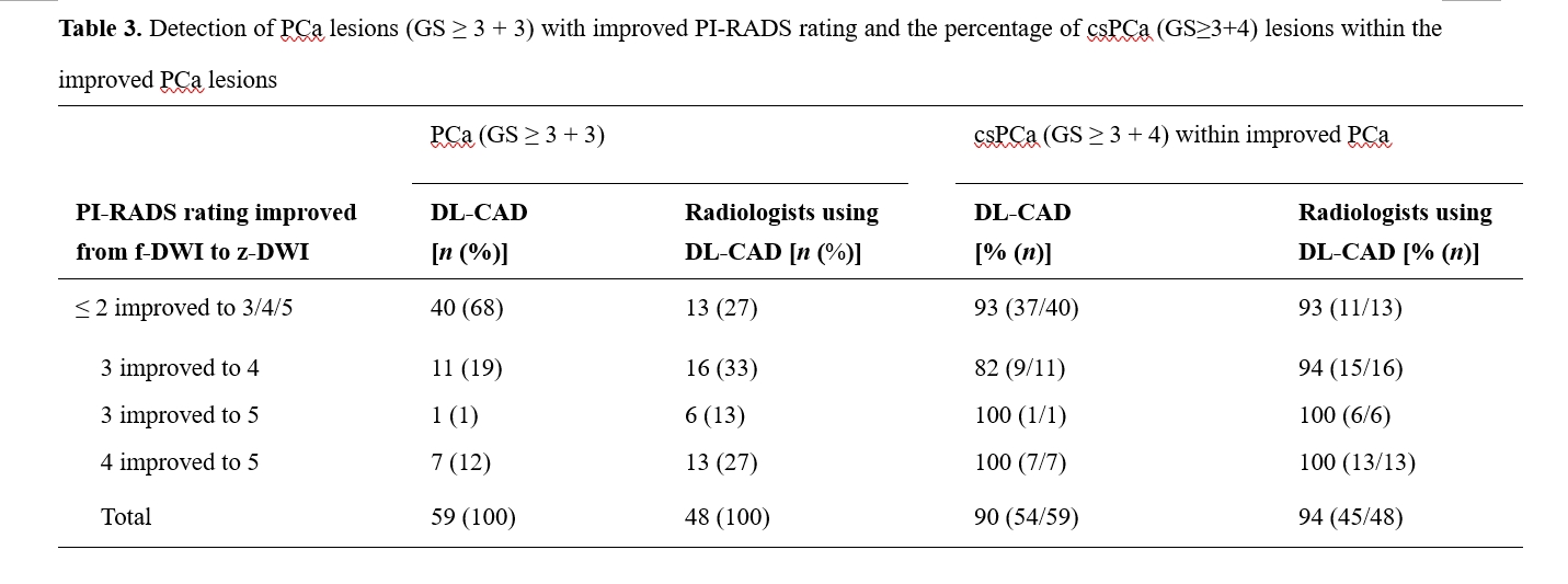
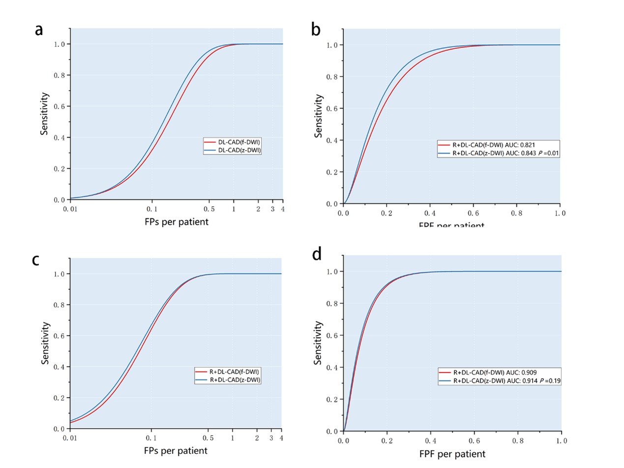
Compared with DL-CAD (f-DWI), DL-CAD (z-DWI) demonstrated a lower false-positive rate per patient at the same sensitivity and improved performance for detecting PCa lesions [AUC: 0.843 (95% CI: 0.823-0.860) vs 0.821 (95%CI: 0.797-0.842); P =.01]. Radiologists using DL-CAD (z-DWI) demonstrated enhanced diagnostic performance over those using DL-CAD (f-DWI), but with no statistically significant difference [AUC: 0.914 (95% CI: 0.900-0.925) vs 0.908 (95% CI: 0.90-0.92); P =.19] (see detailed results in Table 2, and the FROC and AFROC curves in Figure 1).Furthermore, z-DWI improved the PI-RADS rating by DL-CAD of 59 PCa lesions compared with f-DWI, with 90% of lesions being csPCa. Radiologists using DL-CAD (z-DWI) improved PI-RADS categories of 45 PCa lesions compared with those using DL-CAD (f-DWI), with 94% of lesions being csPCa (see Table 3).
Discussion
This preliminary study applied z-DWI in the evaluation of PCa by DL-CAD. The results suggested that z-DWI not only enhanced the diagnostic performance of DL-CAD and radiologists using DL-CAD but also improved the PI-RADS rating of PCa lesions, particularly for csPCa. However, no statistical significance was found between the performance of radiologists using DL-CAD based on different DWI technology. This might be due to the extensive experience of the radiologists. Hambrock et al. reported that the improvement in diagnostic performance brought by CAD is mainly reflected in inexperienced observers [11]. A precise PI-RADS rating of PCa lesions, especially for csPCa, prompts timely biopsy and treatment while reducing unnecessary biopsies.Conclusion
Zoomed DWI improved the PI-RADS rating of PCa lesions in DL-CAD based on superior diagnostic performance compared with conventional DWI. This can lead to a more precise assessment of PCa, thus prompting timely biopsy and treatment for patients while reducing unnecessary biopsies.Acknowledgements
This study was supported by the National Natural Science Foundation of China (82302130).References
1. Hiremath, A., et al., Test-retest repeatability of a deep learning architecture in detecting and segmenting clinically significant prostate cancer on apparent diffusion coefficient (ADC) maps. Eur Radiol, 2021. 31(1): p. 379-391.
2. Michaely, H.J., et al., Current Value of Biparametric Prostate MRI with Machine-Learning or Deep-Learning in the Detection, Grading, and Characterization of Prostate Cancer: A Systematic Review. Diagnostics (Basel), 2022. 12(4).
3. Winkel, D.J., et al., A Novel Deep Learning Based Computer-Aided Diagnosis System Improves the Accuracy and Efficiency of Radiologists in Reading Biparametric Magnetic Resonance Images of the Prostate: Results of a Multireader, Multicase Study. Invest Radiol, 2021. 56(10): p. 605-613.
4. Schiavina, R., et al., State-of-the-art imaging techniques in the management of preoperative staging and re-staging of prostate cancer. Int J Urol, 2019. 26(1): p. 18-30.
5. Turkbey, B., et al., Prostate Imaging Reporting and Data System Version 2.1: 2019 Update of Prostate Imaging Reporting and Data System Version 2. Eur Urol, 2019. 76(3): p. 340-351.
6. Kozlowski, P., S.D. Chang, and S.L. Goldenberg, Diffusion-weighted MRI in prostate cancer -- comparison between single-shot fast spin echo and echo planar imaging sequences. Magn Reson Imaging, 2008. 26(1): p. 72-6.
7. Hu, L., et al., Advanced zoomed diffusion-weighted imaging vs. full-field-of-view diff usion-weighted imaging in prostate cancer detection: a radiomic featur es study. European radiology. 31(3): p. 1760-1769.
8. Hu, L., et al., Better lesion conspicuity translates into improved prostate cancer detection: comparison of non-parallel-transmission-zoomed-DWI with conventional-DWI. Abdom Radiol (NY), 2021. 46(12): p. 5659-5668.
9. Finsterbusch, J., Improving the performance of diffusion-weighted inner field-of-view echo-planar imaging based on 2D-selective radiofrequency excitations by tilting the excitation plane. J Magn Reson Imaging, 2012. 35(4): p. 984-92.
10. DeLong, E.R., D.M. DeLong, and D.L. Clarke-Pearson, Comparing the areas under two or more correlated receiver operating characteristic curves: a nonparametric approach. Biometrics, 1988. 44(3): p. 837-45.
11. Hambrock, T., et al., Prostate cancer: computer-aided diagnosis with multiparametric 3-T MR imaging--effect on observer performance. Radiology, 2013. 266(2): p. 521-30.
Figures