4806
The value of QSM combined with free water imaging in the differentiation of early multisystem atrophy from Parkinson's disease1Shandong Provincial Hospital Affiliated to Shandong First Medical University, Jinan, China, 2Shanghai Key Laboratory of Magnetic Resonance, School of Physics and Electronic Science, East China Normal University, Shanghai, China, 3Shandong University, Jinan, China, 4Philips Healthcare, Shanghai, China
Synopsis
Keywords: Parkinson's Disease, Parkinson's Disease, Parkinson's disease; Multiple system atrophy; QSM; Free-water imaging
Motivation: We hope to provide some clinical assistance in distinguishing early Parkinson's disease from early multisystem atrophy.
Goal(s): In this study, we investigated alterations in iron deposition in deep grey matter nuclei and free water indicators in patients with early Parkinson's disease and early multisystem atrophy.
Approach: We manually sketched the putamen (PT), substantia nigra (SN) and dentate nucleus (DN) to obtain magnetic susceptibility values and free water imaging metrics for each nucleus.
Results: We found that DN plays an important role in identifying patients with early PD and early MSA.
Impact: This study suggests that QSM and free-water imaging are complementary tools to reveal pathophysiological differences in iron deposition and microstructural changes between early PD and early MSA. Therefore, the combination of both techniques may support differential clinical diagnosis further.
Introduction
Multiple system atrophy(MSA) and parkinson's disease (PD)are both alpha synucleinopathies with similar clinical presentations.This study aimed to examine the clinical value of quantitative susceptibility mapping (QSM) combined with free-water imaging in assessing the diagnostic efficacy of deep grey matter nuclei in the early stages of MSA and PD .Method
The study population consisted of 23 individuals with early MSA and 40 with early PD diagnosed in the last two years,alongside 33 age-matched healthy controls (HCs). T1-weighted, QSM, and diffusion tensor imaging (DTI) sequences were applied, respectively, and regions of interest (ROIs) for the putamen (PT), substantia nigra (SN), and dentate nucleus (DN) were manually outlined and compared for average susceptibility and diffusion coefficient.Results
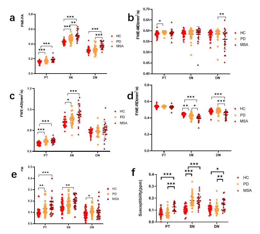
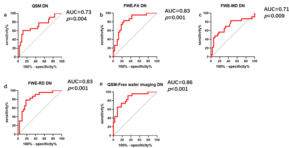
QSM data showed that SN susceptibility was higher in both early PD (0.1831±0.0495 p<0.001) and MSA (0.1797±0.0546 p<0.001) patients than HCs (0.1057±0.0328). In addition, magnetic susceptibility values in the PT (0.1263±0.0319) and DN (0.1502±0.0493) were higher in patients with early MSA than in HCs (PT: 0.0775±0.0321 p<0.001; DN: 0.1117±0.0328, p=0.025). However, magnetic susceptibility levels in the PT and DN were similar in the PD and HC groups. Magnetic susceptibility levels in the PT (0.1263±0.0319) and DN (0.1502±0.0493) were higher in MSA than in PD (PT: 0.075±0.0315, p<0.001; DN: 0.1112±0.0390, p=0.006). Free-water imaging data showed that free water corrected fractional anisotropy (FWE-FA) levels in the SN and DN were higher in early MSA cases (SN: 0.5168±0597; DN: 0.3839±0.0441) compared with early PD (SN: 0.4688±0.0440, p=0.006; DN: 0.3228±0.0506, P<0.001). FW-corrected mean diffusivity (FWE-MD) (0.5600±0.04687, p=0.009) and FW-corrected radial diffusivity (FWE-RD) (0.4395±0.0405, p<0.001) levels in the DN were lower in MSA compared with early PD (FWE-MD: 0.5853±0.02628; FWE-RD: 0.4829±0.03212). In addition, FWE-RD in the SN was higher in early PD cases (0.4234±0.0294) compared with early MSA patients (0.3985±0.0371, p=0.041). In contrast, no significant difference was found in the PT. Neither QSM nor Free-water imaging metrics differed based on disease stage. ROC curve analysis showed that combining QSM with free-water imaging revealed the DN as a good marker to differentiate early PD from MSA (p<0.001 AUC=0.856), and the diagnostic efficacy of the combination was greater than that of either sequence alone.Discussion
The excessive iron deposition induces oxidative stress and the production of free radicals, leading to neuronal cell death and demyelination, which may further enhance microglia proliferation and macrophage infiltration1-2. Our results suggest that a large amount of iron is deposited in the SN in both early PD and early MSA patients. In addition, excessive iron deposition was also observed in the striatal pathway region of the SN in some patients, which may further lead to apoptosis of dopamine neurons and neurotransmitter transport, inducing motor symptoms in patients with Parkinson's syndrome (PDS)3-4. Although MSA was not assessed in this study, higher iron deposition was still found in the PT and DN in early MSA compared with early PD, which is consistent with pathological findings and may further complement the specificity of these two grey matter nuclei in MSA5-7. In addition, according to previous studies8, neuroinflammatory demyelination may induce cell proliferation, and elevated FA and decreased RD in grey matter regions may represent a sign of gliosis9. Therefore, we hypothesized that increased FWE-FA and decreased FWE-RD in patients with early MSA may be associated with gliosis in these areas. However, similar changes were not found in the PT, while previous reports9-10 demonstrated that gliosis and demyelination in white matter regions may reduce FA values. The PT contains a large number of white matter fiber tracts, and significant demyelination is only detected in MSA among all types of Parkinson's syndrome. Because the α-synuclein of MSA is found mostly in oligodendrocytes as opposed to PD, which is found mainly in neurons, oligodendrocytes are the main factor affecting the formation of myelin11. Therefore, we speculate that the absence of significant alterations in the PT of patients with early MSA and PD may result from a complex interaction of grey and white matter gliosis and demyelination. Finally, no significant difference in FW was found between early PD and MSA in any of the three grey matter nuclei. This may be because patients with early disease were examined in this study.Conclusion
This study suggests that QSM and free-water imaging are complementary tools to reveal pathophysiological differences in iron deposition and microstructural changes between early PD and early MSA. Therefore, the combination of both techniques may provide further supporting references for differential clinical diagnosis.Acknowledgements
No acknowledgement found.References
1. Dusek, P., Hofer, T., Alexander, J., Roos, P. M. & Aaseth, J. O. Cerebral Iron Deposition in Neurodegeneration. Biomolecules 12, 714 (2022).
2. Ward, R. J., Dexter, D. T. & Crichton, R. R. Iron, Neuroinflammation and Neurodegeneration. IJMS 23, 7267 (2022).
3. Zecca, L., Zucca, F. A., Albertini, A., Rizzio, E. & Fariello, R. G. A proposed dual role of neuromelanin in the pathogenesis of Parkinson’s disease. Neurology 67, S8–S11 (2006).
4. Zucca, F. A. et al. Neuromelanin of the Human Substantia Nigra: An Update. Neurotox Res 25, 13–23 (2014)
5. Jellinger, K. A. Neuropathological spectrum of synucleinopathies. Mov Disord. 18, 2–12 (2003).
6. Dexter, D. T. et al. Alterations in the levels of iron, ferritin and other trace metals in Parkinson's disease and other neurodegenerative diseases affecting the basal ganglia. Brain 114, 1953–1975 (1991).
7. Matsusue, E. et al. Cerebellar Lesions in Multiple System Atrophy: Postmortem MR Imaging−Pathologic Correlations. AJNR Am J Neuroradiol 30, 1725–1730 (2009).
8. Wang, Y. et al. Quantification of increased cellularity during inflammatory demyelination. Brain 134, 3590–3601 (2011).
9. Budde, M. D., Janes, L., Gold, E., Turtzo, L. C. & Frank, J. A. The contribution of gliosis to diffusion tensor anisotropy and tractography following traumatic brain injury: validation in the rat using Fourier analysis of stained tissue sections. Brain 134, 2248–2260 (2011)
10. Yang, H. et al. Application of diffusion tensor imaging in multiple system atrophy: the involvement of pontine transverse and longitudinal fibers. International Journal of Neuroscience 125, 18–24 (2015).
11. Nasrabady, S. E., Rizvi, B., Goldman, J. E. & Brickman, A. M. White matter changes in Alzheimer’s disease: a focus on myelin and oligodendrocytes. acta neuropathol commun 6, 22 (2018).
Figures