4792
Comparison of multi-excitation and multi-frequency MR elastography to estimate anisotropic mechanical properties of the human brain.1Biomedical Engineering, University of Delaware, Newark, DE, United States, 2Département de Génie Mécanique, Université de Sherbrooke, Sherbrooke, QC, Canada, 3Thayer School of Engineering, Dartmouth College, Hanover, NH, United States, 4McKelvey School of Engineering, Washington University in St. Louis, St. Louis, MO, United States, 5Dartmouth-Hitchcock Medical Center, Lebanon, NH, United States
Synopsis
Keywords: Elastography, Elastography
Motivation: MR elastography can estimate anisotropic mechanical properties of fibrous white matter, traditionally using multi-excitation approaches. Multi-frequency elastography from a single driver is more common and could expand measurements of anisotropy.
Goal(s): Our goal was to compare mechanical anisotropy from multi-frequency and multi-excitation reconstructions.
Approach: Transversely isotropic parameters were reconstructed using simulated and in vivo multi-frequency wave data, then compared between approaches and against ground truth maps. Adolescent and adult measurements were compared in white matter regions.
Results: Multi-frequency elastography performed comparably with the multi-excitation approach in simulations. Higher shear anisotropy was observed in adults compared to adolescents, with no differences in tensile anisotropy.
Impact: This study demonstrates that multi-frequency magnetic resonance elastography can reliably estimate anisotropic mechanical properties from single driver data, enabling broader application. Quantifying developmental changes in anisotropy of white matter provides new insights into brain mechanics during maturation.
Introduction
Magnetic resonance elastography (MRE) noninvasively estimates mechanical properties of brain tissue that are sensitive to aging and disease1,2. We developed anisotropic MRE to estimate the properties of fibrous white matter tracts in the brain using transversely isotropic nonlinear inversion (TI-NLI)3-5. Traditionally we used a multi-excitation MRE approach (me-MRE)6–8 to generate waves for repeatable anisotropic inversions9–11. However, me-MRE requires using multiple drivers within the constraints of an MRI head coil. Alternatively, we can use multi-frequency MRE (mf-MRE) with a single driver to produce diverse wave fields for anisotropic property estimation12, as previously demonstrated by Guo et al. in leg muscles13. Here we aim to quantitatively compare the performance of TI-NLI with mf-MRE and me-MRE using a realistic brain MRE data simulation14 to confirm the ability to recover anisotropic parameters. We will apply mf-MRE with TI-NLI to publicly available datasets and calculate anisotropic properties for incorporation into brain biomechanics models15, allowing for comparison between adolescents (14-21 years) and adults (22-50 years) for the first time.Methods
Image AcquisitionMulti-frequency MRE data for adolescents (n = 18, age: 18.4±2.6) and adults (n = 18, age: 30.2±7.8) from a publicly available dataset15 included anterior-posterior (AP) direction displacements at 30/50/70 Hz with 1.5 mm3 resolution16,17. Auxiliary scans for all data sets included T1-weighted imaging at 0.9 mm3, and diffusion tensor imaging at 1.5 mm3 from which the primary diffusion direction was estimated.
In vivo Multi-frequency and TI-NLI Inversion
For in vivo TI-NLI, we combined AP wave motion fields from mf-MRE and the primary eigenvector from diffusion, which informs the local fiber direction, to recover the complex shear modulus (G*=G’+iG”), shear anisotropy (φ=G1/G2-1), and tensile anisotropy (ζ=E1/E2–1). Anisotropy maps were averaged in the corpus callosum body, forceps major and minor, corona radiata, and the corticospinal tract. We analyzed group differences within each region via two-sample t-tests.
FE Brain Simulation
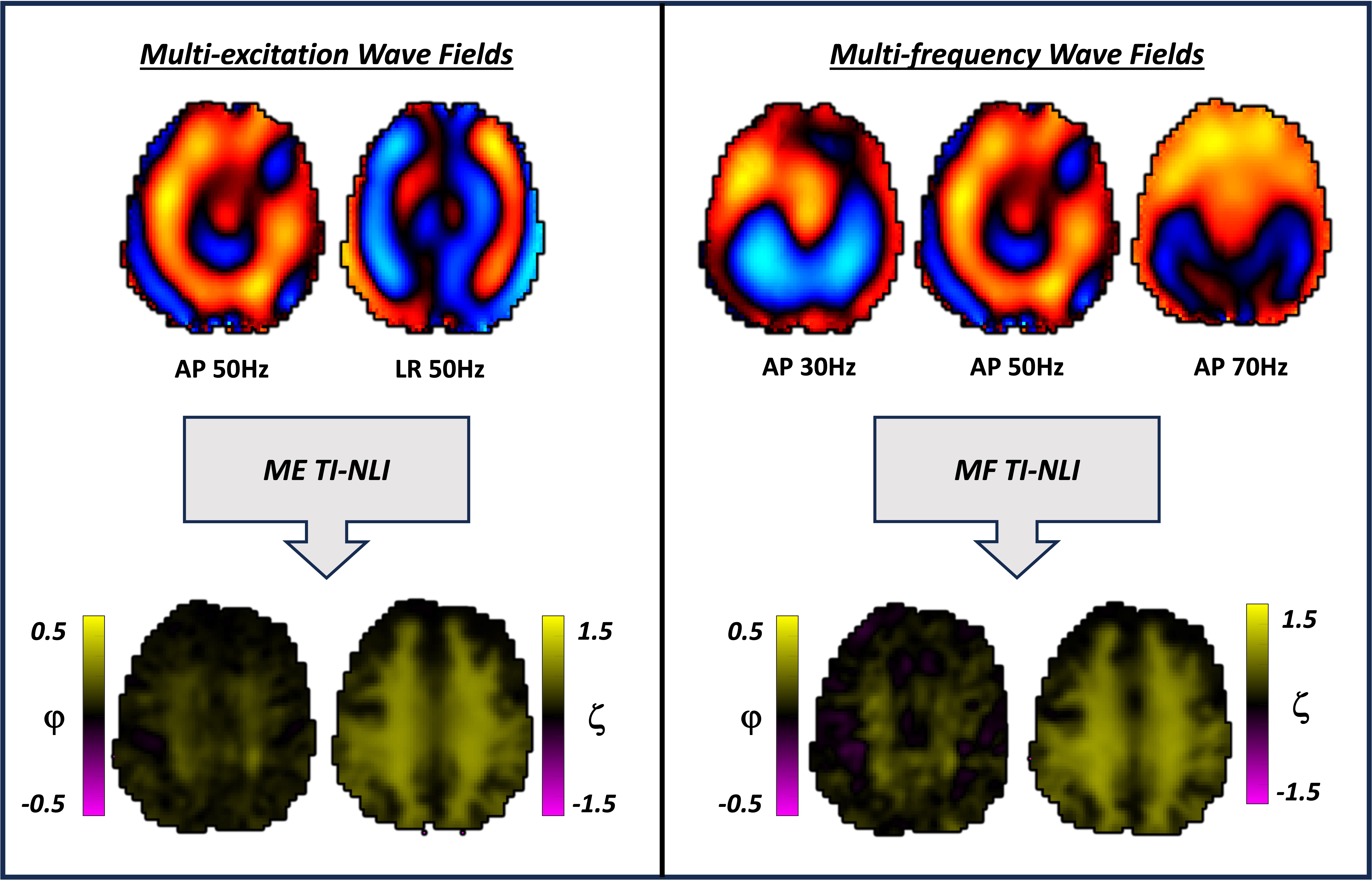
A FE simulation platform14 was adapted to compare the reconstruction accuracy of TI-NLI using mf-MRE versus me-MRE. A participant dataset including MRE at 30, 50, and 70 Hz in the AP direction and 50 Hz in the left-right (LR) direction was used to generate distinct realistic wavefields from assigned properties in brain regions of interest: whole brain gray matter (GM) and white matter (WM), ventricles, CSF, subcortical GM, and WM tracts. Regions were assigned values for shear moduli, shear anisotropy (φ), and tensile anisotropy (ζ) from adult MRE data at 50 Hz9. Shear moduli for 30/70 Hz were interpolated via linear power law18. A forward simulation of the nearly incompressible TI model produced wave fields for TI-NLI (Figure 1A-B). Differences between mf-MRE and me-MRE reconstructions, and ground truth maps, were estimated as the median of the absolute difference between images.
Results
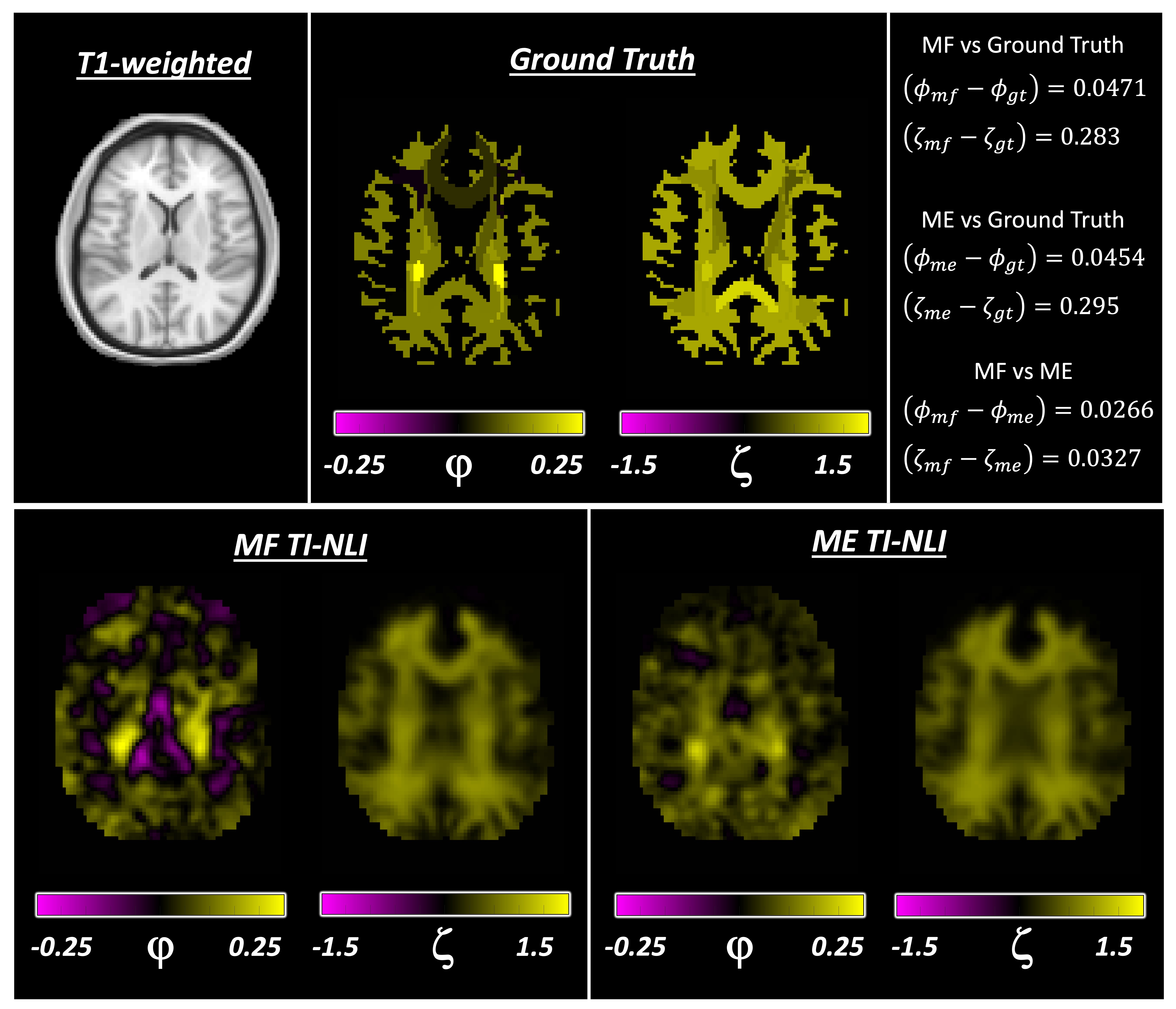
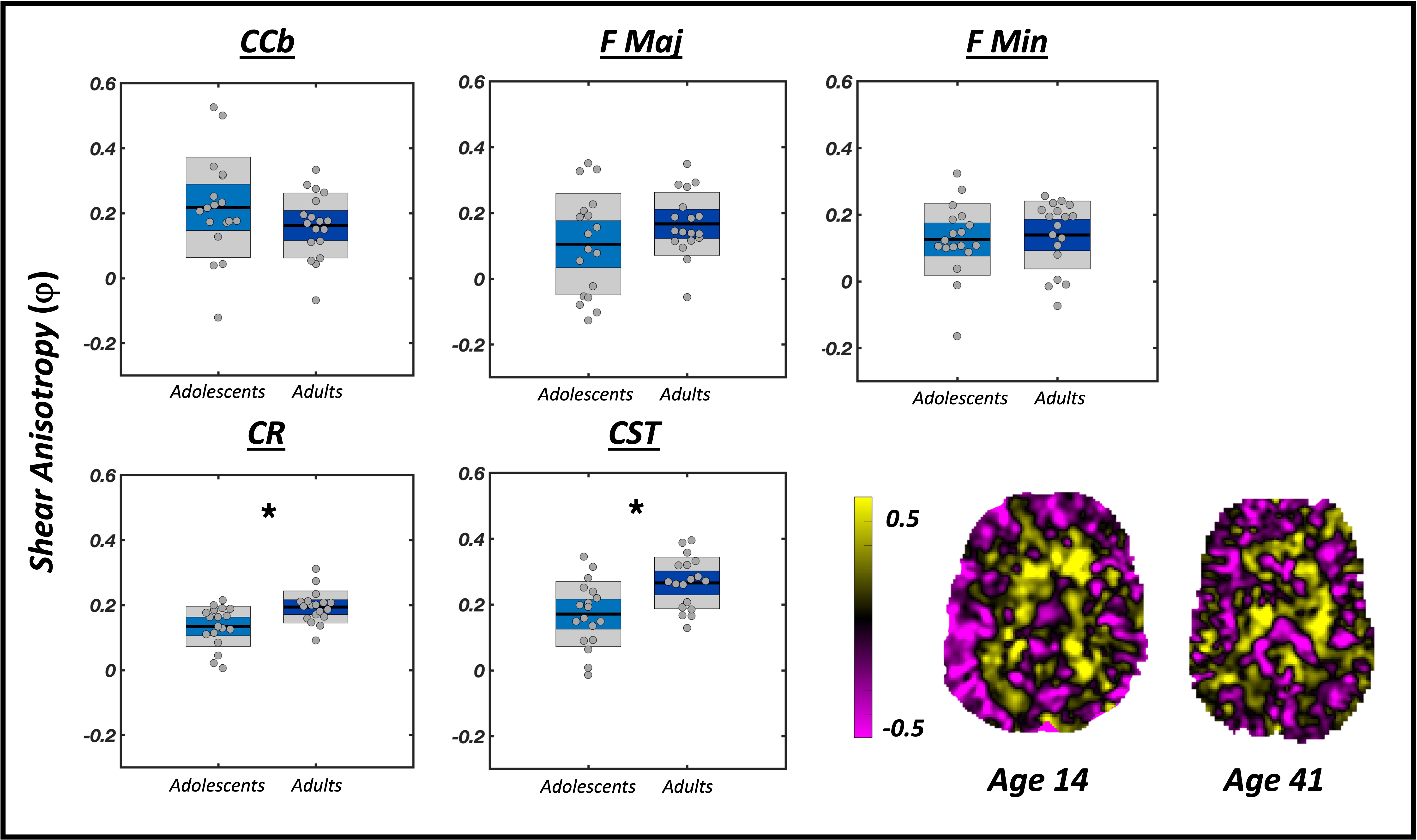
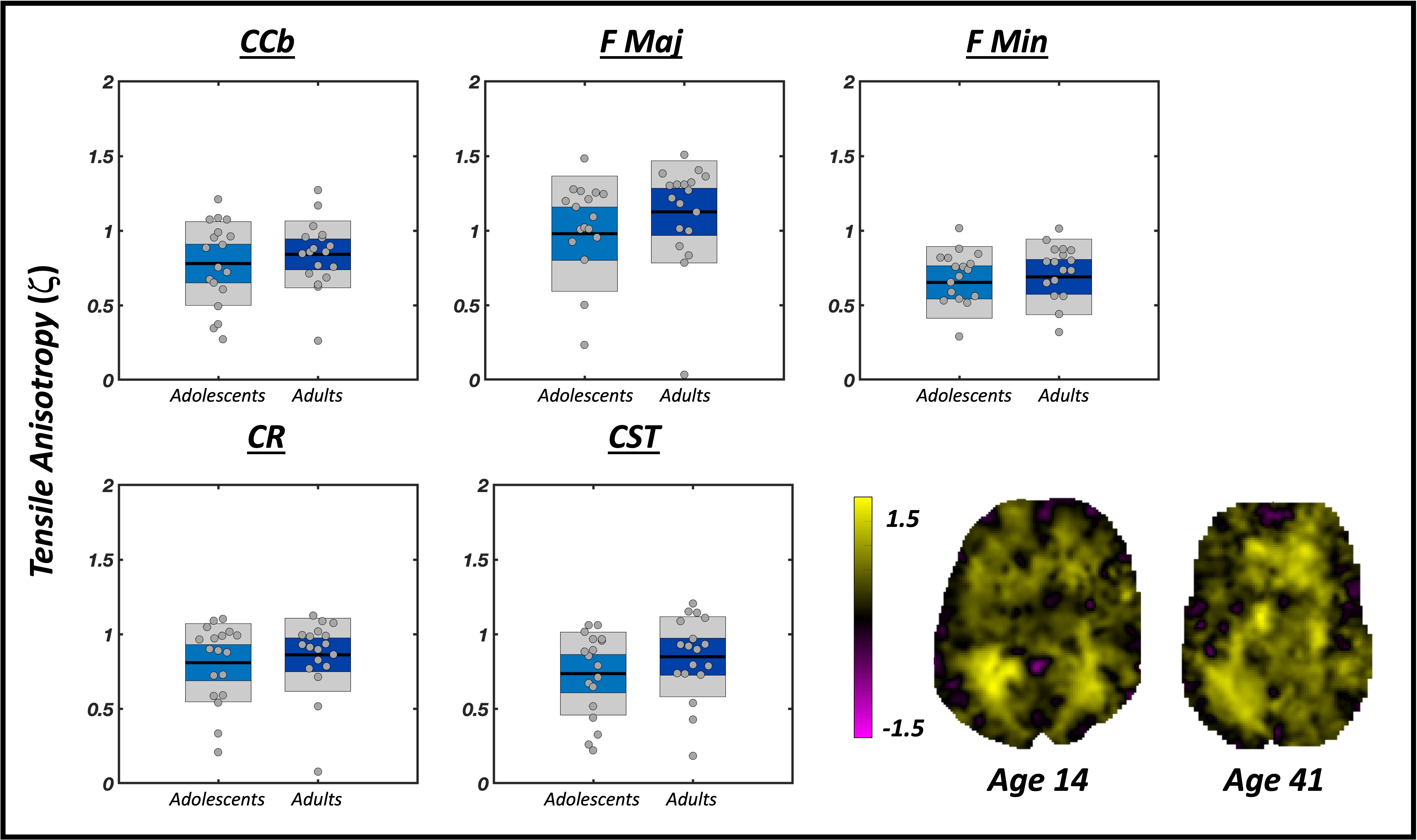
Multi-frequency MRE inversion of simulated wave fields produced TI property maps consistent with me-MRE inversion (Figure 2) yielding positive shear and tensile anisotropy throughout the white matter. Compared to ground truth, simulated mf-MRE reconstructions had a median difference of 0.0417 and 0.283 for φ and ζ respectively, while me-MRE reconstructions had median differences of 0.0454 and 0.295 for φ and ζ respectively. In vivo mf-MRE reconstructions of shear anisotropy in the corona radiata and corticospinal tract were significantly higher in adults (φadults = CR: 0.19±0.05, CST: 0.27±0.08) compared to adolescents (φadolescents = CR: 0.14±0.06, CST: 0.17±0.09) (Figure 3). Tensile anisotropy was not significantly different between groups (Figure 4).Discussion and Conclusions
Wave data at multiple frequencies from a single driver was used to recover anisotropic mechanical properties in the human brain using TI-NLI, with comparable accuracy to me-MRE. Mechanical anisotropy parameters from in vivo adolescent and adult mf-MRE scans were within the range of previous measurements in young adults9. Existing measurements of shear stiffness in adolescents show differences in their mechanical properties and microstructure compared to adults19. We observed significant age-related differences in shear anisotropy in the corona radiata and corticospinal tract. Higher shear anisotropy in adulthood could reflect changes related to development in oligodendrocyte density and myelination, likely driving increased shear stiffness changes along the fiber direction. Conversely, no measured differences in tensile anisotropy may indicate primary sensitivity of this parameter to only axonal changes in microstructure due to development. In longitudinal porcine brain measurements, tensile anisotropy declined partially with age, which was not observed in our group measurements11. Interestingly, our previous work found the greatest reductions in tensile anisotropy in older adults10, highlighting different mechanical dynamics across the lifespan. Overall, mf-MRE enables reliable estimation of anisotropic mechanical properties, facilitating these measurements at more sites without customized driver hardware.Acknowledgements
This research was supported by NIH grants R01-EB027577, R01-AG058853, U01-NS112120, and Office of Naval Research grant N00014-22-1-2198.
References
1. Hiscox L V, Johnson CL, Barnhill E, et al. Magnetic resonance elastography (MRE) of the human brain: technique, findings and clinical applications. Phys Med Biol. 2016;61(24):R401-R437. doi:10.1088/0031-9155/61/24/R401.
2. Murphy MC, Huston J 3rd, Ehman RL. MR elastography of the brain and its application in neurological diseases. Neuroimage. 2019;187:176-183. doi:10.1016/j.neuroimage.2017.10.008
3. McGarry MDJ, Van Houten EEW, Sowinski DR, et al. Model-Based Heterogeneous Transverse Isotropic MR Elastography Inversion for Brain Tissue with Aligned Fiber Tracts. 28th Annu Meet Int Soc Magn Reson Med.
4. McGarry MDJ, Van Houten E, Guertler C, et al. A heterogenous, time harmonic, nearly incompressible transverse isotropic finite element brain simulation platform for MR elastography. Phys Med Biol. June 2020. doi:10.1088/1361-6560/ab9a84
5. McGarry M, Van Houten E, Sowinski D, et al. Mapping Heterogeneous Anisotropic Tissue Mechanical Properties with Transverse Isotropic Nonlinear Inversion MR Elastography. Med Image Anal. 2022;78:102432. doi:10.1016/j.media.2022.102432
6. Anderson AT, Van Houten EEW, McGarry MDJ, et al. Observation of direction-dependent mechanical properties in the human brain with multi-excitation MR elastography. J Mech Behav Biomed Mater. 2016;59:538-546. doi:10.1016/j.jmbbm.2016.03.005
7. Smith D, Guertler CA, Okamoto RJ, Romano A, Bayly P V, Johnson CL. Multi-Excitation MR Elastography of the Brain: Wave Propagation in Anisotropic White Matter. J Biomech Eng. 2020;142(7):710051-710059. doi:10.1115/1.4046199
8. Caban-Rivera DA, Smith DR, Kailash K, et al. Multi-Excitation Actuator Design for Anisotropic Brain MRE. 29th Annu Meet Int Soc Magn Reson Med May 15-20, 2021. 2021.
9. Smith DR, Caban-Rivera DA, Mcgarry MDJ, et al. Anisotropic mechanical properties in the healthy human brain estimated with multi-excitation transversely isotropic MR elastography. Brain Multiphysics. 2022;3(June):100051. doi:10.1016/j.brain.2022.100051
10. Caban-Rivera DA, Smith DR, McGarry MD, et al. Anisotropic Mechanical Properties of White Matter Tracts in Aging via Transversely Isotropic MR Elastography. 30th Annu Meet Int Soc Magn Reson Med. 2022.
11. Wang S, Guertler CA, Okamoto RJ, Johnson CL, McGarry MDJ, Bayly P V. Mechanical stiffness and anisotropy measured by MRE during brain development in the minipig. Neuroimage. 2023;277(June):120234. doi:10.1016/j.neuroimage.2023.120234
12. Tweten DJ, Okamoto RJ, Bayly P V. Requirements for accurate estimation of anisotropic material parameters by magnetic resonance elastography: A computational study. Magn Reson Med. 2017;78(6):2360-2372. doi:10.1002/mrm.26600
13. Guo J, Hirsch S, Scheel M, Braun J, Sack I. Three-parameter shear wave inversion in MR elastography of incompressible transverse isotropic media: Application to in vivo lower leg muscles. Magn Reson Med. 2016;75(4):1537-1545. doi:10.1002/MRM.25740
14. McGarry M, Houten E Van, Guertler C, et al. A heterogenous, time harmonic, nearly incompressible transverse isotropic finite element brain simulation platform for MR elastography. Phys Med Biol. 2021;66(5):1-19. doi:10.1088/1361-6560/ab9a84
15. Bayly P V., Alshareef A, Knutsen AK, et al. MR Imaging of Human Brain Mechanics In Vivo: New Measurements to Facilitate the Development of Computational Models of Brain Injury. Ann Biomed Eng. 2021;49(10):2677-2692. doi:10.1007/s10439-021-02820-0
16. Johnson CL, Holtrop JL, Anderson AT, Sutton BP. Brain MR Elastography with Multiband Excitation and Nonlinear Motion-Induced Phase Error Correction. 24th Annu Meet.
17. McIlvain G, Cerjanic AM, Christodoulou AG, McGarry MDJ, Johnson CL. OSCILLATE: A low-rank approach for accelerated magnetic resonance elastography. Magn Reson Med. 2022;88(4):1659-1672. doi:10.1002/MRM.29308
18. Testu J, McGarry MDJ, Dittmann F, et al. Viscoelastic power law parameters of in vivo human brain estimated by MR elastography. J Mech Behav Biomed Mater. 2017;74:333-341. doi:10.1016/J.JMBBM.2017.06.027
19. McIlvain G, Schwarb H, Cohen NJ, Telzer EH, Johnson CL. Mechanical properties of the in vivo adolescent human brain. Dev Cogn Neurosci. 2018;34:27-33. doi:10.1016/j.dcn.2018.06.001
Figures